báo cáo hóa học:" Vascular endothelial growth factor regulates osteoblast survival – evidence for an autocrine feedback mechanism" pptx
Bạn đang xem bản rút gọn của tài liệu. Xem và tải ngay bản đầy đủ của tài liệu tại đây (725.39 KB, 13 trang )
BioMed Central
Page 1 of 13
(page number not for citation purposes)
Journal of Orthopaedic Surgery and
Research
Open Access
Research article
Vascular endothelial growth factor regulates osteoblast survival –
evidence for an autocrine feedback mechanism
John Street*
1,2
and Brian Lenehan
1,2
Address:
1
Department of Orthopedic Surgery, National University of Ireland, Cork, Ireland and
2
Combined Neurosurgical and Orthopedic Spine
Program, University of British Columbia, Vancouver, BC, Canada
Email: John Street* - ; Brian Lenehan -
* Corresponding author
Abstract
Background: Apoptosis of osteoblasts and osteoclasts regulates bone homeostasis. Skeletal injury
in humans results in 'angiogenic' responses primarily mediated by vascular endothelial growth
factor(VEGF), a protein essential for bone repair in animal models. Osteoblasts release VEGF in
response to a number of stimuli and express receptors for VEGF in a differentiation dependent
manner. This study investigates the putative role of VEGF in regulating the lifespan of primary
human osteoblasts(PHOB) in vitro.
Methods: PHOB were examined for VEGF receptors. Cultures were supplemented with VEGF(0–
50 ng/mL), a neutralising antibody to VEGF, mAB VEGF(0.3 ug/mL) and Placental Growth Factor
(PlGF), an Flt-1 receptor-specific VEGF ligand(0–100 ng/mL) to examine their effects on mineralised
nodule assay, alkaline phosphatase assay and apoptosis The role of the VEGF specific antiapoptotic
gene target BCl2 in apoptosis was determined.
Results: PHOB expressed functional VEGF receptors. VEGF 10 and 25 ng/mL increased nodule
formation 2.3- and 3.16-fold and alkaline phosphatase release 2.6 and 4.1-fold respectively while 0.3
ug/mL of mAB VEGF resulted in approx 40% reductions in both. PlGF 50 ng/mL had greater effects
on alkaline phosphatase release (103% increase) than on nodule formation (57% increase). 10 ng/
mL of VEGF inhibited spontaneous and pathological apoptosis by 83.6% and 71% respectively, while
PlGF had no significant effect. Pretreatment with mAB VEGF, in the absence of exogenous VEGF
resulted in a significant increase in apoptosis (14 vs 3%). VEGF 10 ng/mL increased BCl2 expression
4 fold while mAB VEGF decreased it by over 50%.
Conclusion: VEGF is a potent regulator of osteoblast life-span in vitro. This autocrine feedback
regulates survival of these cells, mediated via a non flt-1 receptor mechanism and expression of
BCl2 antiapoptotic gene.
Introduction
Bone is a complex, dynamic and highly specialized tissue
that undergoes continuous regeneration and remodeling
throughout life. Deposition and resorption of mineral-
ized matrix occurs during development and growth, dur-
ing physiological adult skeletal remodeling and during
repair of surgically or traumatically injured bone. Appro-
priate blood supply, and intricate coupling of the vascula-
Published: 16 June 2009
Journal of Orthopaedic Surgery and Research 2009, 4:19 doi:10.1186/1749-799X-4-19
Received: 26 February 2009
Accepted: 16 June 2009
This article is available from: />© 2009 Street and Lenehan; licensee BioMed Central Ltd.
This is an Open Access article distributed under the terms of the Creative Commons Attribution License ( />),
which permits unrestricted use, distribution, and reproduction in any medium, provided the original work is properly cited.
Journal of Orthopaedic Surgery and Research 2009, 4:19 />Page 2 of 13
(page number not for citation purposes)
ture with osteoblasts and osteoclasts is a prerequisite to
regulation of this formation and removal of bone. Blood
vessel formation, angiogenesis, and blood vessel removal,
pruning, are strictly coordinated to facilitate the ever-
changing demands of the skeleton. Within the temporary
functioning structure of the basic multicellular unit
(BMU), osteoblasts mediate bone formation, osteoclasts
bone resorption, while both cells share intimate proxim-
ity with the vascular endothelium and haemopoietic and
stromal cells of the bone marrow. These BMU's represent
the spatial and temporal orchestration of the strictly con-
trolled activities of osteoblasts, osteoclasts and cells of the
vascular tree. The function of these cells is regulated by a
number of systemic and local factors that modulate bone
metabolism and vasclarization [1]. The systemic factors
include parathyroid hormone, growth hormone, Vitamin
D3, glucocorticoids, calcitonin and numerous vasoactive
peptides. Local soluble factors known to enhance the for-
mation of mineralized matrix include the insulin-like
growth factors (IGF-I and -II), transforming growth factor
beta (TGFβ), platelet derived growth factor (PDGF) and
basic fibroblast growth factor (bFGF). Cytokines that
enhance osteoclast function and bone resorption include
interleukin-1 (IL-1), interleukin-6 (IL-6) and tumor
necrosis factor alpha (TNFα) [2]. The principle 'ang-
iogenic' cytokines that regulate blood vessel formation are
vascular endothelial growth factor (VEGF), bFGF, PDGF,
TGFβ, TNFα and angiopoietin-1 (Ang-I). Clearly the activ-
ities of many of these factors are common to the regula-
tion of bone forming, bone resorbing and endothelial
cells. Of these factors, vascular endothelial growth has
been the focus of most recent interest [3]. This dimeric
glycoprotein, with a molecular weight range from 17 to 22
kDa, has several isoforms with very similar biological
activities. For a long time, VEGF was considered endothe-
lial cell specific, however recent reports have confirmed
the presence of VEGF receptors, flt-1 and/or KDR on
numerous other cell types, including osteoblasts [4]. Pla-
centa Growth Factor is another angiogenic protein specif-
ically of the VEGF family. This protein is known to bind to
Flt-1 receptor with high affinity but fails to bind the KDR
VEGF receptor [5]. Recent studies have demonstrated that
the mitogenic and antiapoptotic effects of the VEGF pro-
teins on endothelial cells are mediated through specific
receptors [5]. We have reported that isolated skeletal
injury in humans results in local and systemic 'angiogenic'
responses primarily mediated by VEGF [6,7]. VEGF has
been identified as essential for bone repair in animal
models [8], and is a prerequisite to hypertrophic cartilage
removal and ossification during murine skeletal growth
[3,5,9]. Osteoblasts may release VEGF in response to a
number of stimuli, including myriad bone derived
cytokines and hypoxia, simulating bone injury [[10-
15]ejost]. Osteoblasts also express receptors for VEGF in a
differentiation dependent manner [4]. Meanwhile osteo-
clasts express VEGF receptors and osteoclast differentia-
tion and bone resorption is enhanced by VEGF in
osteopetrotic mice in the absence of macrophage colony
stimulating factor (MCSF) [16]. Whether VEGF has any
direct effects on osteoblast activity or life span, and which
receptors may be specific for this signal transduction is
unknown.
The life-span of a BMU far exceeds that of the composite
cells and so continuous turnover of these cells is manda-
tory for skeletal homeostasis [1,2]. The average bone
forming life-span of an osteoblast is 10 – 14 weeks, at
which time the alternative two fates are either to become
buried within the lacunae of mineralized matrix as an
osteocyte, or to become an elongated lining cell on the
quiescent unmineralized surface of bone. Examination of
human bone reveals that approximately 65% of the oste-
oblasts initially present within a BMU cannot be
accounted for after enumeration of lining cells and osteo-
cytes. These cells have most likely died by apoptosis, or
programmed cell death, and been rapidly phagocytosed,
and are thus 'missing' [2]. Indeed apoptotic cell death of
osteoclasts and osteoblasts is a key regulator of the bal-
ance between bone formation and resorption in an active
BMU [17]. While the rate of osteoblast programmed cell
death in active seams of normal human bone is extremely
uncommon, significantly increased osteoblast and osteo-
cyte apoptosis characterize various pathological condi-
tions e.g. postmenopausal osteoporosis, glucocorticoid
induced osteopenia, rheumatoid and septic periarticular
osteoporosis and avascular necrosis [2,18-23]. Increased
turnover of bone forming cells is also seen within normal
fracture callus [24], a temporally dependent phenomenon
which can be modulated by the exogenous administration
of IL-1β and TGFβ. As each of these physiological and
pathological conditions of bone are inexorably linked
with alterations and perturbations in skeletal vasculariza-
tion one could conceptualize at least a role for the vascu-
lature, and for angiogenic cytokines in particular, in
modulating osteoblast life-span within the BMU.
The aim of this study, therefore, was to elucidate the
mechanisms of apoptosis in primary human osteoblasts
and to examine the effects of numerous angiogenic
cytokines on osteoblast life-span. In particular we investi-
gate the direct effects of vascular endothelial growth factor
and its Flt-1 specific mutant PlGF on osteoblast differenti-
ation, bone formation and apoptosis.
Methods
Study Design
Primary human osteoblast cultures are examined for
expression of VEGF receptors and subsequently supple-
mented with VEGF 165, a neutralising antibody to VEGF
and PlGF to examine the direct mitogenic effects of these
Journal of Orthopaedic Surgery and Research 2009, 4:19 />Page 3 of 13
(page number not for citation purposes)
angiogenic proteins by mineralised nodule and alkaline
phosphatase assays. The effects of these receptor specific
VEGF family members on steoblast apoptosis is then
determined and their mechanism of 'survival protection'
elucidated. The role of the VEGF specific antiapoptotic
gene target BCl2 is then determined by osteoblast cell
transfection.
Primary Human Osteoblast Cultures
Primary normal human osteoblasts were cultured from
trabecular bone explants obtained at the time of ortho-
paedic procedures performed on consenting young adults
who had no evidence of metabolic bone disease. The bone
fragments were washed extensively and repeatedly with
culture medium to remove adherent marrow cells and to
expose the trabecular surface of the bone. Small bone
chips (1 × 1 × 1 mm) were then placed in culture flasks
(75 cm
2
), each containing 15 mls α modified Earle's
medium supplemented with 10% heat inactivated fetal
calf serum, penicillin (100 U/mL), streptomycin (50 μg/
mL; α MEM – 10% FCS) and cultured at 37°C in a humid-
ified atmosphere with 5% CO
2
. Cell outgrowth from the
trabecular bone surfaces was apparent after 5 days, and
the osteoblast-like cells became confluent after 10 – 14
days of culture. Verification of osteoblast lineage was per-
formed by mineralised bone nodule formation assay,
alkaline phosphatase activity of the cell lysate using
sodium p-nitrophenyl phosphate substrate and by FACS
analysis for osteocalcin. Cell passages were performed by
incubating confluent cells 0.25% trypsin diluted in cal-
cium and magnesium free phosphate buffered saline.
Experiments were performed on osteoblasts subcultured
to passage 3 – 6.
Human osteoblast cell line culture
The primary human osteoblast cultures described above
were used for n = 3 experiments, each in triplicate, for each
step of the study design. Their use was limited because of
the difficulty in obtaining specimens and of the technical
difficulty in harvesting and culture. For the remainder of
the experiments the primary human osteoblast cell line
NHOst (Clonetics, San Diego, California, USA) was used.
These cells have not been transformed and so have a lim-
ited lifespan in culture. Preliminary studies confirmed
that there were no significant differences in activity, recep-
tor expression, cytokine release and response to ang-
iogenic factors between the two cultures of osteoblasts.
The NHOst cell line were cultured in osteoblast basal
medium (OBM™) supplemented with ascorbic acid, fetal
bovine serum and gentamicin/amphotericin-B as per the
manufacturers protocol. Cell passages were performed by
incubating confluent cells 0.25% trypsin diluted in cal-
cium and magnesium free phosphate buffered saline.
Experiments were performed on osteoblasts subcultured
to passage 3 – 6.
Reagents, assay kits and recombinant proteins and antibodies
All reagents were purchased from Sigma Chemical Co. (St.
Louis, Missouri, USA) unless otherwise stated. Adult nor-
mal human osteoblasts (NHOst) and NHOst culture
media and detatchement kits were purchased from
Clonetics (Walkersville, Maryland, USA). Recombinant
human proteins vascular endothelial growth factor, basic
fibroblast growth factor, insulin-like growth factor-1,
platelet derived growth factor, placenta growth factor and
tumor necrosis factor alpha were purchased from R&D
Systems (Minneapolis, Minnesota, USA). Neutralising
antibody to VEGF and the isotype control antibody and
the Human VEGF Biotinylated Fluorokine kit were also
purchased from R&D Systems (Minneapolis, Minnesota,
USA). The CD 95 ligand anti-APO-1/Fas monoclonal anti-
body was purchased from Boehringer Ingelheim Bioprod-
ucts Partnership (Heidelberg, Germany).
Analysis Of VEGF binding by Osteoblasts
Primary human osteoblasts were trypsinized from 75
mm
2
flasks (Falcon) and returned in round bottomed
polypropelene tubes (Falcon) to 37°C for 6 hours to
allow regeneration of cell surface receptors (recovery
period). Cells were harvested by centrifugation at 500 × g
for 5 minutes and then washed twice with PBS to remove
any residual growth factors that may be present in the cul-
ture medium. Cells were resuspended in PBS to a final
concentration of 4 × 10
6
cells/mL. 10 μL of biotinylated
VEGF reagent (4.5 μg/mL) was added to 25 μL of the
washed cell suspension in a 12 × 75 mm tube. As a nega-
tive control, an identical sample of cells was stained with
10 μL of biotinylated negative control reagent (soybean
trypsin inhibitor at 5 μg/mL). The cells were then incu-
bated at 4°C for 2 hours at which time 10 μL of avidin-
FITC reagent was added. The cell suspension was further
incubated at 4°C in the dark for 30 minutes. The cells
were then washed twice with 2 mL of buffered saline-pro-
tein solution to remove unbound avidin-fluorescein and
resuspended in 200 μL of buffered saline-protein solution
for flow cytometric analysis of VEGF binding. This assay
quantitatively determines the percentage of osteoblasts
expressing biologically functional VEGF receptors within
a population and estimates the receptor density for VEGF
on cell surfaces.
Bone Nodule Formation
Human Osteoblasts were seeded in 6-well plates at a den-
sity of 1 × 10⁵ cells/mL and cultured as described above.
Upon confluence (48–72 hours after plating), 50 ug/mL
ascorbic acid was added to the cultures. The cell cultures
were then supplemented with recombinant human VEGF
165 (0 – 50 ng/mL), PlGF (0 – 100 ng/mL) or a mono-
clonal mouse anti-human VEGF neutralizing antibody
(0.3 ug/mL). The treated medium was replenished daily.
Mineralised nodules began to appear by 3–8 days at
Journal of Orthopaedic Surgery and Research 2009, 4:19 />Page 4 of 13
(page number not for citation purposes)
which time the medium was further supplemented with
[beta]- glycerol phosphate and ascorbic acid to further
stimulate osteogenic differentiation. After 18 days in cul-
ture, the number of mineralized bone nodules was quan-
tified by von Kossa staining. All cultures were performed
in triplicate and six fields per culture well were counted.
Alkaline Phosphatase Assay
Alkaline phosphatase activity in the osteoblast culture sys-
tem was determined by measuring cell supernatant
hydrolysis of p-nitrophenyl phosphate, yielding p-nitro-
phenol, which when alkaline is converted to a yellow
complex easily measured by spectophotometric analysis
at 400–420 nm (Sigma Diagnostics).
Annexin V- Fluorescein Isothiocyanate Labeling
Primary human osteoblasts were plated into six well
dishes in serum free culture medium and allowed to
become adherent for 8 hours. Cell cultures were then
treated for 6 hours with TNFα (0 – 1000 ng/mL) in the
presence and absence of anti Fas IgM (1000 ng/mL) or an
isotype control IgM. These experiments were repeated in
the presence of VEGF (0 – 1000 ng/mL), PlGF (0 – 100 ng/
mL), bFGF (0 – 100 ng/mL), IGF-1 (0 – 200 ng/mL) or
PDGF (0 – 100 ng/mL). Following trypsinization and
washing (with annexin buffer, 10 mM HEPES and 0.5%
bovine serum albumin), 2 × 10
5
cells/100 μL annexin
buffer were incubated with 25 μg/mL of fluorescein iso-
thiocyanate-labeled annexin V. Cells were then incubated
at 4°C for 60 minutes, washed with and resuspended in
annexin buffer and analyzed by flow cytometry. The per-
centage of cells staining positive for annexin V was deter-
mined using Cell Quest software (Becton Dickinson).
Quantification of DNA fragmentation
Primary human osteoblasts were plated into six well
dishes in serum free culture medium and allowed to
become adherent for 8 hours. Cell cultures were then
treated for 6 hours with TNFα (0 – 1000 ng/mL) in the
presence and absence of anti Fas IgM (1000 ng/mL) or an
isotype control IgM. These experiments were repeated in
the presence of VEGF (0 – 1000 ng/mL), PlGF (0 – 100 ng/
mL), bFGF (0 – 100 ng/mL), IGF-1 (0 – 200 ng/mL) or
PDGF (0 – 100 ng/mL). Following trypsinization and
washing, 2 × 10
6
cells were gently resuspended in 1.0 mLs
of hypotonic fluorochrome solution (50 μg/mL propid-
ium iodide (PI), 3.4 mM sodium citrate, 1.0 mM Tris, 0.1
mM ethyelenediamine tetraacetic acid, 0.1% Triton X-
100), and incubated in the dark at 4°C for 2 hours before
they were analyzed by a FACScan flow cytometer (Becton
Dickinson). The forward scatter and side scatter of cell
particles were simultaneously measured. The PI fluores-
cence of individual nuclei with an acquisition of FL2 was
plotted against forward scatter, and the data was registered
on a logarithmic scale. The minimum number of 5,000
events were collected and analysed using Cell Quest soft-
ware. Apoptotic cell nuclei were distinguished by their
hypodiploid DNA content from the diploid content of
normal cell nuclei. Cell debris was excluded from analysis
by raising the forward threshold. All measurements were
performed under the same instrument settings.
Western Immunoblotting Analysis for BCl2 Protein
Primary human osteoblasts were plated into six well
dishes in serum free culture medium and allowed to
become adherent for 8 hours. Cell cultures were then
treated for 6 hours with TNFα (0 – 1000 ng/mL) in the
presence and absence of anti Fas IgM (1000 ng/mL) or an
isotype control IgM. These experiments were repeated in
the presence of VEGF (0 – 1000 ng/mL) or PlGF (0 – 100
ng/mL). Following trypsinization the cells were harvested
by centrifugation, washed twice in PBS and resuspended
as 6 × 10
6
cells in 2 mLs of lysis buffer (50 mM Tris, pH
7.4, 5 mM EDTA, 250 mM NaCl, 50 mM NaF, 0.1% Triton
X-100, 10 μg/mL leupeptin, and PMSF). Cell lysis was
achieved after 10 minutes on ice. Protein concentrations
were measured by the Bradford assay and normalized to
50 μg/lane on 12.5% SDS-polyacrylamide gel. An internal
control, beta actin, was utilized to ensure that the loading
quantity of protein was equal in all lanes. The gel was
blotted for 150 minutes at 300 mA onto a Hybond-ECL
nitrocellulose filter. The filter was washed twice with Tris
buffered saline containing 0.1% Tween-20, and then non-
specific binding sites were blocked by incubation, under
constant agitation, in 5% bovine serum albumin/Tris
buffered saline/0.1% Tween-20 for one hour at room tem-
perature. The filter was then incubated, under constant
agitation, for two hours at room temperature with the spe-
cific rabbit anti-human polyclonal antibody to BCl2
(1:500 dilution) diluted in 3% BSA/TBS-Tween-20. The
nitrocellulose filter was washed twice and detection per-
formed for 2 hours at room temperature using horserad-
ish peroxidaze-conjugated goat antirabbit (1:10,000
dilution) secondary antibody.
Statistical Analysis
The following data represents the mean +/- standard error
of the mean (s.e.m) in all cases. All determinations were
performed in triplicate, and n = 6 experiments in each
case. Single factor analysis of variance (ANOVA) was per-
formed to determine statistical significance, and a p value
< 0.05, or a confidence interval of 95% was considered
significant.
Results
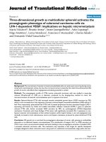
Primary Human Osteoblasts express VEGF receptors
(see Figure 1) Osteoblast rich cultures from trabecular
bone explants demonstrated no significant differences in
activity, receptor expression, cytokine release or response
to angiogenic cytokines from the commercially available
Journal of Orthopaedic Surgery and Research 2009, 4:19 />Page 5 of 13
(page number not for citation purposes)
human osteoblast cell line. As shown, 97.8% of cells in
the culture system expressed VEGF receptors. This bioti-
nylated VEGF binding assay does not distinguish the
VEGF receptor isotypes involved but rather indicates the
functional biological activity of VEGF receptor expression
in the cell population.
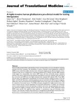
Exogenous VEGF induces alkaline phosphatase release
(Figure 2)
The concentration of endogenously produced VEGF was
measured in the cultures. The range was 0.8 – 2.25 ng/mL
following 72 hours of incubation. This was within the
range of exogenously administered VEGF that produced
equivalent results. After 48 hours in culture, recombinant
human VEGF 165 increased alkaline phosphatase release
in a dose dependant manner. VEGF concentrations of 5,
10 and 25 ng/mL were sufficient to increase nodule for-
mation 1.3-(not significant), 2.6 and 4.1-fold, over that of
cultures replete of exogenous VEGF. Daily administration
of 0.3 ug/mL of mAB VEGF again resulted in a significant
decrease (39% reduction) in nodule formation in the cul-
tures replete of exogenous VEGF, again highlighting the
importance of this positive feedback loop. PlGF was
slightly more efficacious at 25 ng/mL (66% increase) and
50 ng/mL (103% increase) in its effects on alkaline phos-
phatase release. This data suggests that ligation and activa-
tion of the specific VEGF receptor types has differential
effects on its various mitogenic activities.
Expression of functional VEGF receptors on Primary Human OsteoblastsFigure 1
Expression of functional VEGF receptors on Primary Human Osteoblasts. Mean Channel Fluorescence is measured
using flow cytometric analysis of avidin- FITC labelling of osteoblast rich cultures treated with a biotinylated VEGF or negative
control antibody for 2 hours. The flow cytometric image shown is representative in each case. Receptor expression was per-
formed in six separate experiments, with triplicates in each experiment.
Journal of Orthopaedic Surgery and Research 2009, 4:19 />Page 6 of 13
(page number not for citation purposes)
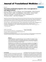
Exogenous VEGF induces bone nodule formation in
primary human osteoblasts (Figure 3)
After 18 days in culture, recombinant human VEGF 165
increased mineralized nodule formation in a dose
dependant manner. VEGF concentrations of 5, 10 and 25
ng/mL were sufficient to increase nodule formation 1.6-,
2.3- and 3.16-fold respectively, over that of cultures
replete of exogenous VEGF. Daily administration of 0.3
ug/mL of a neutralising antibody to VEGF (mAB VEGF)
resulted in a significant decrease (44% reduction) in nod-
ule formation in the cultures replete of exogenous VEGF.
Addition of a control monoclonal antibody had no effect
on the culture system. This data demonstrates that endog-
enously released VEGF is involved in a positive feedback
loop that serves to stimulate osteoblast bone formation
When compared to VEGF 165, Placental Growth Factor
(PlGF) had only a minimal effect on mineralised nodule
formation at 25 ng/mL (30% increase) and 50 ng/mL
(57% increase) concentrations. This demonstrates that
the Flt-1 receptor plays little role in the effects of VEGF on
osteoblast formation of mineralised nodules
The effect of angiogenic cytokines on osteoblast apoptosis
(Figure 4)
For this series of experiments serum free conditions were
used in order to examine the effects of the various ang-
The effect of a neutralizing monoclonal antibody and of VEGF receptor-specific ligands on Primary Human Osteoblast alkaline phosphatase release in vitroFigure 2
The effect of a neutralizing monoclonal antibody and of VEGF receptor-specific ligands on Primary Human
Osteoblast alkaline phosphatase release in vitro. Bone nodule formation was assessed by von Kossa staining and alkaline
phosphatase release by p-nitrophenyl phosphate hydrolysis as described in Materials and Methods. mAB: neutralizing mono-
clonal antibody to VEGF 165 (0.3 ug/mL), VEGF 165: vascular endothelial growth factor isotype 165, PlGF: placental like
growth factor. Data illustrates mean +/- standard error of the mean in each case. The results were derived from six separate
experiments, with triplicates performed in each experiment. # p < 0.05 represents statistically significant differences compared
to control, * p < 0.05 represents statistically significant differences compared to VEGF 165.
Journal of Orthopaedic Surgery and Research 2009, 4:19 />Page 7 of 13
(page number not for citation purposes)
iogenic cytokines in isolation. Control osteoblast apopto-
sis under these conditions was 25.6%, as compared to 3%
in the presence of serum (see Figure 4). Cell cultures were
treated with anti Fas-IgM (1000 ng/mL) and TNF alpha
(100 ng/mL) to simulate 'pathological' apoptosis in con-
ditions of excessive bone loss. This resulted in reproduci-
ble rates of programmed cell death of 68%. We found that
TNF alpha, in the absence of Fas IgM, did not induce
apoptosis of primary human osteoblasts but rather served
to increase expression of Fas receptor. Thus increasing
concentrations of TNF alpha resulted in increased rates of
'pathological' apoptosis (Data not shown here). Addition
of 40 ng/mL of IGF-1 inhibited spontaneous and patho-
logical apoptosis by 73.6% and 53% respectively. Treat-
ment of the cultures with 10 ng/mL of VEGF inhibited
spontaneous and TNF/Fas induced programmed cell
death by 83.6% and 71% respectively. Thus VEGF
afforded significantly better protection from apoptosis
under both normal and particularly pathological condi-
tions than the 'gold standard' osteotropic cytokine IGF-1.
PlGF had no significant effect on the rate of osteoblast
apoptosis demonstrating that the Flt-1 receptor was not
involved in the survival activity of VEGF. Both bFGF (46%
reduction in spontaneous and 27% reduction in patho-
logical apoptosis) and PDGF (34% reduction in sponta-
neous and 23.5% reduction in pathological apoptosis) at
The effect of a neutralizing monoclonal antibody and of VEGF receptor-specific ligands on Primary Human Osteoblast Bone Nodule Formation in vitroFigure 3
The effect of a neutralizing monoclonal antibody and of VEGF receptor-specific ligands on Primary Human
Osteoblast Bone Nodule Formation in vitro. Bone nodule formation was assessed by von Kossa staining and alkaline
phosphatase release by p-nitrophenyl phosphate hydrolysis as described in Materials and Methods. mAB: neutralizing mono-
clonal antibody to VEGF 165 (0.3 ug/mL), VEGF 165: vascular endothelial growth factor isotype 165, PlGF: placental like
growth factor. Data illustrates mean +/- standard error of the mean in each case. The results were derived from six separate
experiments, with triplicates performed in each experiment.
#
p < 0.05 represents statistically significant differences compared
to control, * p < 0.05 represents statistically significant differences compared to VEGF 165.
Journal of Orthopaedic Surgery and Research 2009, 4:19 />Page 8 of 13
(page number not for citation purposes)
25 ng/mL attenuated osteoblast apoptosis, but to a signif-
icantly lesser extent. These concentrations of VEGF, bFGF
and PDGF were used, as they were found to be compara-
ble in induction of endothelial cell proliferation (an in
vitro measure of angiogenesis) in preliminary studies.
Endogenous VEGF and BCl2 regulate osteoblast apoptosis.
(Figure 5)
The percentage of cells staining positive for annexin V was
analyzed by flow cytometry. Spontaneous apoptosis at 24
hours in the absence of serum is 25.6%. Pretreatment of
the osteoblasts with VEGF 10 ng/mL (4.7% apoptosis
rate) was almost as effective as culture in the presence of
10% serum (3% apoptosis rate) in inhibiting spontane-
ous programmed cell death Pretreatment of the cultures
with a neutralising monoclonal antibody to VEGF (mAB
VEGF), in the absence of exogenous VEGF resulted in a
spontaneous apoptosis rate of 14%. This indicates that
VEGF released in culture by primary human osteoblasts is
integral to the regulation of the rate of programmed cell
death. PlGF had no effect on apoptosis (23.8%), again
demonstrating that the Flt-1 receptor was not involved in
the survival activity of VEGF.
The effects of osteotropic and angiogenic cytokines on Primary Human Osteoblast apoptosis in vitroFigure 4
The effects of osteotropic and angiogenic cytokines on Primary Human Osteoblast apoptosis in vitro. Osteob-
last apoptosis was determined by Annexin V- Fluorescein Isothiocyanate labelling and hypodiploid DNA measurement as
described in Materials and Methods. IGF-1: insulin like growth factor -1, VEGF 165: vascular endothelial growth factor isotype
165, PlGF: placental like growth factor, bFGF: basic fibroblast growth factor, PDGF: platelet derived growth factor. Data illus-
trates mean +/- standard error of the mean in each case. The results were derived from six separate experiments, with tripli-
cates performed in each experiment. * p < 0.05 represents statistically significant differences compared to control,
&
p < 0.05
represents statistically significant differences compared to IGF-1, PlGF, bFGF and PDGF.
Journal of Orthopaedic Surgery and Research 2009, 4:19 />Page 9 of 13
(page number not for citation purposes)
VEGF attenuates osteoblast apoptosis by enhancing
expression of BCl2 gene (Figures 6 and 7)
Western Immunoblotting confirms that pretreatment of
the osteoblast cultures with exogenous VEGF 10 ng/mL,
results in up-regulation in expression of the anti-apoptotic
gene BCl2, reflecting a decrease in the rates of pro-
grammed cell death (Figure 5). This is true for both spon-
taneous and pathological (TNF alpha/anti Fas-IgM
induced) apoptosis, with relative band density increases
of 4.9 and 2.8 respectively. Treatment of the cultures with
a neutralising monoclonal antibody to VEGF 0.3 ug/mL
(mAB VEGF), in the absence of any exogenous VEGF,
resulted in a downregulation of BCl2 expression with rel-
ative band density decreases of 0.43 and 0.31 for sponta-
neous and pathological apoptosis respecively. These
decreases in anti-apoptotic gene expression reflect the
increased apoptotic rates of these cultures as seen in Figure
4. These data demonstrate that the significant protective
effect of VEGF on primary human osteoblasts is mediated
by expression of the critical antiapoptotic gene BCl2.
Discussion
Bone formation and resorption is the function of the basic
multicellular unit (BMU), where osteoblasts and osteo-
clasts interact with one another and with haemopoietic
and stromal cells of the bone marrow [1]. Regulation of
the numbers and activities of bone cells is essential for
skeletal homeostasis while mismatch between formation
and resorption is largely responsible for most systemic
and localised bone diseases. The rate of bone formation is
largely determined by the number of osteoblasts, which in
turn is determined by the rate of replication of progenitors
and the life-span of the mature cells, reflecting timing of
cell death by apoptosis [2]. Current evidence suggests that
apoptosis is the fate of the majority of osteoblasts, and
that changes in the prevalence of osteoblast apoptosis
alter the balance of skeletal homeostasis [1,2,23,25-29].
Glucocorticoid induced osteopenia is characterised by
increased osteoblast apoptosis, a phenomenon which is
reversible by estrogen administration in vitro and in vivo
[30]. Bisphosphanates and parathyroid hormone increase
The effects of 10% serum, a neutralizing monoclonal antibody and of VEGF receptor-specific ligands on spontaneous Primary Human Osteoblast apoptosis in vitroFigure 5
The effects of 10% serum, a neutralizing monoclonal antibody and of VEGF receptor-specific ligands on spon-
taneous Primary Human Osteoblast apoptosis in vitro. Osteoblast apoptosis was determined by Annexin V- Fluores-
cein Isothiocyanate Labeling and hypodiploid DNA measurement as described in Materials and Methods. mAB VEGF:
neutralising monoclonal antibody to vascular endothelial growth factor isotype 165 (0.3 ug/mL), VEGF: vascular endothelial
growth factor, PlGF: placental like growth factor. Data illustrates mean +/- standard error of the mean in each case. The results
were derived from six separate experiments, with triplicates performed in each experiment. * p < 0.05 represents statistically
significant differences compared to control,
#
p < 0.05 represents statistically significant differences compared to 10% serum
alone.
Journal of Orthopaedic Surgery and Research 2009, 4:19 />Page 10 of 13
(page number not for citation purposes)
bone formation by prevention of osteoblast apoptosis,
suggesting novel therapeutic strategies for osteoporsis.
Active and coordinated apoptosis of cells within the post
traumatic bone callus is thought to regulate local release
of cellular agents that modulate fracture repair [13,24].
Disuse osteopenia, periprosthetic and infection related
bone loss are all associated with increases in 'pathological'
osteoblast apoptosis, enhanced cell suicide mediated by
proinflammatory cytokines such as TNF alpha, anti CD
95-IgM, Il-1β and Il-6 [2,17-20,22]. The vasculature of
bone is an essential component of bone repair, remode-
ling and growth, however the precise interactions between
vascular cells and bone forming cells are still unclear [28].
The bone loss of osteoporosis is characterised by reduced
capillary compartments, distraction osteogenesis
increases bone formation with a parallel increase in capil-
lary number and vascularised bone grafts are far superior
to conventional ones. Bone chamber models using intra-
vital microscopy and microangiographic studies have
demonstrated that neovascularization temporally pre-
cedes neo-osteogenesis [28]. Endothelial cells cocultured
with fetal rat calvaria induce bone formation. Following
bone injury the developing osteoprogenitor cells and
osteoblasts are in intimate contact with the basement
membrane of the invading capillaries. During fracture
healing ossification by osteoblasts is spatially associated
with sites of capillary penetration into the callus [8]. Nor-
mal remodeling of mature bone occurs in discrete vascu-
larised elongated structures, termed osteons, in which old
bone is resorbed by osteoclasts at the cutting cone, and the
defect is filled with new bone by trailing osteoblasts. Dur-
ing skeletogenesis vascular endothelial growth factor
(VEGF) mediated blood vessel invasion of the growth
plate coincides with mineralisation of the extracellular
matrix (ECM), apoptosis of hypertrophic chondrocytes
and bone formation [9]. We have previously reported that
musculoskeletal injury results in systemic and fracture site
angiogenic responses in the human, that are primarily
mediated by vascular endothelial growth factor (VEGF)
[6,7]. We have also demonstrated that VEGF is essential
for both intramembranous and endochondral bone for-
mation, and exogenous enhances fracture repain in a
number of animal models [8]. As outlined earlier, osteob-
lasts release VEGF in the setting of bone injury. They
The effect of exogenous VEGF 165 on primary human osteoblast BCl2 expression during spontaneous and 'pathological' apop-tosis in vitroFigure 6
The effect of exogenous VEGF 165 on primary human osteoblast BCl2 expression during spontaneous and
'pathological' apoptosis in vitro. Expression of the antiapoptotic gene BCl2 was determined using Western immunoblot-
ting as described in Materials and Methods. Spontaneous apoptosis was measured for cells in serum free conditions. Pathologi-
cal apoptosis was induced by treatment of the cultures with TNFa and anti-Fas IgM. BCl2: antiapoptotic gene, VEGF: vascular
endothelial growth factor 165, TNFa: tumour necrosis factor alpha (100 ng/mL), anti-Fas IgM: anti Fas receptor immunoglobulin
(1000 ng/mL). The immunoblot shown is representative in each case. Western Immunoblotting was performed for six separate
experiments, with triplicates in each experiment.
Journal of Orthopaedic Surgery and Research 2009, 4:19 />Page 11 of 13
(page number not for citation purposes)
express receptors for VEGF in a differentiation dependent
manner [4], while osteoclasts express VEGF receptors and
osteoclast differentiation and bone resorption is
enhanced by VEGF [16]. Thus VEGF may represent a prin-
cipal regulator of the activities of the BMU, under normal
and indeed pathological conditions. The aim of this study
was to determine the role of VEGF on the activity and life
span of primary human osteoblasts in vitro.
We utilised both osteoblast rich cultures from human
trabecular bone explants and commercially available pri-
mary human osteoblasts in this study. We could not deter-
mine any significant differences in activity, receptor
expression or patterns of apoptosis between the two pop-
ulations. Previous studies have examined some of the fea-
tures of osteoblast apoptosis, but these have used
transformed cell lines e.g. murine MC3T3-E1 and human
MG-63 [13,14,29]. As these cell lines have been immortal-
ised accurate conclusions cannot be drawn based on their
responses to pro and/or antiapoptotic stimuli. It has pre-
viously been reported that the murine pre-osteoblast cell
line KS483 express VEGF receptor isotypes in a differenti-
ation dependant manner [4]. In order to examine the
effects of VEGF on primary human osteoblasts we first
demonstrated that these cells express functionally active
VEGF receptors. Using a biotinylated VEGF binding assay
we were able to show that 97.8% of the osteoblast-rich
cell population expressed VEGF receptors. Thus we could
anticipate a response of these cells to exogenously admin-
istered VEGF to the culture system. Performing parallel
experiments with Placental Growth Factor allowed us to
examine the relative roles of KDR and Flt-1 receptors in
the mitogenic and antiapoptotic effects of VEGF. As PlGF
has high affinity for Flt-1 and does not bind KDR, it's
effects reflect specific activation of the Flt-1 receptor. Dif-
ferences in the effects of VEGF 165 and PlGF, at compara-
ble concentrations, can therefore be attributed to
activation of KDR or perhaps neuropillin receptors. Using
a variety of VEGF selective mutants Gerber et al have
reported that the antiapoptotic effects of VEGF on
endothelial cells are mediated primarily by KDR receptor
through the activation of P-13 kinase [5]. VEGF mRNA
expression in osteoblasts is increased by several factors
e.g. PGE 1 and 2, IGF-1Vit D3, TGFβ and hypoxia
[10,12,13,15,30]. However, to date it was not known if
VEGF had a direct effect on osteoblasts themselves. Our
present data demonstrates that primary human osteob-
lasts are stimulated by exogenously administered VEGF
165 to increase mineralised nodule formation and alka-
line phosphatase release. Therefore VEGF has direct oste-
otropic effects independant of a prevailing vasculature.
The concentrations of VEGF that were used in this experi-
ment are similar to those measured in plasma of patients
with isolated long bone fractures, those relesased by stim-
ulated osteoblasts in vitro, and to those shown to enhance
osteoclastic bone resorption and survival of mature osteo-
clasts. In that report using purified mature rabbit osteo-
clasts the specific VEGF receptor isoform involved was not
examined [16]. Our data clearly demonstrates that the
mitogenic effects of VEGF on human osteoblasts are not
mediated by the flt-1 receptor and so are mediated by
either KDR or neuropillin. The results of treatment with
PlGF show that Flt-1 has little or no role in mediating
mineralisation but some limited role in activation of alka-
line posphatase release. While this appears contradictory,
it must be remembered that the process of mineralisation
is far more involved than just release of a single protein.
KDR activation on the osteoblast appears to signal all the
coordinated processes required to lay down bone, while
Flt-1 activation does not achieve this level of cell mitogen-
esis. Neutralisation of endogenous VEGF in our cell cul-
ture system had a significant effect on mineralisation and
alkaline phosphatase release. Thus osteoblasts release
VEGF in an autocrine fashion regulating their own activ-
ity. These data taken together, it is likely that VEGF can
mediate either bone formation or resorption, the ultimate
balance depending on cell receptor expression, differenti-
ation state, and the cytokine, biophysical and biochemical
milieu of the basic multicellular unit. Perhaps this may
The effect of neutralisation of endogenous VEGF on primary human osteoblast BCl2 expression during spontaneous apop-tosis in vitroFigure 7
The effect of neutralisation of endogenous VEGF on
primary human osteoblast BCl2 expression during
spontaneous apoptosis in vitro. Expression of the antiap-
optotic gene BCl2 was determined using Western immunob-
lotting as described in Materials and Methods. Spontaneous
apoptosis was measured for cells in serum free conditions.
BCl2: antiapoptotic gene, mAB VEGF: neutralising mono-
clonal antibody to vascular endothelial growth factor isotype
165 (0.3 ug/mL), control antibody: isotype control antibody
with no biological activity in vitro. The immunoblot shown is
representative in each case. Western Immunoblotting was
performed for six separate experiments, with triplicates in
each experiment.
Journal of Orthopaedic Surgery and Research 2009, 4:19 />Page 12 of 13
(page number not for citation purposes)
explain why the degree and nature of vascularisation must
be optimal for the formation of bone. Osteonecrosis is
characterised by relative avascularity, failure to reestablish
appropriate blood supply signals atrophic fracture non-
union, while excessive bone resorption is associated with
large, disorganised vascular channels. Systemic disorders
such as polyostotic Pagets disease, with simultaneous
pathological bone formation and resorption, may well
represent a critical breakdown in the coupling of skeletal
homeostasis and angiogenesis as mediated by various fac-
tors including VEGF.
Abnormalities in cell death control contribute to a variety
of diseases including cancer, autoimmunity, degenerative
disorders and osteoporosis [1,2,17,23]. Signaling for
apoptosis occurs through multiple independent pathways
that are initiated either from triggering events within the
cell e.g. mitochondrial membrane depolarisation or from
outside the cell, e.g. ligation of the death receptors
[25,26]. The ligation of the Fas or CD 95 receptor by its
ligand initiates programmed cell death in a number of
normal cell types [31-33]. This mechanism is implicated
as the principle pathway for excessive apoptosis in many
pathological conditions. Ligation of Fas with the Fas
receptor activates an intracellular cascade of cysteine pro-
teases (caspases) that ultimately dismantles the cell and
facilitates phagocytosis by neighboring cells. While cas-
pase 3 is the common terminal protease to all pathways of
apoptosis, caspase 6 and 8 are known to specifically signal
the activation of the death receptor pathway. Our data
herein demonstrates that TNF alpha of itself does not
induce apoptosis of primary human osteoblasts but rather
increases the expression of the death receptor Fas. Tsuboi
et al have previously reported comparable data for the
human osteoblast cell line MG63 [33]. Thus in the pres-
ence of Fas ligand, as in inflammatory and septic condi-
tions of bone, high levels of TNF alpha may contribute to
the associated osteolysis. Treatment of the cells with IETD-
FMK, a specific inhibitor of caspases 6 and 8 completely
reverses the effect of Fas receptor activation, confirming
the specificity of this pathway in pathological apoptosis of
primary human osteoblasts. Using mouse calvarial oste-
oblasts Hill et al demonstrated that IGF-1 was the most
potent inhibitor of apoptoss of 20 growth factors tested,
however, not including VEGF [19]
Our study demonstrates that VEGF released in culture by
primary human osteoblasts is integral to the regulation of
the rate of programmed cell death. PlGF had no effect on
apoptosis demonstrating that the Flt-1 receptor was not
involved in the survival activity of VEGF. Transfection of
the osteoblasts with the antiapoptotic gene BCl2 was suf-
ficient to inhibit osteoblast apoptosis induced by serum
starvation, demonstrating that BCl2 levels are critical for
regulation of osteoblast life-span.
Western Immunoblotting confirmed that pretreatment of
the osteoblast cultures with exogenous VEGF resulted in
up-regulation in expression of the anti-apoptotic gene
BCl2 and a decrease in the rates of programmed cell
death. Treatment of the cultures with a neutralising mon-
oclonal antibody to VEGF, in the absence of any exoge-
nous VEGF, resulted in a downregulation of BCl2
expression and a parallel increase in apoptotic rates of
these cultures. These data demonstrate that the significant
protective effect of VEGF on primary human osteoblasts is
mediated by expression of the critical antiapoptotic gene
BCl2.
In conclusion, our data demonstrates that VEGF is a
potent regulator of osteoblast life-span in vitro, attenuat-
ing both spontaneous and pathological programmed cell
death. This autocrine feedback mechanism is critical to
the survival of these cells and is mediated primarily via
non flt-1 receptor mediation and expression of BCl2
antiapoptotic gene.
Competing interests
The authors declare that they have no competing interests.
Authors' contributions
Both authors, JS and BL were involved in the design and
execution of the experimental studies described in this
manuscript. Both authors are responsible for writing and
editing the manuscript. Both authors have read and
approved the final manuscript for publication.
References
1. Jilka RL: Biology of the basic multicellular unit and the patho-
physiology of osteoporosis. Med Pediatr Oncol 2003, 41(3):182-5.
2. Manolagas SC: Birth and death of bone cells: basic regulatory
mechanisms and implications for the pathogenesis and
treatment of osteoporosis. Endocr Rev 2000, 21(2):115-37.
3. Gerber HP, Hillan KJ, Ryan AM, Kowalski J, Keller GA, Rangell L,
Wright BD, Radtke F, Aguet M, Ferrara N: VEGF is required for
growth and survival in neonatal mice. Development 1999,
126(6):1149-59.
4. Deckers MM, Karperien M, Bent C van der, Yamashita T, Papapoulos
SE, Lowik CW: Expression of vascular endothelial growth fac-
tors and their receptors during osteoblast differentiation.
Endocrinology 2000, 141(5):1667-74.
5. Gerber HP, McMurtrey A, Kowalski J, Yan M, Keyt BA, Dixit V, Fer-
rara N: Vascular endothelial growth factor regulates
endothelial cell survival through the phosphatidylinositol 3'-
kinase/Akt signal transduction pathway. Requirement for
Flk-1/KDR activation. J Biol Chem 1998, 273(46):30336-43.
6. Street J, Winter D, Wang JH, Wakai A, McGuinness A, Redmond HP:
Is human fracture hematoma inherently angiogenic? Clin
Orthop 2000:224-37.
7. Street JT, Wang JH, Wu QD, Wakai A, McGuinness A, Redmond HP:
The angiogenic response to skeletal injury is preserved in the
elderly. J Orthop Res 2001, 19(6):1057-66.
8. Street J, Bao M, deGuzman L, Bunting S, Peale FV Jr, Ferrara N, Stein-
metz H, Hoeffel J, Cleland JL, Daugherty A, van Bruggen N, Redmond
HP, Carano RA, Filvaroff EH: Vascular endothelial growth factor
stimulates bone repair by promoting angiogenesis and bone
turnover. Proc Natl Acad Sci USA 2002, 99(15):9656-61.
9. Gerber HP, Vu TH, Ryan AM, Kowalski J, Werb Z, Ferrara N: VEGF
couples hypertrophic cartilage remodeling, ossification and
Publish with BioMed Central and every
scientist can read your work free of charge
"BioMed Central will be the most significant development for
disseminating the results of biomedical research in our lifetime."
Sir Paul Nurse, Cancer Research UK
Your research papers will be:
available free of charge to the entire biomedical community
peer reviewed and published immediately upon acceptance
cited in PubMed and archived on PubMed Central
yours — you keep the copyright
Submit your manuscript here:
/>BioMedcentral
Journal of Orthopaedic Surgery and Research 2009, 4:19 />Page 13 of 13
(page number not for citation purposes)
angiogenesis during endochondral bone formation. Nat Med
1999, 5(6):623-8.
10. Goad DL, Rubin J, Wang H, Tashjian AH Jr, Patterson C: Enhanced
expression of vascular endothelial growth factor in human
SaOS-2 osteoblast-like cells and murine osteoblasts induced
by insulin-like growth factor I. Endocrinology 1996,
137(6):2262-8.
11. Harada S, Nagy JA, Sullivan KA, Thomas KA, Endo N, Rodan GA,
Rodan SB: Induction of vascular endothelial growth factor
expression by prostaglandin E2 and E1 in osteoblasts. J Clin
Invest 1994, 93(6):2490-6.
12. Saadeh PB, Mehrara BJ, Steinbrech DS, Dudziak ME, Greenwald JA,
Luchs JS, Spector JA, Ueno H, Gittes GK, Longaker MT: Transform-
ing growth factor-beta1 modulates the expression of vascu-
lar endothelial growth factor by osteoblasts. Am J Physiol 1999,
277(4 Pt 1):C628-37.
13. Schlaeppi JM, Gutzwiller S, Finkenzeller G, Fournier B: 1,25-Dihy-
droxyvitamin D3 induces the expression of vascular
endothelial growth factor in osteoblastic cells. Endocr Res
1997, 23(3):213-29.
14. Steinbrech DS, Mehrara BJ, Saadeh PB, Greenwald JA, Spector JA,
Gittes GK, Longaker MT: VEGF expression in an osteoblast-like
cell line is regulated by a hypoxia response mechanism. Am J
Physiol Cell Physiol 2000, 278(4):C853-60.
15. Street JT, Lenehan B, Wang JH, Wakai A, Redmond HP: Hypoxia
couples the paracrine interaction between osteoblasts and
endothelial cells. European Journal of Orthopaedic Surgery and Trau-
matology 2005, 15(3):214-225.
16. Niida S, Kaku M, Amano H, Yoshida H, Kataoka H, Nishikawa S,
Tanne K, Maeda N, Nishikawa S, Kodama H: Vascular endothelial
growth factor can substitute for macrophage colony-stimu-
lating factor in the support of osteoclastic bone resorption.
J Exp Med 1999, 190(2):293-8.
17. Clohisy DR: Could apoptosis be responsible for localized
imbalances in bone cell homeostasis? J Lab Clin Med 1999,
134(3):190-1.
18. Alexander EH, Rivera FA, Marriott I, Anguita J, Bost KL, Hudson MC:
Staphylococcus aureus – induced tumor necrosis factor –
related apoptosis – inducing ligand expression mediates
apoptosis and caspase-8 activation in infected osteoblasts.
BMC Microbiol
2003, 3(1):5.
19. Hill PA, Tumber A, Meikle MC: Multiple extracellular signals
promote osteoblast survival and apoptosis. Endocrinology 1997,
138(9):3849-58.
20. Hock JM: Stemming bone loss by suppressing apoptosis. J Clin
Invest 1999, 104(4):371-3.
21. Hock JM, Krishnan V, Onyia JE, Bidwell JP, Milas J, Stanislaus D: Oste-
oblast apoptosis and bone turnover. J Bone Miner Res 2001,
16(6):975-84.
22. Jilka RL, Weinstein RS, Bellido T, Parfitt AM, Manolagas SC: Osteob-
last programmed cell death (apoptosis): modulation by
growth factors and cytokines. J Bone Miner Res 1998,
13(5):793-802.
23. Landry P, Sadasivan K, Marino A, Albright J: Apoptosis is coordi-
nately regulated with osteoblast formation during bone
healing. Tissue Cell 1997, 29(4):413-9.
24. Olmedo ML, Landry PS, Sadasivan KK, Albright JA, Marino AA: Pro-
grammed cell death in post-traumatic bone callus. Cell Mol
Biol (Noisy-le-grand) 2000, 46(1):89-97.
25. Adams CS, Mansfield K, Perlot RL, Shapiro IM: Matrix regulation of
skeletal cell apoptosis. Role of calcium and phosphate ions. J
Biol Chem 2001, 276(23):20316-22.
26. Adams CS, Shapiro IM: Mechanisms by which extracellular
matrix components induce osteoblast apoptosis. Connect Tis-
sue Res 2003, 44(Suppl 1):230-9.
27. Li G, Dickson GR, Marsh DR, Simpson H: Rapid new bone tissue
remodeling during distraction osteogenesis is associated
with apoptosis. J Orthop Res 2003, 21(1):28-35.
28. Glowacki J: Angiogenesis in fracture repair. Clin Orthop 1998,
355(Suppl):S82-9.
29. Pascher E, Perniok A, Becker A, Feldkamp J: Effect of
1alpha,25(OH)2-vitamin D3 on TNF alpha-mediated apop-
tosis of human primary osteoblast-like cells in vitro.
Horm
Metab Res 1999, 31(12):653-6.
30. Forsythe JA, Jiang BH, Iyer NV, Agani F, Leung SW, Koos RD,
Semenza GL: Activation of vascular endothelial growth factor
gene transcription by hypoxia-inducible factor 1. Mol Cell Biol
1996, 16(9):4604-13.
31. Fuller K, Wong B, Fox S, Choi Y, Chambers TJ: TRANCE is neces-
sary and sufficient for osteoblast-mediated activation of
bone resorption in osteoclasts. J Exp Med 1998,
188(5):997-1001.
32. Chua CC, Chua BH, Chen Z, Landy C, Hamdy RC: TGF-beta1
inhibits multiple caspases induced by TNF-alpha in murine
osteoblastic MC3T3-E1 cells. Biochim Biophys Acta 2002,
1593(1):1-8.
33. Tsuboi M, Kaku M, Amano H, Yoshida H, Kataoka H, Nishikawa S,
Tanne K, Maeda N, Nishikawa S, Kodama H: Tumor necrosis fac-
tor-alpha and interleukin-1beta increase the Fas-mediated
apoptosis of human osteoblasts. J Lab Clin Med 1999,
134(3):222-31.