Báo cáo hóa học: " Down-regulation of cell surface CXCR4 by HIV-1" pdf
Bạn đang xem bản rút gọn của tài liệu. Xem và tải ngay bản đầy đủ của tài liệu tại đây (1.02 MB, 10 trang )
BioMed Central
Page 1 of 10
(page number not for citation purposes)
Virology Journal
Open Access
Research
Down-regulation of cell surface CXCR4 by HIV-1
Bongkun Choi*
1,4
, Paul J Gatti
1,5
, Cesar D Fermin
2
, Sandor Vigh
3
,
Allyson M Haislip
1
and Robert F Garry
1
Address:
1
Department of Microbiology and Immunology, Tulane University Health Sciences Center, New Orleans, LA 70112, USA,
2
College of
Veterinary Medicine, Nursing & Allied Health (CVMNAH), Tuskegee University, Tuskegee, AL 36088, USA,
3
Department of Structural & Cellular
Biology, Tulane University Health Sciences Center, New Orleans, LA 70112, USA,
4
Departments of Environmental Medicine, Pathology, and
Medicine, New York University School of Medicine, Tuxedo, NY 10987, USA and
5
Biocompare, Inc., 395 Oyster Point Blvd, South San Francisco,
CA 94080, USA
Email: Bongkun Choi* - ; Paul J Gatti - ; Cesar D Fermin - ;
Sandor Vigh - ; Allyson M Haislip - ; Robert F Garry -
* Corresponding author
Abstract
Background: CXC chemokine receptor 4 (CXCR4), a member of the G-protein-coupled
chemokine receptor family, can serve as a co-receptor along with CD4 for entry into the cell of T-
cell tropic X4 human immunodeficiency virus type 1 (HIV-1) strains. Productive infection of T-
lymphoblastoid cells by X4 HIV-1 markedly reduces cell-surface expression of CD4, but whether
or not the co-receptor CXCR4 is down-regulated has not been conclusively determined.
Results: Infection of human T-lymphoblastoid cell line RH9 with HIV-1 resulted in down-
regulation of cell surface CXCR4 expression. Down-regulation of surface CXCR4 correlated
temporally with the increase in HIV-1 protein expression. CXCR4 was concentrated in intracellular
compartments in H9 cells after HIV-1 infection. Immunofluorescence microscopy studies showed
that CXCR4 and HIV-1 glycoproteins were co-localized in HIV infected cells. Inducible expression
of HIV-1 envelope glycoproteins also resulted in down-regulation of CXCR4 from the cell surface.
Conclusion: These results indicated that cell surface CXCR4 was reduced in HIV-1 infected cells,
whereas expression of another membrane antigen, CD3, was unaffected. CXCR4 down-regulation
may be due to intracellular sequestering of HIV glycoprotein/CXCR4 complexes.
Background
Chemokine receptors are seven-transmembrane G-pro-
tein-coupled receptors that upon ligand binding transmit
signals, such as calcium flux, resulting in chemotactic
responses [1-3]. Chemokine receptors are divided into
four families that reflect differential binding of the CXC,
CC, CX3C and XC subfamilies of chemokines [4]. Several
members of the chemokine receptor family function as
coreceptors with the primary receptor CD4 to allow entry
of various strains of human immunodeficiency virus type
1 (HIV-1) into the cells [5-8]. T-cell-tropic X4 HIV-1 use
CD4 and chemokine receptor CXCR4 for entry into target
cells, whereas macrophage-tropic R5 HIV-1 use CD4 and
chemokine receptor CCR5. Dual-tropic strains can use
either CCR5 and CXCR4 as co-receptors. In addition,
CCR3, CCR2, CXCR6 (Bonzo/STLR6) among other chem-
okine receptors can function as coreceptors and support
infection by a more restricted subset of macrophage-tropic
or dual-tropic HIV strains [9,10,5,11,12].
Published: 11 January 2008
Virology Journal 2008, 5:6 doi:10.1186/1743-422X-5-6
Received: 21 December 2007
Accepted: 11 January 2008
This article is available from: />© 2008 Choi et al; licensee BioMed Central Ltd.
This is an Open Access article distributed under the terms of the Creative Commons Attribution License ( />),
which permits unrestricted use, distribution, and reproduction in any medium, provided the original work is properly cited.
Virology Journal 2008, 5:6 />Page 2 of 10
(page number not for citation purposes)
CXCL12 (stromal derived factor 1 α/β, SDF-1α/β) is the
natural ligand for CXCR4, whereas CC chemokines, CCL3
(macrophage inflammatory factor 1α, MIP-1α/chemok-
ine LD78α), CCL3-L1 (LD78β), CCL4 (MIP-1β), and
CCL5 (RANTES), are ligands for CCR5 [13-16]. CXCL12,
CCL3, CCL4 and CCL5 as well as other natural and syn-
thetic chemokine receptor ligands are able to inhibit cell
fusion and infection by various strains of HIV-1, depend-
ent or independent of co-receptor usage [17-21]. These
findings have encouraged the development of antiHIV
therapeutics targeting chemokine receptors [22-25].
Productive infection of CD4+ cells with HIV-1 markedly
reduces cell-surface expression of CD4, which follows a
classic mechanism for retroviral interference [26,27].
Down-regulation of CD4 by HIV-1 has been attributed to
the formation of intracellular complexes consisting of
HIV-1 envelope glycoproteins and CD4 receptors [28],
although other mechanisms may also be involved in a cell
type dependent manner [29,30]. Chemokine receptors,
including CCR5 and CXCR4, can be down-regulated after
binding of their respective chemokine ligands by a mech-
anism involving endocytosis of the complex [31-33]. The
envelope glycoproteins of HIV-1 competitively antago-
nize signaling by coreceptors CXCR4 and CCR5 [34,35].
Exogenously added recombinant soluble HIV-1 surface
glycoprotein (SU, gp120) can be coprecipitated from the
cell surface into a complex with CD4 and CXCR4, that
may lead to the formation of a trimolecular complex
between HIV SU, CD4 and CXCR4 [36,37]. However,
prior studies have suggested that although CCR5 corecep-
tors are down-modulated during infection by R5 HIV-1,
CXCR4 co-receptor is not down-regulated after productive
X4 HIV-1 infection [38]. CXCR4 was shown to be selec-
tively down-regulated from the cell surface by HIV-2/vcp
in the context of CD4-independent infection [39] or from
cells infected with CD4-independent HIV-1 isolate that
enters directly via CXCR4 [40]. Furthermore, exogenous
expression of the HIV-1 Nef protein reduced cell surface
levels of CCR5 or CXCR4 [41,42]. Here, we examine
whether or not productive infection by HIV-1 alters the
cell surface expression of CXCR4. Our results indicate that
CXCR4 is down-regulated from the surface of CD4+ T-
lymphoblastoid cells infected by HIV-1 and that HIV-1
Env and CXCR4 are colocalized in infected cells.
Results
HIV-1 infection down-regulates surface expression of
CXCR4 in RH9 cells
To determine whether HIV infection alters cell surface
CXCR4 levels, RH9 T-lymphoblastoid cells were infected
with HIV-1
LA1
at a MOI of 4 or mock-infected. At 1, 4 and
7 days post infection (PI), the level of cell surface CXCR4
on RH9 cells and HIV-1-infected RH9 cells were deter-
mined by flow cytometric analysis using CXCR4 mono-
clonal antibody (MAb) 12G5 [39]. Relative binding of
12G5 monoclonal antibody was significantly reduced
compared to uninfected cells at 4, and 7 days postinfec-
tion, respectively (Fig. 1A). As a control, we also deter-
mined the effect of HIV infection on CD3 in RH9 cells. H9
cells infected with HIV maintained surface CD3 expres-
sion at a similar level to that of uninfected H9 cells (Fig.
1B). To determine the relationship between the expres-
sion of surface CXCR4 and HIV-1 protein expression, HIV-
1 production by infected cells was quantified by a antigen-
capture enzyme-linked immunosorbant assay (Ag-capture
ELISA; Abbott Laboratories) and the number of HIV-1
antigen expressing cells were measured by indirect
immunofluorescence microscopy. The decline in CXCR4
expression was accompanied by a rapid increase in HIV-1
protein expression in infected RH9 cells.
The results of these flow cytometric analyses were con-
firmed by immunofluorescence microscopy (Fig. 2). RH9
T-lymphoblastoid cells were infected with HIV-1
LA1
at a
MOI of 4 or mock-infected. At 4 days after infection cells
were labeled with the CXCR4 12G5 MAb, followed by a
FITC-conjugated secondary antibody and analyzed by
indirect immunofluorescence microscopy. Whereas iso-
type-matched control antibody showed no reactivity (Fig.
2A, B), all control cells expressed CXCR4. The CXCR4-spe-
cific MAb displayed cell surface membrane fluorescence in
100% of mock-infected cells (Fig. 2C, D). Most cells in the
HIV-1-infected cultures (>90%) showed markedly
decreased surface CXCR4 staining (Fig. 2E–H), reflective
of the flow cytometry results. The distribution of CXCR4
on the minor population of cells (<10%) with surface
CXCR was similar to that of uninfected cells (Fig. 2G, H).
HIV infection had no significant effect on the cell surface
expression of CD3 indicating that decreased expression of
CXCR4 is not a non-specific consequence of HIV-1 infec-
tion (not shown).
HIV-1 infection induces internalization of CXCR4 in RH9
cells
Down-regulation of surface CD4 by envelope glycopro-
teins from the plasma membrane has been attributed at
least in part to the formation of intracellular complexes
consisting of HIV-1 envelope molecules and CD4 recep-
tors [26,43,44]. The potential internalization of CXCR4 in
permeabilized HIV-infected H9 cells was investigated by
immunofluorescence microscopy. RH9 T-lymphoblastoid
cells were infected with HIV-1
LA1
at a MOI of 4 or mock-
infected. After 4 days PI, the cells were fixed, permeabi-
lized by incubation with 0.05% saponin in PBS for 15
min to allow the entry of antibody and incubated with
CXCR4 MAb followed by a FITC-conjugated second anti-
body. No fluorescence was observed in cells incubated
with control antibodies (Fig. 3A, B). CXCR4-specific MAb
12G5 stained the surface of uninfected control cells (Fig.
Virology Journal 2008, 5:6 />Page 3 of 10
(page number not for citation purposes)
3C, D). A weak additional intracellular signal observed in
some control cells may be attributed to newly synthesized
CXCR4 molecules in intracellular compartments of secre-
tory pathways. In cultures productively infected with HIV-
1, intracellular CXCR4 staining was markedly increased in
approximately 50% of the cells, with a redistribution of
the staining that is consistent with the intracellular accu-
mulation of the receptor (Fig. 3E–H).
HIV-1 SU and CXCR4 are colocalized in HIV-1
productively-infected RH9 cells
Exogenously added HIV SU or SU expressed from recom-
binant vectors can form a complex with CD4 and chem-
okine receptor [36,37]. Double labeling was used to
determine if an analogous complex of CXCR4 and HIV-1
glycoprotein can be detected in HIV-1 productively
infected cells. RH9 T-lymphoblastoid cells were infected
with HIV-1
LA1
at a MOI of 4 or mock-infected. After 4 days
PI, the cells were fixed, permeabilized with saponin and
incubated with 12G5 CXCR4 MAb followed by a FITC-
conjugated second antibody. For staining of HIV-1 glyco-
proteins, cells were incubated with rhodamine-conju-
gated antibodies to the HIV-1 proteins and double-
fluorescence analysis was performed. A phase contrast
micrograph of a multinucleated HIV-1 infected cell is
shown in Figure 4A. Figure 4C and Figure 4D represent
staining for anti-HIV-1 proteins (red) and anti-CXCR4
(green) MAb, respectively. Superpositions of the two color
channels appear in yellow representing the degree of colo-
calization of CXCR4 and HIV-1 proteins (Fig. 4B). Similar
results were observed in nonsyncytial cells expressing
HIV-1 proteins. These results suggest that HIV-1 SU and
CXCR4 are colocalized in HIV-1 productively-infected
RH9 cells.
Inducible expression of HIV-1 Env down-regulates cell
surface CXCR4 expression
HIV-1 Env have been suggested to play a role in down-reg-
ulation of surface CD4 molecules from the plasma mem-
brane [28,45,46]. The effect of inducible expression of the
HIV-1 envelope protein (strain HXB2) on CXCR4 expres-
sion was analyzed in CD4+ Jurkat lymphocytes with a
well-characterized tetracycline inducible expression sys-
tem [47,48]. Env expression was monitored by syncitial
formation and immunofluorecence staining for Env pro-
teins. In the presence of tetracycline, no fluorescence was
observed in Jurkat cells, indicating that Env expression
was repressed. When Jurkat cells were cultured in the
absence of tetracycline to induce Env expression, >95% of
cells stained positive for HIV-1 Env. In the presence of tet-
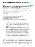
Flow cytometry analysis demonstrating reduced CXCR4 expression in HIV-1 infected RH9 cellsFigure 1
Flow cytometry analysis demonstrating reduced CXCR4 expression in HIV-1 infected RH9 cells. Panel A: RH9 T-lymphoblast-
oid cells infected with HIV-1
LA1
. On days 1, 4, and 7 postinfection cells were fixed with 4% paraformaldehyde, stained with
mouse MAb 12G5 anti-CXCR4 (10 μg/ml) or isotype-matched control antibody followed by fluorescein isothiocyanate (FITC)-
conjugated goat anti-mouse immunoglobulin G, and analyzed by flow cytometry. Median fluorescence intensity was calculated
as an indicator of the level of cell surface CXCR4 expression. Data are presented as single-color histograms with FITC fluores-
cence (CD3 expression) along the horizontal axis and relative cell number along the vertical axis. RH9 cells (control cells),
heavy solid line: H9 cells infected with HIV, dotted line; H9 with an isotype-matched control antibody, thin solid line. Panel B:
Analysis of surface CD3 expression in HIV-1 and mock infected RH9 cells by FACS analyzed on day 7 post-infection.
Virology Journal 2008, 5:6 />Page 4 of 10
(page number not for citation purposes)
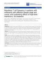
Immunofluorescence microscopy demonstrating reduced cell surface expression of CXCR4 in HIV-1 infected RH9 cellsFigure 2
Immunofluorescence microscopy demonstrating reduced cell surface expression of CXCR4 in HIV-1 infected RH9 cells. Panel
A: Immunofluorescence staining control with isotype-matched monoclonal antibody. Panel C: CXCR4 immunofluorescence
staining of H9 cells. Panels E and G: CXCR4 immunofluorescence staining of H9 cells acutely infected by HIV-1. Panels B, D, F
and H show phase contrast images of the same fields of cells shown in left panels. The fluorescent syncytial cell in panel G is
representative of a minor population of cells in the infected culture (<10%) with a CXCR4 surface distribution similar to unin-
fected cells.
Virology Journal 2008, 5:6 />Page 5 of 10
(page number not for citation purposes)
Immunofluorescence microscopy analysis of CXCR4 expression in permeabilized HIV-1 and mock infected RH9 cellsFigure 3
Immunofluorescence microscopy analysis of CXCR4 expression in permeabilized HIV-1 and mock infected RH9 cells. Four
days after HIV-1 infection, cells were fixed, permeabilized with saponin and labeled with a mouse monoclonal antibody to
CXCR4 (12G5) and a secondary, FITC-conjugated anti-mouse antibodies for observation with a fluorescence microscopy.
Panel A: Immunofluorescence staining control with isotype-matched monoclonal antibody. Panel C: CXCR4 immunofluores-
cence staining of H9 cells. Panels E and G, CXCR4 immunofluorescence staining of HIV-1 infected H9 cells. Panels B, D, F and
H show phase contrast images of the same fields of cells shown in left panels.
Virology Journal 2008, 5:6 />Page 6 of 10
(page number not for citation purposes)
racycline, i.e, no Env expression, cells expressed a similar
amount of CXCR4 as Jurkat cells without the Env expres-
sion plasmid (Fig. 5A–D). In contrast, a decrease in the
level of CXCR4 expression was seen in >95% of Jurkat
cells expressing Env proteins (Fig 5E–G), indicating that
Env expression leads to down-regulation of cell surface
CXCR4 expression. There was a strong correlation
between a lack of Env expression and expression of
CXCR4 in cells of the induced cultures. The distribution of
CXCR4 on the minor population of induced Jurkat cells
(<5%) with surface CXCR4 was similar to that of unin-
duced cells (Fig. 2G, H).
Discussion
Cellular receptors for viruses are often down-regulated
from the plasma membrane following productive infec-
tion, making infected cells refractory to superinfection by
other viruses that use the same receptor for entry [49-
51,27,52]. The decrease in surface expression may be
caused in part by the formation of a complex between the
viral receptor binding protein and cellular receptors in
intracellular compartments. Both HIV-1 and simian
immunodeficiency virus down-regulate cell surface
expression of CD4, their primary receptor [26,53]. Several
mechanisms have been proposed to account for the
down-regulation of CD4 following primate lentivirus
infection [26,28,54,55]. Internalization of CD4 can occur
upon binding of HIV-1 envelope glycoproteins [45,46].
Down-regulation of CD4 may also be mediated by the
HIV-1 Nef and Vpu accessory proteins [55]. Nef is
expressed early and Vpu late preventing CD4 expression
throughout the HIV-1 replication cycle. Nef links CD4 to
components of clathrin-dependent trafficking pathways
resulting in internalization and delivery of CD4 to lyso-
somes for degradation [56-59]. Vpu links CD4 to a ubiq-
uitin ligase thereby facilitating degradation of CD4 in the
endoplasmic reticulum [60].
Here we demonstrate that during productive acute cyto-
pathic infection of CD4+ T-lymphoblastoid cells by HIV-
1 there is an extensive down-regulation of cell surface
CXCR4 expression, which correlated with the increase in
HIV-1 protein expression. CXCR4 appears to be concen-
trated in intracellular compartments in H9 cells after HIV-
1 infection. Colocalization of both CXCR4 and HIV-1
glycoproteins was detected in HIV-1 infected cells.
Epitope masking is unlikely to be responsible for the loss
of CXCR4 surface staining since intracellular complexes
were readily detected. Down-regulation of the CXCR4
coreceptor during productive infection by CD4-depend-
ent X4 HIV-1 strains was not observed in a previous study
by Chenine and coworkers [38]. In contrast to results with
the X4 HIV-1 strains they tested, Chenine and coworkers
observed a complete loss of CCR5 staining on the surface
of cells chronically infected with R5 viruses [38]. Further-
more, it has been shown that CXCR4 is down-regulated by
HIV-2 isolates that use CXCR4 as their primary receptor
[39]. CXCR4 is also down-regulated in cells infected with
CD4-independent X4 HIV-1 isolate m7NDK [40]. How-
ever, another CD4-independent HIV-1 isolate, HIV-1/
IIIBx, failed to down-regulate CXCR4 on chronically
infected cells [61].
There are several plausible explanations for the differences
in the results we obtained in the current study with those
obtained previously by Chenine et al.[38]. As with the two
CD4-independent HIV-1 isolates tested that differ in
CXCR4 down-regulation [40,61], it is possible that Env of
the two X4 strains of HIV-1 we used (LA1, HXB2) differ in
their ability to down-modulate CXCR4 from the Env of
the X4 viruses (HX10, MN) used by Chenine and cowork-
ers. HIV-1 strain LA1 grows to high titers and the Tet-Off
system in Jurkat cells produces significant amounts of
HXB2 Env. LA1 is highly cytopathic and significant CPE is
observed in the inducible HXB2 Env expression system
[48]. In contrast, "little syncytium formation and cell
Co-localization CXCR4 and HIV-1 glycoprotein in HIV-1 infected H9 cellsFigure 4
Co-localization CXCR4 and HIV-1 glycoprotein in HIV-1
infected H9 cells. Four days after HIV-1 infection, cells were
fixed and permeabilized with saponin. Cells were then
labeled with a human monoclonal antibody that interact with
SU and then rhodamine-conjugated goat anti-human antibod-
ies (Panel C: red fluorescence) and with 12G5 mAb followed
by fluorescein-conjugated goat anti-mouse antibodies (Panel
D:green fluorescence). Panel A: phase contrast image. Panel
B represents a superposition of green and red fluorescence,
with costained regions appearing in yellow. Yellow regions in
panel B indicate the colocalization of chemokine receptor
CXCR4 and HIV-1 proteins.
Virology Journal 2008, 5:6 />Page 7 of 10
(page number not for citation purposes)
CXCR4 expression is reduced in Jurkat cells after induction of HIV-1 Env expressionFigure 5
CXCR4 expression is reduced in Jurkat cells after induction of HIV-1 Env expression. After 4 days induction of HIV-1 Env pro-
teins, non-induced and induced cells were fixed and labeled with a mouse MAb to CXCR4 (12G5) and a secondary FITC-con-
jugated anti-mouse antibodies for observation with a fluorescence microscopy. Panels A and C: CXCR4 staining of non-
induced Jurakt cells. Panel E and G: CXCR4 staining of induced Jurkat cells. The fluorescent cell in panel G is representative of
a minor population of cells in the induced culture (<5%) with a CXCR4 surface distribution similar to uninduced cells. Panels B,
D, F and H show phase contrast images of the same fields of cells shown in left panels.
Virology Journal 2008, 5:6 />Page 8 of 10
(page number not for citation purposes)
death" was observed in the X4 HIV-1 infected cultures
used by Chenine and coworkers [38]. The CD4 independ-
ent HIV-2 strain that down-regulates CXCR4 used by
Endres et al. (1996) was also highly cytopathic. However,
it is unlikely that cytopathic effects are responsible for the
decrease in surface CXCR4 by simply selecting for cells in
the culture with a low level of CXCR4. CXCR4 is uni-
formly present on the cells in the RH9 and Jurkat cultures.
It is possible that other strains of HIV-1, which grow to
lower titers than LA1 or produce less HIV-1 Env than the
HXB2 inducible expression system, may have a smaller
impact on cell surface CXCR4 for stochastic reasons. The
Env of the strains used here may also have a higher affin-
ities for CXCR4 than certain other X4 viruses, allowing
direct CXCR4-Env complexing intracellularly. It is also
possible that differences in the ability to down-regulate
CXCR4 are cell specific. However, we used two different
cell lines, RH9 and Jurkat, in the current studies and
observed HIV-1 induced CXCR4 down-regulation in both.
We also observed a partial down-regulation of CXCR4 in
primary human peripheral blood mononuclear cells after
infection of HIV-1 (not shown).
Alteration in CXCR4 expression after infection by HIV-1
could result from sequestration of CXCR4 intracellularly
or from the direct effects of other HIV-1 proteins on the
synthesis of CXCR4 or its transport to the cell surface. Sev-
eral studies have shown that HIV-1 SU can displace chem-
okines from their receptors [34,35]. Interactions between
SU, CD4, and CXCR4 have also been well established
[62,36]. Previous studies demonstrated that treatment
with the HIV-1 SU increased colocalization of CD4 with
CXCR4 and cocapping of the gp120-CD4-CXCR4 com-
plexes resulted in the cointernalization of a proportion of
the gp120-CXCR4 complexes into intracellular vesicles
[37]. We did observe down-regulation of surface CXCR4
in an inducible system for Env (and Rev) in which acces-
sory proteins Nef and Vpu are not expressed. However,
given other studies suggesting that Nef and Vpu may be
able to down-regulate CXCR4 independently of Env, the
role these proteins should be considered in future work.
HHV-6 and HHV-7 induce down-regulation of CXCR4
[63]. These viruses do not use CXCR4 for cell entry, and
induce a markedly decreased level of CXCR4 gene tran-
scription without any significant alteration of the post-
transcriptional stability of CXCR4 mRNA. Reduced levels
of CXCR4 mRNA transcripts were observed in cells
infected with CD4-independent HIV-1 isolate [26]. Fur-
thermore, the modulation of CCR5 expression by the R5
viruses is at the level of transcription [38]. Further experi-
ments will be needed to determine the mechanisms of
down-modulation of surface CXCR4 by HIV-1.
Conclusion
The amount of surface CXCR4 was greatly reduced in T-
lymphoblastoid cells infected with HIV-1 strain LA1, but
expression of another membrane antigen, CD3, was unaf-
fected. CXCR4 was concentrated in intracellular compart-
ments in RH9 cells after HIV-1 infection.
Immunofluorescence microscopy studies showed that
CXCR4 and HIV-1 glycoproteins were co-localized in HIV-
1 infected cells. Inducible expression of HIV-1 envelope
glycoproteins also resulted in down-regulation of CXCR4
from the cell surface. CXCR4 down-regulation may be due
in part to intracellular sequestering of HIV glycoprotein/
CXCR4 complexes.
Methods
Cells and virus
Cells of the RH9 subclone of the CD4+ human T-lym-
phoblastoid cell line RH9 were the kind gift of Dr. Suraiya
Rasheed (University of Southern California), and were
maintained in RPMI 1640 supplemented with 10% fetal
bovine serum (GIBCO, Long Island, NY), penicillin (100
U/ml) and streptomycin (100 μg/ml). Joseph Sodroski
(Harvard University) kindly provided the Env-inducible
Jurkat cell line [48].
Flow cytometry and immunofluorescence microscopy
RH9 T-lymphoblastoid cells were infected with HIV-1
LA1
at a MOI of 4 or mock-infected. At various times after the
addition of virus, cells were fixed in 4% paraformalde-
hyde for 15 min at room temperature, washed and stained
with the mouse MAb 12G5 (10 μg/ml) against human
CXCR4 followed by fluorescein isothiocyanate (FITC)-
conjugated goat anti-mouse immunoglobulin G (Sigma).
In some experiments cells were permeabilized by incuba-
tion with 0.05% saponin in PBS for 15 min prior to addi-
tion of antibody. CXCR4 monoclonal antibody 12G5
derived by Dr. James Hoxie [39] was obtained through the
AIDS Research and Reference Reagent Program, Division
of AIDS, NIAID, NIH. Mouse isotype-matched antibodies
(Sigma) were used as a negative control for the gating of
those cells staining negative for a cell surface marker. Flow
cytometry was performed on a Coulter EPICS fluores-
cence-activated flow cytometer (Coulter Electronics,
Hialeah, Fla.). For immunofluorescence microscopy cells
were analyzed with a Nikon microscope equipped for epi-
fluorescence. Fluorescent images were acquired with an
Olympus microscope, a 100 W UV source, appropriate
exciter and blocking filters, captured with a CCD, and
processed with Adobe PhotoShop.
Competing interests
The author(s) declare that they have no competing inter-
ests.
Virology Journal 2008, 5:6 />Page 9 of 10
(page number not for citation purposes)
Authors' contributions
BC performed all experiments with substantial help from
PJG and AH. RFG, SV and CDF provided guidance, exper-
tise, equipment, and funding for these experiments. All
authors have read and approved this manuscript.
Acknowledgements
This research was supported by Public Health Service grants AI054238,
AI054626 and AI068230 from the National Institute of Allergy and Infec-
tious Diseases. We thank Drs. Rasheed, Sodroski and Hoxie for making
materials available.
References
1. Murphy PM: Viral exploitation and subversion of the immune
system through chemokine mimicry. Nat Immunol 2001,
2(2):116-122.
2. Baggiolini M, Dewald B, Moser B: Human chemokines: an update.
Annu Rev Immunol 1997, 15:675-705.
3. Allen SJ, Crown SE, Handel TM: Chemokine: receptor structure,
interactions, and antagonism. Annu Rev Immunol 2007,
25:787-820.
4. Bacon K, Baggiolini M, Broxmeyer H, Horuk R, Lindley I, Mantovani
A, Maysushima K, Murphy P, Nomiyama H, Oppenheim J, Rot A,
Schall T, Tsang M, Thorpe R, Van Damme J, Wadhwa M, Yoshie O,
Zlotnik A, Zoon K: Chemokine/chemokine receptor nomen-
clature. J Interferon Cytokine Res 2002, 22(10):1067-1068.
5. Feng Y, Broder CC, Kennedy PE, Berger EA: HIV-1 entry cofactor:
functional cDNA cloning of a seven-transmembrane, G pro-
tein-coupled receptor [see comments]. Science 1996,
272(5263):872-877.
6. Moore JP: Coreceptors: implications for HIV pathogenesis
and therapy. Science 1997, 276(5309):51-52.
7. Berger EA, Murphy PM, Farber JM: Chemokine receptors as HIV-
1 coreceptors: roles in viral entry, tropism, and disease. Annu
Rev Immunol 1999, 17:657-700.
8. Bjorndal A, Deng H, Jansson M, Fiore JR, Colognesi C, Karlsson A,
Albert J, Scarlatti G, Littman DR, Fenyo EM: Coreceptor usage of
primary human immunodeficiency virus type 1 isolates var-
ies according to biological phenotype. J Virol 1997,
71(10):7478-7487.
9. Dragic T, Litwin V, Allaway GP, Martin SR, Huang Y, Nagashima KA,
Cayanan C, Maddon PJ, Koup RA, Moore JP, Paxton WA: HIV-1
entry into CD4+ cells is mediated by the chemokine recep-
tor CC- CKR-5 [see comments]. Nature 1996,
381(6584):667-673.
10. Alkhatib G, Combadiere C, Broder CC, Feng Y, Kennedy PE, Murphy
PM, Berger EA: CC CKR5: a RANTES, MIP-1alpha, MIP-1beta
receptor as a fusion cofactor for macrophage-tropic HIV-1.
Science 1996, 272(5270):1955-1958.
11. He J, Chen Y, Farzan M, Choe H, Ohagen A, Gartner S, Busciglio J,
Yang X, Hofmann W, Newman W, Mackay CR, Sodroski J, Gabuzda
D: CCR3 and CCR5 are co-receptors for HIV-1 infection of
microglia. Nature
1997, 385(6617):645-649.
12. Choe H, Farzan M, Sun Y, Sullivan N, Rollins B, Ponath PD, Wu L,
Mackay CR, LaRosa G, Newman W, Gerard N, Gerard C, Sodroski J:
The beta-chemokine receptors CCR3 and CCR5 facilitate
infection by primary HIV-1 isolates. Cell 1996,
85(7):1135-1148.
13. Bleul CC, Farzan M, Choe H, Parolin C, Clark-Lewis I, Sodroski J,
Springer TA: The lymphocyte chemoattractant SDF-1 is a lig-
and for LESTR/fusin and blocks HIV-1 entry. Nature 1996,
382(6594):829-833.
14. Combadiere C, Ahuja SK, Tiffany HL, Murphy PM: Cloning and
functional expression of CC CKR5, a human monocyte CC
chemokine receptor selective for MIP-1(alpha), MIP-1(beta),
and RANTES. J Leukoc Biol 1996, 60(1):147-152.
15. Samson M, Labbe O, Mollereau C, Vassart G, Parmentier M: Molec-
ular cloning and functional expression of a new human CC-
chemokine receptor gene. Biochemistry 1996, 35(11):3362-3367.
16. Blanpain C, Migeotte I, Lee B, Vakili J, Doranz BJ, Govaerts C, Vassart
G, Doms RW, Parmentier M: CCR5 binds multiple CC-chemok-
ines: MCP-3 acts as a natural antagonist. Blood 1999,
94(6):1899-1905.
17. Jansson M, Popovic M, Karlsson A, Cocchi F, Rossi P, Albert J, Wigzell
H: Sensitivity to inhibition by beta-chemokines correlates
with biological phenotypes of primary HIV-1 isolates. Proc
Natl Acad Sci U S A 1996, 93(26):15382-15387.
18. Oravecz T, Pall M, Norcross MA: Beta-chemokine inhibition of
monocytotropic HIV-1 infection. Interference with a post-
binding fusion step. J Immunol 1996, 157(4):1329-1332.
19. Capobianchi MR, Abbate I, Antonelli G, Turriziani O, Dolei A, Dian-
zani F: Inhibition of HIV type 1 BaL replication by MIP-1alpha,
MIP-1beta, and RANTES in macrophages. AIDS Res Hum Ret-
roviruses 1998, 14(3):233-240.
20. Stantchev TS, Broder CC: Consistent and significant inhibition
of human immunodeficiency virus type 1 envelope-mediated
membrane fusion by beta-chemokines (RANTES) in primary
human macrophages. J Infect Dis 2000, 182(1):68-78.
21. Pugach P, Marozsan AJ, Ketas TJ, Landes EL, Moore JP, Kuhmann SE:
HIV-1 clones resistant to a small molecule CCR5 inhibitor
use the inhibitor-bound form of CCR5 for entry. Virology
2007,
361(1):212-228.
22. Simmons G, Clapham PR, Picard L, Offord RE, Rosenkilde MM,
Schwartz TW, Buser R, Wells TNC, Proudfoot AE: Potent inhibi-
tion of HIV-1 infectivity in macrophages and lymphocytes by
a novel CCR5 antagonist. Science 1997, 276(5310):276-279.
23. Clapham PR, Reeves JD, Simmons G, Dejucq N, Hibbitts S, McKnight
A: HIV coreceptors, cell tropism and inhibition by chemok-
ine receptor ligands. Mol Membr Biol 1999, 16(1):49-55.
24. Simmons G, Reeves JD, Hibbitts S, Stine JT, Gray PW, Proudfoot AE,
Clapham PR: Co-receptor use by HIV and inhibition of HIV
infection by chemokine receptor ligands. Immunol Rev 2000,
177:112-126.
25. Trkola A, Ketas TJ, Nagashima KA, Zhao L, Cilliers T, Morris L,
Moore JP, Maddon PJ, Olson WC: Potent, broad-spectrum inhi-
bition of human immunodeficiency virus type 1 by the CCR5
monoclonal antibody PRO 140. J Virol 2001, 75(2):579-588.
26. Hoxie JA, Alpers JD, Rackowski JL, Huebner K, Haggarty BS, Cedar-
baum AJ, Reed JC: Alterations in T4 (CD4) protein and mRNA
synthesis in cells infected with HIV. Science 1986,
234(4780):1123-1127.
27. Potash MJ, Volsky DJ: Viral interference in HIV-1 infected cells.
Rev Med Virol 1998, 8(4):203-211.
28. Crise B, Buonocore L, Rose JK: CD4 is retained in the endoplas-
mic reticulum by the human immunodeficiency virus type 1
glycoprotein precursor. J Virol 1990, 64(11):5585-5593.
29. Hoxie JA, Rackowski JL, Haggarty BS, Gaulton GN: T4 endocytosis
and phosphorylation induced by phorbol esters but not by
mitogen or HIV infection. J Immunol 1988, 140(3):786-795.
30. Geleziunas R, Bour S, Wainberg MA: HIV-1 associated down-
modulation of CD4 gene expression is differentially
restricted in lymphocytic and monocytic cell lines. J Leukoc
Biol 1994, 55(5):589-595.
31. Amara A, Gall SL, Schwartz O, Salamero J, Montes M, Loetscher P,
Baggiolini M, Virelizier JL, Arenzana-Seisdedos F: HIV coreceptor
downregulation as antiviral principle: SDF-1alpha- depend-
ent internalization of the chemokine receptor CXCR4 con-
tributes to inhibition of HIV replication. J Exp Med 1997,
186(1):139-146.
32. Aramori I, Ferguson SS, Bieniasz PD, Zhang J, Cullen B, Cullen MG:
Molecular mechanism of desensitization of the chemokine
receptor CCR-5: receptor signaling and internalization are
dissociable from its role as an HIV-1 co-receptor. Embo J 1997,
16(15):4606-4616.
33. Brandt SM, Mariani R, Holland AU, Hope TJ, Landau NR: Associa-
tion of chemokine-mediated block to HIV entry with core-
ceptor internalization. J Biol Chem 2002, 277(19):17291-17299.
34. Madani N, Kozak SL, Kavanaugh MP, Kabat D: gp120 envelope
glycoproteins of human immunodeficiency viruses competi-
tively antagonize signaling by coreceptors CXCR4 and
CCR5. Proc Natl Acad Sci U S A 1998, 95(14):8005-8010.
35. Wang JM, Ueda H, Howard OM, Grimm MC, Chertov O, Gong X,
Gong W, Resau JH, Broder CC, Evans G, Arthur LO, Ruscetti FW,
Oppenheim JJ: HIV-1 envelope gp120 inhibits the monocyte
response to chemokines through CD4 signal-dependent
chemokine receptor down-regulation. J Immunol 1998,
161(8):4309-4317.
Publish with BioMed Central and every
scientist can read your work free of charge
"BioMed Central will be the most significant development for
disseminating the results of biomedical research in our lifetime."
Sir Paul Nurse, Cancer Research UK
Your research papers will be:
available free of charge to the entire biomedical community
peer reviewed and published immediately upon acceptance
cited in PubMed and archived on PubMed Central
yours — you keep the copyright
Submit your manuscript here:
/>BioMedcentral
Virology Journal 2008, 5:6 />Page 10 of 10
(page number not for citation purposes)
36. Lapham CK, Ouyang J, Chandrasekhar B, Nguyen NY, Dimitrov DS,
Golding H: Evidence for cell-surface association between fusin
and the CD4-gp120 complex in human cell lines [see com-
ments]. Science 1996, 274(5287):602-605.
37. Ugolini S, Moulard M, Mondor I, Barois N, Demandolx D, Hoxie J,
Brelot A, Alizon M, Davoust J, Sattentau QJ: HIV-1 gp120 induces
an association between CD4 and the chemokine receptor
CXCR4. J Immunol 1997, 159(6):3000-3008.
38. Chenine AL, Sattentau Q, Moulard M: Selective HIV-1-induced
downmodulation of CD4 and coreceptors. Arch Virol 2000,
145(3):455-471.
39. Endres MJ, Clapham PR, Marsh M, Ahuja M, Turner JD, McKnight A,
Thomas JF, Stoebenau-Haggarty B, Choe S, Vance PJ, Wells TN,
Power CA, Sutterwala SS, Doms RW, Landau NR, Hoxie JA: CD4-
independent infection by HIV-2 is mediated by fusin/CXCR4.
Cell 1996, 87(4):745-756.
40. Valente ST, Chanel C, Dumonceaux J, Olivier R, Marullo S, Briand P,
Hazan U: CXCR4 is down-regulated in cells infected with the
CD4-independent X4 human immunodeficiency virus type 1
isolate m7NDK. J Virol 2001, 75(1):439-447.
41. Michel N, Allespach I, Venzke S, Fackler OT, Keppler OT: The Nef
protein of human immunodeficiency virus establishes super-
infection immunity by a dual strategy to downregulate cell-
surface CCR5 and CD4. Curr Biol 2005, 15(8):714-723.
42. Venzke S, Michel N, Allespach I, Fackler OT, Keppler OT: Expres-
sion of Nef downregulates CXCR4, the major coreceptor of
human immunodeficiency virus, from the surfaces of target
cells and thereby enhances resistance to superinfection. J
Virol 2006, 80(22):11141-11152.
43. Cefai D, Ferrer M, Serpente N, Idziorek T, Dautry-Varsat A, Debre
P, Bismuth G: Internalization of HIV glycoprotein gp120 is
associated with down-modulation of membrane CD4 and
p56lck together with impairment of T cell activation. J Immu-
nol 1992, 149(1):285-294.
44. Bour S, Boulerice F, Wainberg MA: Inhibition of gp160 and CD4
maturation in U937 cells after both defective and productive
infections by human immunodeficiency virus type 1. J Virol
1991, 65(12):6387-6396.
45. Fujita K, Omura S, Silver J: Rapid degradation of CD4 in cells
expressing human immunodeficiency virus type 1 Env and
Vpu is blocked by proteasome inhibitors. J Gen Virol 1997, 78 (
Pt 3):619-625.
46. Su SB, Ueda H, Howard OM, Grimm MC, Gong W, Ruscetti FW,
Oppenheim JJ, Wang JM: Inhibition of the expression and func-
tion of chemokine receptors on human CD4+ leukocytes by
HIV-1 envelope protein gp120. Chem Immunol 1999, 72:141-160.
47. Gossen M, Bujard H: Studying gene function in eukaryotes by
conditional gene inactivation. Annu Rev Genet 2002, 36:153-173.
48. Cao J, Park IW, Cooper A, Sodroski J: Molecular determinants of
acute single-cell lysis by human immunodeficiency virus type
1. J Virol 1996, 70(3):1340-1354.
49. Vogt PK, Ishizaki R: Patterns of viral interference in the avian
leukosis and sarcoma complex. Virology 1966, 30(3):368-374.
50. Temin HM: Mechanisms of cell killing/cytopathic effects by
nonhuman retroviruses. Rev Infect Dis 1988, 10(2):399-405.
51. Weller SK, Joy AE, Temin HM: Correlation between cell killing
and massive second-round superinfection by members of
some subgroups of avian leukosis virus. J Virol 1980,
33(1):494-506.
52. Nethe M, Berkhout B, van der Kuyl AC: Retroviral superinfection
resistance. Retrovirology 2005, 2:52.
53. Salmon P, Olivier R, Riviere Y, Brisson E, Gluckman JC, Kieny MP,
Montagnier L, Klatzmann D: Loss of CD4 membrane expression
and CD4 mRNA during acute human immunodeficiency
virus replication. J Exp Med 1988, 168(6):1953-1969.
54. Crise B, Rose JK: Human immunodeficiency virus type 1 glyc-
oprotein precursor retains a CD4-p56lck complex in the
endoplasmic reticulum. J Virol 1992, 66(4):2296-2301.
55. Lindwasser OW, Chaudhuri R, Bonifacino JS: Mechanisms of CD4
downregulation by the Nef and Vpu proteins of primate
immunodeficiency viruses. Curr Mol Med 2007, 7(2):171-184.
56. Gama Sosa MA, DeGasperi R, Kim YS, Fazely F, Sharma P, Ruprecht
RM: Serine phosphorylation-independent downregulation of
cell-surface CD4 by nef. AIDS Res Hum Retroviruses 1991,
7(11):859-860.
57. Kim YH, Chang SH, Kwon JH, Rhee SS: HIV-1 Nef plays an essen-
tial role in two independent processes in CD4 down-regula-
tion: dissociation of the CD4-p56(lck) complex and targeting
of CD4 to lysosomes. Virology 1999, 257(1):208-219.
58. Stoddart CA, Geleziunas R, Ferrell S, Linquist-Stepps V, Moreno ME,
Bare C, Xu W, Yonemoto W, Bresnahan PA, McCune JM, Greene
WC: Human immunodeficiency virus type 1 Nef-mediated
downregulation of CD4 correlates with Nef enhancement of
viral pathogenesis. J Virol 2003, 77(3):2124-2133.
59. Chaudhuri R, Lindwasser OW, Smith WJ, Hurley JH, Bonifacino JS:
Downregulation of CD4 by human immunodeficiency virus
type 1 Nef is dependent on clathrin and involves direct inter-
action of Nef with the AP2 clathrin adaptor. J Virol 2007,
81(8):3877-3890.
60. Willey RL, Maldarelli F, Martin MA, Strebel K: Human immunode-
ficiency virus type 1 Vpu protein induces rapid degradation
of CD4. J Virol 1992, 66(12):7193-7200.
61. Hoxie JA, LaBranche CC, Endres MJ, Turner JD, Berson JF, Doms
RW, Matthews TJ: CD4-independent utilization of the CXCR4
chemokine receptor by HIV-1 and HIV-2. J Reprod Immunol
1998, 41(1-2):197-211.
62. Trkola A, Dragic T, Arthos J, Binley JM, Olson WC, Allaway GP,
Cheng-Mayer C, Robinson J, Maddon PJ, Moore JP: CD4-depend-
ent, antibody-sensitive interactions between HIV-1 and its
co- receptor CCR-5 [see comments]. Nature 1996,
384(6605):184-187.
63. Yasukawa M, Hasegawa A, Sakai I, Ohminami H, Arai J, Kaneko S,
Yakushijin Y, Maeyama K, Nakashima H, Arakaki R, Fujita S: Down-
regulation of CXCR4 by human herpesvirus 6 (HHV-6) and
HHV-7. J Immunol 1999, 162(9):5417-5422.