Báo cáo hóa học: " Hepatitis C virus NS5A protein binds the SH3 domain of the Fyn tyrosine kinase with high affinity: mutagenic analysis of residues within the SH3 domain that ?" doc
Bạn đang xem bản rút gọn của tài liệu. Xem và tải ngay bản đầy đủ của tài liệu tại đây (1.05 MB, 9 trang )
BioMed Central
Page 1 of 9
(page number not for citation purposes)
Virology Journal
Open Access
Research
Hepatitis C virus NS5A protein binds the SH3 domain of the Fyn
tyrosine kinase with high affinity: mutagenic analysis of residues
within the SH3 domain that contribute to the interaction
Holly Shelton
1,2
and Mark Harris*
1
Address:
1
Institute of Molecular and Cellular Biology, Faculty of Biological Sciences and Astbury Centre for Structural Molecular Biology,
University of Leeds, Leeds LS2 9JT, UK and
2
Department of Microbiology, School of Biological Sciences, The University of Reading, Whiteknights,
RG6 6AJ, Reading, UK
Email: Holly Shelton - ; Mark Harris* -
* Corresponding author
Abstract
Background: The hepatitis C virus (HCV) non-structural 5A protein (NS5A) contains a highly
conserved C-terminal polyproline motif with the consensus sequence Pro-X-X-Pro-X-Arg that is
able to interact with the Src-homology 3 (SH3) domains of a variety of cellular proteins.
Results: To understand this interaction in more detail we have expressed two N-terminally
truncated forms of NS5A in E. coli and examined their interactions with the SH3 domain of the Src-
family tyrosine kinase, Fyn. Surface plasmon resonance analysis revealed that NS5A binds to the
Fyn SH3 domain with what can be considered a high affinity SH3 domain-ligand interaction (629
nM), and this binding did not require the presence of domain I of NS5A (amino acid residues
32–250). Mutagenic analysis of the Fyn SH3 domain demonstrated the requirement for an acidic
cluster at the C-terminus of the RT-Src loop of the SH3 domain, as well as several highly conserved
residues previously shown to participate in SH3 domain peptide binding.
Conclusion: We conclude that the NS5A:Fyn SH3 domain interaction occurs via a canonical SH3
domain binding site and the high affinity of the interaction suggests that NS5A would be able to
compete with cognate Fyn ligands within the infected cell.
Background
Hepatitis C virus is an enveloped RNA virus that is esti-
mated to infect 2% of the global population, 123 million
individuals [1]. The virus has a positive sense RNA
genome of 9.5 kb that comprises a single open reading
frame encoding a ~3000 residue polyprotein, flanked by
5' and 3' untranslated regions. The polyprotein is cleaved
into 10 individual polypeptides by a combination of host-
cell and viral proteases, the N-terminal one-third of the
polyprotein produces the four structural proteins (Core,
E1, E2 and p7), whereas the C-terminal two-thirds com-
prises the six non-structural proteins (NS2, NS3, NS4A,
NS4B, NS5A and NS5B). Use of a sub-genomic replicon
system has demonstrated that five of these (NS3-NS5B)
are necessary and sufficient to replicate an RNA molecule
containing the 5' and 3' untranslated regions of the viral
genome. However, apart from the RNA-dependent RNA
polymerase (NS5B), the precise details of the roles of each
of the non-structural proteins in the process of RNA repli-
cation remain undefined.
Published: 11 February 2008
Virology Journal 2008, 5:24 doi:10.1186/1743-422X-5-24
Received: 8 January 2008
Accepted: 11 February 2008
This article is available from: />© 2008 Shelton and Harris; licensee BioMed Central Ltd.
This is an Open Access article distributed under the terms of the Creative Commons Attribution License ( />),
which permits unrestricted use, distribution, and reproduction in any medium, provided the original work is properly cited.
Virology Journal 2008, 5:24 />Page 2 of 9
(page number not for citation purposes)
NS5A is a 448 amino acid phosphoprotein that interacts
with a plethora of cellular proteins and has been reported
to have multiple effects on cell physiology, for review see
[2]. At the N-terminus of NS5A is a 31 residue amphip-
athic helix that mediates association of the protein with
cytoplasmic membranes [3], this is followed by three
domains, separated by short flexible linker regions [4]
(Figure 1a). The three-dimensional structure of domain I
has been determined and it has been shown to complex
with a zinc ion and (at least in the crystal structure) exists
as a dimer [5]. The structures of domains II and III remain
undetermined but of particular interest is the observation
that the flexible linker between these domains contains
two motifs with the consensus sequence Pro-X-X-Pro-X-
Arg/Lys. We, and others, have shown that these motifs
(termed PP2.1 and PP2.2) bind to the Src homology 3
(SH3) domains of a range of cellular proteins. In particu-
lar, work has focussed on the PP2.2 motif which is con-
served throughout all HCV isolates of all genotypes
(unlike the PP2.1 motif which is only conserved in geno-
type 1 isolates). The PP2.2 motif binds to the SH3
domains of the Src-family kinases Fyn, Lyn, Hck and Lck
[6], as well as the adaptor proteins Grb2 [7] and
amphiphysin II (also known as BinI) [8,9]. Mutation of
the PP2.2 motif also abrogated the ability of NS5A to
inhibit activation of the Ras-Erk MAPK pathway [10,11]
thus implicating a role for NS5A:SH3 domain interactions
in this process. However, the role of the PP2.2 motif in
virus replication is controversial; although a mutation of
this motif in the context of the genotype 1b sub-genomic
replicon had variously either no effect [11], or exhibited a
minimal reduction [8,9] in viral RNA replication, it was
also reported that a full-length infectious genome con-
taining the same mutation was unable to establish an
infection in a chimpanzee [9].
A number of other viral proteins interact with host cell
SH3 domains – the best characterised of these is the HIV-
1 Nef protein. The interaction between Nef and the SH3
domain of the Src-family kinase, Hck, is reported as one
of the strongest interactions between an SH3 domain and
its ligand (K
D
= 250 nM) [12], results in activation of the
kinase and has been shown to be required for viral patho-
genesis in vivo [13]. The affinity of a Nef derived PxxPxR-
containing peptide for the Hck SH3 domain was much
lower than that of the intact protein (K
D
= 91 μM), sug-
gesting that the interaction between Nef and the Hck SH3
domain involved additional intermolecular interactions.
We were therefore interested to determine the molecular
details of the NS5A:SH3 domain interaction. As an initial
approach to this question we used the crystal structure of
Nef complexed with a mutated form of the Fyn tyrosine
kinase SH3 domain (R96I) [14] as the basis for a molecu-
lar modelling study to predict the residues involved in the
interaction between the NS5A PP2.2 motif and the Fyn
SH3 domain [11]. The results of this study predicted that
NS5A would interact with the SH3 domain in a very sim-
ilar fashion to Nef, and the work presented here was
designed to evaluate this prediction using both surface
plasmon resonance and mutagenesis of the Fyn SH3
domain. The data confirm the predictions, and further-
more show that NS5A interacts with the Fyn SH3 domain
with a similar affinity to that exhibited by the Nef:SH3
domain interaction. We conclude that NS5A binds to SH3
domains with high affinity, and such interactions could
occur in the context of an HCV infected cell.
Results and discussion
We, and others, have previously shown that a conserved
C-terminal polyproline motif in NS5A interacts with the
SH3 domains of a range of cellular proteins. Although the
functional consequences of these interactions remain to
be elucidated, recent evidence suggests that this motif may
be important for virus replication [9], and thus represents
a valid target for antiviral drug development. We therefore
performed a detailed biochemical and biophysical analy-
sis of the interaction between NS5A and SH3 domains. To
facilitate this analysis we expressed two N-terminally
deleted forms of NS5A in E. coli – firstly NS5A(Δ32), in
which the membrane anchoring amphipathic helix was
removed to aid solubility [15]. Secondly, as we had previ-
ously shown that the N-terminal 270 residues were dis-
pensable for SH3 domain binding (Andrew Macdonald,
PhD thesis, University of Leeds), we expressed
NS5A(Δ250) in which both the amphipathic helix and
domain I [4] were deleted. Both proteins were expressed
with an N-terminal hexahistidine tag to aid purification.
Figure 1a shows a schematic of the expressed proteins and
figure 1b western blot analysis and Coomassie Blue stain-
ing of various stages in the purification process. We estab-
lished a two stage purification protocol in which NS5A
was first purified via the hexahistidine tag by immobilised
metal affinity chromatography and further purified by gel
filtration (lanes 10). Using this protocol, both forms of
NS5A could be purified to approximately 80% purity as
judged by Coomassie blue staining (lanes 11). The two
forms of NS5A migrated on SDS-PAGE with apparent
molecular masses of 55 kDa (Δ32) and 40 kDa (Δ250). To
confirm that the expressed proteins were the correct
molecular mass they were subjected to slow crystallisation
mass spectrometry [16], figure 1c demonstrates that the
actual molecular masses were in close agreement with pre-
dicted masses. Of note, the apparent molecular masses of
each NS5A derived protein species (as indicated by the
aberrant migration of the protein on SDS-PAGE) were sig-
nificantly higher than the actual molecular masses, this is
most likely due to the high proline content of NS5A (11%
in the intact protein).
Virology Journal 2008, 5:24 />Page 3 of 9
(page number not for citation purposes)
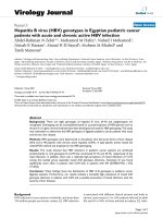
Expression and purification of N-terminally truncated forms of NS5AFigure 1
Expression and purification of N-terminally truncated forms of NS5A. (a) Schematic of the structure of NS5A and
the expressed truncated forms showing the locations of the N-terminal amphipathic helix and the three domains. (b) Anti-
NS5A western blot analysis and Coomassie staining of the purification of NS5A(Δ32) (left) and NS5A(Δ250) (right). Lanes: 1;
uninduced lysate, 2; induced lysate, 3; lysate clarified by centrifugation and applied to the Ni-NTA column, 4; flow-through, 5–8;
washes, 9; 200 mM imidazole elution, 10; gel filtration eluate, 11; Coomassie stain of gel filtration eluate. (c) Slow crystallization
mass spectrometry of NS5A(Δ32) (left) and NS5A(Δ250) (right). The peak at ~69 kDa on the left represents the E. coli chaper-
one protein DnaK. The measured molecular masses correspond closely to the predicted values: NS5A(Δ32): 47,222 Da, and
NS5A(Δ250): 26,500 Da. (d) Interaction of truncated NS5A forms with the Fyn SH3 domain. Purified NS5A(Δ32) (left) and
NS5A(Δ250) (right) were subjected to GST pulldown analysis using GA-beads alone (lanes 2), GST (lanes 3) or GST-FynSH3
(lanes 4). Samples were blotted for NS5A, lanes 1 show 20% of input protein, lanes 2–4 show bound protein eluted by compe-
tition with 20 mM reduced glutathione. The lower panel shows a Coomassie stained SDS-PAGE of the purified GST-FynSH3
domain fusion proteins.
Virology Journal 2008, 5:24 />Page 4 of 9
(page number not for citation purposes)
We had previously shown that NS5A bound to the SH3
domain of the Fyn tyrosine kinase and that in the context
of the intact kinase this interaction led to Fyn activation.
As Fyn is expressed in Huh7 cells [6] that are permissive
for HCV replication we chose to use the Fyn SH3 domain
as a basis for our investigation. We first confirmed that the
bacterially expressed NS5A was able to bind a GST-Fyn
SH3 domain fusion protein in vitro (figure 1d), in agree-
ment with our previous data both forms of NS5A (Δ32
and Δ250) bound equally well. This observation also
demonstrated that eucaryotic post-translational modifica-
tions were not required for the NS5A-SH3 domain inter-
action.
To obtain quantitative data about the affinity of the inter-
action between NS5A and the Fyn SH3 domain we uti-
lised surface plasmon resonance. We first confirmed that
we could detect no binding of either form of NS5A to
either sensor chips loaded with anti-GST antibody alone,
or anti-GST antibody with the addition of GST (data not
shown). Purified NS5A was then flowed over sensor chips
on to which were immobilised GST-FynSH3, as shown in
figures 2 and 3 this analysis revealed that both forms of
NS5A bound the Fyn SH3 domain with high affinity – for
NS5A(Δ32) the K
D
was calculated at 629 nM, for
NS5A(Δ250) 556 nM. The individual values of k
d
and k
a
used to calculate the K
D
values are presented in Table 1.
This data confirmed that domain I of NS5A was not
involved in interactions with the SH3 domain, however
we cannot rule out the possibility that domain I might
mediate interactions with the intact kinase. Interestingly,
the affinity of NS5A for Fyn SH3 was similar to that deter-
mined for the HIV-1 Nef protein binding to either Hck
SH3 or a mutant Fyn SH3 (R96I) (250 nM). In the case of
Nef it has been shown that the high affinity of the interac-
tion was mediated not only by interactions between resi-
dues in the SH3 domain and the polyproline motif, but
also by other interactions, for example involving the RT
loop of the SH3 domain.
Based on the crystal structure of the Nef-Fyn(R96I)SH3
domain complex we had previously predicted that both
Nef and NS5A would make the same intermolecular con-
tacts with the SH3 domain [11]. Four residues in the SH3
domain, namely D100, W119, N136 and Y137, were pre-
dicted to make major contributions to the binding energy
of the NS5A-SH3 domain interaction. To test this hypoth-
esis we constructed the corresponding site-directed
mutants of the Fyn SH3 domain in the context of a GST-
FynSH3 domain fusion, replacing the indicated residues
with alanine. These were purified and tested for binding
to NS5A both by GST-pulldown and ELISA (figure 4a). As
expected from the molecular modelling analysis, both
W119A and the double mutant N136A/Y137A abrogated
binding, however D100A had no effect. This was unex-
pected as our previous study had predicted that D100
would form a salt bridge with residue R356 in NS5A, how-
ever, as D100 was preceded by an additional acidic resi-
due (D99) it was plausible that R356 could form a salt
bridge with either acidic residue. To test this hypothesis
we constructed two further mutants, D99A and a double
substitution D99A/D100A. Gratifyingly, this analysis
revealed that D99A had no effect, however the double
mutant dramatically reduced binding (figure 4a) – thus it
appears that there is some flexibility in the interaction as
R356 within NS5A can form a salt bridge with either of
two adjacent acidic residues in the SH3 domain. Interest-
ingly the side chain of residue D100 has also been shown
to make an intramolecular hydrogen bond with R96
within the RT-Src loop of the SH3 domain – it was possi-
ble that the effects of the double mutant were due to the
inability to form this bond – thus perturbing the correct
folding of the SH3 domain. However, a comparison of the
circular dichroism spectra of the four GST-FynSH3 fusions
(wildtype, D99A, D100A and D99A/D100A) revealed no
significant differences in the overall fold of the proteins
(data not shown), suggesting that this is not the case. In
addition we generated two mutants of residues predicted
to make a minor contribution to the interaction – Y91A/
Y93A and Y132A/P134A. Interestingly these mutations
had a significant impact on the binding to NS5A,
although as can be seen in figure 4a these two mutant SH3
domains were somewhat unstable and exhibited degrada-
tion to GST.
Interestingly, of the nine residues predicted to contribute
to the binding energy of the interaction, eight were con-
served between Fyn and c-Src (the only difference being
D99 in the RT-loop – altered to T99 in c-Src). Despite this
both we [6] and others [7] had previously observed that
NS5A was unable to bind to the SH3 domain of Src so to
determine if other residues in the SH3 domain might
make a contribution to the interaction we proceeded to
make a second set of mutations. In order to avoid any
potential structural effects we chose to mutate four resi-
dues in Fyn to their corresponding c-Src residues. Three of
these were non-conservative changes: A95S, E121L and
E129Q, the fourth was a conservative change, L112V. We
therefore made the corresponding mutations within the
Fyn SH3 domain, expressed them as GST-fusion proteins
Table 1: Kinetic parameters for the NS5A:Fyn SH3 domain
interaction.
Protein k
a
(M
-1
s
-1
)k
d
(s
-1
)K
D
(M
-1
)
NS5A(Δ32) 4.93 × 10
3
3.08 × 10
-3
6.29 × 10
-7
(± SD) n = 3 (± 0.66 × 10
3
) (± 0.16 × 10
-3
) (0.59 × 10
-7
)
NS5A(Δ250) 9.05 × 10
3
4.94 × 10
-3
5.56 × 10
-7
(± SD) n = 3 (± 1.62 × 10
3
) (± 0.11 × 10
-3
) (0.59 × 10
-7
)
Virology Journal 2008, 5:24 />Page 5 of 9
(page number not for citation purposes)
and tested these mutant proteins for binding to NS5A by
ELISA (figure 4b). None of these mutations had any sig-
nificant effect on the binding to NS5A.
Our data are consistent with the notion that the PP2.2
polyproline motif of NS5A is a promiscuous and high
affinity SH3 ligand, able to mediate binding of NS5A to a
wide range of SH3 domains. The binding of NS5A to the
Fyn SH3 domain is remarkably resistant to single or mul-
tiple amino acid substitutions, apart from the relatively
well conserved residues – tyrosines 91 and 93 and tryp-
tophan 119 – mutation of which reduced binding to back-
ground levels. These three residues have previously been
shown to play critical roles in peptide binding and are
highly conserved [17], thus the dramatic effect on NS5A
binding is in line with expectations. The affinity of NS5A
for the Fyn SH3 domain is higher than the corresponding
values for most cellular SH3 domains interacting with
their cognate ligands – generally such interactions have
calculated K
D
between 1–50 μM [17], although some have
been reported to have much higher affinities, eg the
amphiphysin:dynamin I interaction was measured at 190
nM [18]. The comparison of binding affinities suggests
that NS5A would be able to effectively compete with cel-
lular ligands for binding to SH3 domains. In this context
it will be of great interest to determine which SH3 domain
containing proteins interact with NS5A in cells infected
with HCV, such experiments are ongoing in our labora-
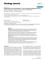
Surface plasmon resonance analysis of the NS5A(Δ32):Fyn SH3 domain interactionFigure 2
Surface plasmon resonance analysis of the NS5A(Δ32):Fyn SH3 domain interaction. SPR analysis was performed as
described for NS5A(Δ32) and GST-FynSH3. The indicated concentrations of NS5A were flowed over a sensor cell on to which
GST-FynSH3 was captured via an anti-GST antibody. The generated fit is shown as black lines in the top panels, the residuals
plotted in the lower panels indicate the relative response difference between the generated fit and the raw data – demonstrat-
ing the closeness of fit.
Virology Journal 2008, 5:24 />Page 6 of 9
(page number not for citation purposes)
tory. It is interesting to note that a recent study [19]
pointed to a key role for Fyn in HCV RNA replication.
However, in apparent contrast to our previous data dem-
onstrating activation of Fyn by NS5A, this study showed
that Fyn activation via phosphorylation mediated by the
upstream kinase, Csk, resulted in inhibition of replicon
replication.
Methods
Protein expression and purification
Coding sequences for the two N-terminally truncated
forms of NS5A were amplified by PCR using the J4 geno-
type 1b clone of HCV [20] as template and cloned into
pET14b. Primer sequences are available upon request.
Protein expression was carried out in E. coli BL21 pLysS
(DE3). Briefly, a single colony was grown in LB containing
100 μg/ml ampicillin, 50 μg/ml chloramphenicol and 1%
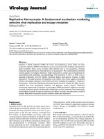
Surface plasmon resonance analysis of the NS5A(Δ250):Fyn SH3 domain interactionFigure 3
Surface plasmon resonance analysis of the NS5A(Δ250):Fyn SH3 domain interaction. SPR analysis was performed
as described for NS5A(Δ250) and GST-FynSH3. The indicated concentrations of NS5A were flowed over a sensor cell on to
which GST-FynSH3 was captured via an anti-GST antibody. The generated fit is shown as black lines in the top panels, the
residuals plotted in the lower panels indicate the relative response difference between the generated fit and the raw data –
demonstrating the closeness of fit.
Virology Journal 2008, 5:24 />Page 7 of 9
(page number not for citation purposes)
Mutational analysis of the NS5A:FynSH3 domain interactionFigure 4
Mutational analysis of the NS5A:FynSH3 domain interaction. The indicated mutations were generated in the Fyn SH3
domain – the numbering refers to the residues in the full length Fyn protein. For reference the Fyn SH3 domain commences at
residue 84 [17]. (a) Top panel – Coomassie stained SDS-PAGE of the purified GST-FynSH3 domain fusion proteins. Lower
panel – GST pulldown analysis. Purified NS5A(Δ32) was subjected to GST pulldown analysis using GA-beads alone (lane 2),
GST (lane 3) or GST-FynSH3 or mutants thereof (lanes 4–11 – corresponding to the GST-FnSH3 mutants in the lanes above).
Samples were blotted for NS5A, lane 1 shows 20% of input protein, lanes 2–11 show bound protein eluted by competition
with 20 mM reduced glutathione. ELISA analysis. GST-FynSH3 domain fusion proteins were immobilised on microplates and
incubated with purified NS5A(Δ32), prior to detection of bound NS5A with sheep anti-NS5A serum. Absorbance values were
normalised to the amount of GST fusion proteins loaded (measured by direct ELISA with an anti-GST antibody). The mean
value determined over 3 independent experiments is indicated on the graph. (b) Coomassie stained SDS-PAGE of the purified
GST-FynSH3 domain fusion proteins and ELISA analysis as in (a).
Virology Journal 2008, 5:24 />Page 8 of 9
(page number not for citation purposes)
(w/v) glucose at 37°C until OD
600
= 0.6. The culture was
chilled at 4°C for 30 minutes prior to induction with 0.2
mM IPTG at 27°C for 5 hours. Bacterial pellets were resus-
pended in buffer A (20 mM disodium orthophosphate,
pH 7.5, 0.5 M NaCl, 5 mM MgCl
2
), containing 1 mg/ml
lysozyme, 2 μg/ml DNase, 1 μg/ml RNase, 0.5% Triton X-
100 and EDTA-free complete protease inhibitor cocktail
(Roche), sonicated, clarified by centrifugation (16,000 × g
at 4°C for 1 hour) and applied to a Ni
2+
-charged NTA-
sepharose column. The column was washed extensively in
buffer A containing 20 mM imidazole and eluted in the
same buffer containing 300 mM imidazole and 0.1% Tri-
ton X-100. NS5A-containing fractions were pooled, dia-
lysed overnight against HBS buffer, clarified by
centrifugation and injected onto an Amersham XK 16/70
size exclusion column packed with Superdex75 (Amer-
sham Biosciences). Fractions containing intact protein
were again pooled and stored at -80°C until use. GST-SH3
domain fusion proteins were expressed and purified as
previously described [6].
GST pulldown assay
GST-SH3 domain fusion proteins were bound to glutath-
ione agarose (GA)-beads at 4°C for 1 h. 5 μg of purified
NS5A protein was applied to the beads and incubated at
4°C on a blood mixer for 3 hours. The beads were washed
twice in GLB (10 mM PIPES-NaOH pH 7.2, 120 mM KCl,
30 mM NaCl, 5 mM MgCl
2
, 1% (v/v) Triton X-100, 10%
(v/v) glycerol) supplemented with 0.5 M KCl, and three
times in GLB only. Bound proteins were eluted from the
GA-beads by incubating in the presence of 20 mM
reduced glutathione in GLB. Samples were analysed by
western blot and Coomassie stained SDS-PAGE to con-
firm equal GST-SH3 domain fusion protein loading onto
the GA-beads.
ELISA assay
1 μg/well of either GST or the appropriate GST-SH3
domain fusion proteins in 50 μl of PBS was coated on
Greiner bio-One PS 96-well microplates overnight at 4°C.
Wells were rinsed with PBS/0.1% (v/v) Tween-20 (PBS-T)
and blocked in PBS-T containing 5% (w/v) dried semi-
skimmed milk powder (PBS-TM) for 2 hours at room tem-
perature. After washing in PBS-T, 0.5 μg/well of purified
NS5A was added in 50 μl of PBS-T and incubated for 2
hours at 4°C. Bound NS5A was detected with a sheep pol-
yclonal anti-NS5A sera (1:5000 dilution) in PBS-TM fol-
lowed by donkey anti-sheep-HRP (Sigma). The ELISA was
developed using OPD, the reaction was stopped with 0.5
M sulphuric acid. The ELISA plate was read at 490 nm (ref-
erenced at 630 nm) using an MRX plate reader (Dynex).
All samples were run in triplicate and an average taken in
each case. To control for differential binding of the GST-
SH3 domains to the 96-well microplate, a GST loading
ELISA was utilised. In this case the primary antibody was
a mouse anti-GST mAb (Serotec), and the secondary was
a goat anti-mouse HRP conjugate (Sigma). The amount of
relative binding for each GST-SH3 fusion protein was ana-
lysed and the signal from the NS5A ELISA normalised
appropriately.
Surface Plasmon Resonance
Experiments were carried out on a Biacore 3000 in HBS
buffer (10 mM Hepes-NaOH pH 7.4, 150 mM NaCl,
0.005% Tween-20) at 25°C. An anti-GST antibody
(Biacore) was amine coupled to a CM5 sensor chip which
allowed the capture and immobilisation of 2 μg of puri-
fied GST-SH3 domains in a uniform orientation to gener-
ate a binding surface. A reference surface containing
antibody only was also prepared. Eight concentrations of
purified NS5A(Δ32) or NS5A(Δ250) (3 μM – 100 nM),
were injected across the binding or reference sensor chip
surface at 30 μl/minute for 8 minutes followed by 8 min-
utes dissociation, in triplicate. A corrected binding profile
was then generated by subtraction of the reference signal
from the binding signal for each concentration. A 1:1
Langmuir binding curve was applied to the concentration
series of each protein and the association (k
a
), dissocia-
tion (k
d
) rates and affinity constant (K
D
) were calculated.
Abbreviations
HCV: hepatitis C virus; NS5A: non-structural 5A protein;
SH3: Src homology 3.
Competing interests
The author(s) declare that they have no competing inter-
ests.
Authors' contributions
MH conceived and supervised the study. HS performed
the experiments. MH wrote the manuscript. Both authors
read and approved the final manuscript.
Acknowledgements
We thank Kalle Saksela (University of Helsinki) for reagents, Andy Baron
and Peter Stockley (University of Leeds) for advice with SPR experiments
and Andrew Macdonald (University of Leeds) for critical reading of this
manuscript. HS was the recipient of a Biomolecular Sciences Committee
PhD studentship from the Biotechnology and Biological Sciences Research
Council. Research in the MH laboratory is supported by the Wellcome
Trust, Medical Research Council and Yorkshire Cancer Research.
References
1. Shepard CW, Finelli L, Alter MJ: Global epidemiology of hepatitis
C virus infection. Lancet Infect Dis 2005, 5:558-567.
2. Macdonald A, Harris M: Hepatitis C virus NS5A: tales of a pro-
miscuous protein. J Gen Virol 2004, 85:2485-2502.
3. Brass V, Bieck E, Montserret R, Wolk B, Hellings JA, Blum HE, Penin
F, Moradpour D: An amino-terminal amphipathic alpha-helix
mediates membrane association of the hepatitis C virus non-
structural protein 5A. J Biol Chem 2002, 277:8130-8139.
4. Tellinghuisen TL, Marcotrigiano J, Gorbalenya AE, Rice CM: The
NS5A protein of hepatitis C virus is a zinc metalloprotein. J
Biol Chem 2004, 279:48576-48587.
Publish with BioMed Central and every
scientist can read your work free of charge
"BioMed Central will be the most significant development for
disseminating the results of biomedical research in our lifetime."
Sir Paul Nurse, Cancer Research UK
Your research papers will be:
available free of charge to the entire biomedical community
peer reviewed and published immediately upon acceptance
cited in PubMed and archived on PubMed Central
yours — you keep the copyright
Submit your manuscript here:
/>BioMedcentral
Virology Journal 2008, 5:24 />Page 9 of 9
(page number not for citation purposes)
5. Tellinghuisen TL, Marcotrigiano J, Rice CM: Structure of the zinc-
binding domain of an essential component of the hepatitis C
virus replicase. Nature 2005, 435:374-379.
6. Macdonald A, Crowder K, Street A, McCormick C, Harris M: The
hepatitis C virus NS5A protein binds to members of the Src
family of tyrosine kinases and regulates kinase activity. J Gen
Virol 2004, 85:721-729.
7. Tan SL, Nakao H, He YP, Vijaysri S, Neddermann P, Jacobs BL, Mayer
BJ, Katze MG: NS5A, a nonstructural protein of hepatitis C
virus, binds growth factor receptor-bound protein 2 adaptor
protein in a Src homology 3 domain/ligand-dependent man-
ner and perturbs mitogenic signaling. Proc Natl Acad Sci USA
1999, 96:5533-5538.
8. Zech B, Kurtenbach A, Krieger N, Strand D, Blencke S, Morbitzer M,
Salassidis K, Cotten M, Wissing J, Obert S, Bartenschlager R, Herget
T, Daub H: Identification and characterization of amphiphysin
II as a novel cellular interaction partner of the hepatitis C
virus NS5A protein. J Gen Virol 2003, 84:555-560.
9. Nanda SK, Herion D, Liang TJ: Src homology 3 domain of hepa-
titis C virus NS5A protein interacts with Bin1 and is impor-
tant for apoptosis and infectivity. Gastroenterology 2006,
130:794-809.
10. Macdonald A, Crowder K, Street A, McCormick C, Saksela K, Harris
M: The hepatitis C virus NS5A protein inhibits activating
protein-1 (AP1) function by perturbing Ras-ERK pathway
signalling. J Biol Chem 2003, 278:17775-17784.
11. Macdonald A, Mazaleyrat S, McCormick C, Street A, Burgoyne NJ,
Jackson RM, Cazeaux V, Shelton H, Saksela K, Harris M: Further
studies on hepatitis C virus NS5A-SH3 domain interactions:
identification of residues critical for binding and implications
for viral RNA replication and modulation of cell signalling. J
Gen Virol 2005, 86:1035-1044.
12. Lee CH, Leung B, Lemmon MA, Zheng J, Cowburn D, Kuriyan J, Sak-
sela K: A single amino acid in the SH3 domain of Hck deter-
mines its high affinity and specificity in binding to HIV-1 Nef
protein. EMBO J 1995, 14:5006-5015.
13. Hanna Z, Weng XD, Kay DG, Poudrier J, Lowell C, Jolicoeur P: The
pathogenicity of human immunodeficiency virus (HIV) type
1 Nef in CD4C/HIV transgenic mice is abolished by mutation
of its SH3-binding domain, and disease development is
delayed in the absence of Hck.
J Virol 2001, 75:9378-9392.
14. Arold S, Franken P, Strub MP, Hoh F, Benichou S, Benarous R, Dumas
C: The crystal structure of HIV-1 Nef protein bound to the
Fyn kinase SH3 domain suggests a role for this complex in
altered T cell receptor signaling. Structure 1997, 5:1361-1372.
15. Huang L, Sineva EV, Hargittai MR, Sharma SD, Suthar M, Raney KD,
Cameron CE: Purification and characterization of hepatitis C
virus non-structural protein 5A expressed in Escherichia
coli. Protein Expr Purif 2004, 37:144-153.
16. Clarke D, Griffin S, Beales L, Gelais CS, Burgess S, Harris M, Rowlands
D: Evidence for the formation of a heptameric ion channel
complex by the hepatitis C virus p7 protein in vitro. J Biol
Chem 2006, 281:37057-37068.
17. Larson SM, Davidson AR: The identification of conserved inter-
actions within the SH3 domain by alignment of sequences
and structures. Protein Sci 2000, 9:2170-2180.
18. Grabs D, Slepnev VI, Songyang Z, David C, Lynch M, Cantley LC, De
Camilli P: The SH3 domain of amphiphysin binds the proline-
rich domain of dynamin at a single site that defines a new
SH3 binding consensus sequence. J Biol Chem 1997,
272:13419-13425.
19. Supekova L, Supek F, Lee J, Chen S, Gray N, Pezacki JP, Schlapbach A,
Schultz PG: Identification of human kinases involved in hepati-
tis C virus replication by small interference RNA library
screening. J Biol Chem 2008, 283:29-36.
20. Yanagi M, StClaire M, Shapiro M, Emerson SU, Purcell RH, Bukh J:
Transcripts of a chimeric cDNA clone of hepatitis C virus
genotype 1b are infectious in vivo. Virology 1998, 244:161-172.