Báo cáo y học: "Hepatitis C Virus Serologic and Virologic Tests and Clinical Diagnosis of HCVRelated Liver Disease"
Bạn đang xem bản rút gọn của tài liệu. Xem và tải ngay bản đầy đủ của tài liệu tại đây (543.43 KB, 6 trang )
Int. J. Med. Sci. 2006, 3
35
International Journal of Medical Sciences
ISSN 1449-1907 www.medsci.org 2006 3(2):35-40
©2006 Ivyspring International Publisher. All rights reserved
Review
Hepatitis C Virus Serologic and Virologic Tests and Clinical Diagnosis of HCV-
Related Liver Disease
Stéphane Chevaliez, Jean-Michel Pawlotsky
Department of Virology, INSERM U635, Henri Mondor Hospital, University of Paris XII, Créteil, France
Corresponding address: Professor Jean-Michel PAWLOTSKY, M.D., Ph.D., Department of Virology, Hôpital Henri Mondor, 51
avenue du Maréchal de Lattre de Tassigny, 94010 Créteil, France. Tel
: +33-1-4981-2827, Fax : +33-1-4981-4831. E-mail : jean-
Received: 2006.01.12; Accepted: 2006.03.15; Published: 2006.04.01
The use of serological and virological tests has become essential in the management of hepatitis C virus (HCV) infection
in order to diagnose infection, guide treatment decisions and assess the virological response to antiviral therapy.
Virological tools include serological assays for anti-HCV antibody detection and serological determination of the HCV
genotype, and molecular assays that detect and quantify HCV RNA and determine the HCV genotype. Anti-HCV
antibody testing and HCV RNA testing are used to diagnose acute and chronic hepatitis C. Only patients with
detectable HCV RNA should be considered for pegylated interferon alfa and ribavirin therapy and the HCV genotype
should be systematically determined before treatment, as it determines the indication, the duration of treatment, the
dose of ribavirin and the virological monitoring procedure. HCV RNA monitoring during therapy is used to tailor
treatment duration in HCV genotype 1 infection, and molecular assays are used to assess the end-of-treatment and,
most importantly the sustained virological response, i.e. the endpoint of therapy.
Key words: Hepatitis C virus, serological and virological tests, HCV RNA
1. INTRODUCTION
Virological testing has become essential in the
management of hepatitis C virus (HCV) infection in order
to diagnose infection, and most importantly guide
treatment decisions and assess the virological response to
antiviral therapy.
2. VIROLOGICAL TOOLS
Serological assays
Anti-HCV antibody detection
The detection of anti-HCV antibodies in plasma or
serum is based on the use of third-generation EIAs, that
detect mixtures of antibodies directed against various
HCV epitopes. Recombinant antigens are used to capture
circulating anti-HCV antibodies onto the wells of
microtiter plates, microbeads, or specific holders adapted
to closed automated devices. The presence of anti-HCV
antibodies is revealed by anti-antibodies labeled with an
enzyme that catalyzes the transformation of a substrate
into a colored compound. The optical density (OD) ratio
of the reaction (sample OD/internal control OD) is
proportional to the amount of antibodies in the serum or
plasma sample [1]. The specificity of third-generation
EIAs for anti-HCV is greater than 99% [2]. Their
sensitivity is more difficult to determine, given the lack of
a gold standard method, but it is excellent in HCV-
infected immunocompetent patients. EIAs can be fully
automated and are well adapted to large volume testing.
Immunoblot tests are nowadays clinically obsolete given
the good performance of third-generation anti-HCV EIAs
[3].
Serological determination of the HCV genotype
The HCV genotype can be determined by seeking for
antibodies directed to genotype-specific HCV epitopes
with a competitive EIA. The currently available assay
(Murex HCV serotyping 1-6 HC02, Abbott Laboratories,
North Chicago, Illinois) identifies the type (1 to 6), but
does not discriminate among the subtypes, and provides
interpretable results in approximately 90% of chronically
infected immunocompetent patients [4]. Mixed serological
reactivities can be observed that could be related to mixed
infection although cross-reactivity or recovery from one
genotype infection and persistence of viremia with
another genotype cannot be ruled out.
Detection and quantification of HCV RNA
Qualitative, non-quantitative HCV RNA detection
Qualitative detection assays are based on the
principle of target amplification using either “classic”
polymerase chain reaction (PCR), “real-time” PCR or
TMA [5]. HCV RNA is extracted and reverse transcribed
into a double stranded complementary DNA (cDNA),
which is subsequently processed into a cyclic enzymatic
reaction leading to the generation of a large number of
detectable copies. Double-stranded DNA copies of HCV
genome are synthesized in PCR-based assays, whereas
single-stranded RNA copies are generated in TMA.
Detection of amplified products is achieved by
hybridizing the produced amplicons onto specific probes
after the reaction in “classic” PCR or TMA techniques [5].
In “real-time” PCR, each round of amplification leads to
the emission of a fluorescent signal and the number of
signals per cycle is proportional to the amount of HCV
RNA in the starting sample [5-7]. Qualitative detection
assays must detect 50 HCV RNA IU/ml or less, and have
equal sensitivity for the detection of all HCV genotypes.
The lower limit of detection of the qualitative, non
quantitative reverse-transcriptase PCR-based assay
Amplicor® HCV v2.0, or of its semi-automated version
Cobas® Amplicor® HCV v2.0 (Roche Molecular Systems,
Pleasanton, California) is 50 IU/ml, whereas that of the
Int. J. Med. Sci. 2006, 3
36
TMA-based assay Versant® HCV RNA Qualitative Assay
(Bayer HealthCare) is 10 IU/ml (Table 1). Real-time PCR
assays, which are also able to quantify HCV RNA, have
lower limits of detection of the order of 5-30 IU/ml when
they are used as purely qualitative, non-quantitative
assays.
Table 1. Characteristics of current HCV RNA assays. RT :
reverse transcriptase, PCR : polymerase chain reaction, TMA :
transcription-mediated amplification, bDNA : “branched DNA“,
NA : not applicable. *for 0.2 ml or 0.5 ml of plasma analyzed,
respectively.
Assay Manufacturer Technique Lower limit
of detection
(qualitative
assay)
Dynamic
range of
quantification
(quantitative
assay)
Amplicor®
HCV v2.0
Roche
Molecular
Systems
Manual RT-
PCR
50 IU/ml
NA
Cobas®
Amplicor®
HCV v2.0
Roche
Molecular
Systems
Semi-
automated
RT-PCR
50 IU/ml NA
Versant®
HCV RNA
Qualitative
Assay
Bayer
HealthCare
Manual
TMA
10 IU/ml NA
Amplicor
HCV
Monitor® v2.0
Roche
Molecular
Systems
Manual RT-
PCR
600 IU/ml 600-500,000
IU/ml
Cobas®
Amplicor
HCV Monitor
v2.0
Roche
Molecular
Systems
Semi-
automated
RT-PCR
600 IU/ml 600-500,000
IU/ml
LCx HCV
RNA
Quantitative
Assay
Abbott
Diagnostic
Semi-
automated
RT-PCR
25 IU/ml 25-2,630,000
IU/ml
Versant®
HCV RNA 3.0
Assay
Bayer
HealthCare
Semi-
automated
bDNA
615 IU/ml 615-7,700,000
IU/ml
Cobas®
TaqMan HCV
Test
Roche
Molecular
Systems
Semi-
automated
real-time
PCR
15 IU/ml 43-69,000,000
IU/ml
Abbott
RealTime
Abbott
Diagnostic
Semi-
automated
real-time
PCR
30 IU/ml or
12 IU/ml*
12-100,000,000
IU/ml
HCV RNA quantification
HCV RNA can be quantified by means of target
amplification techniques (competitive PCR or real-time
PCR) or signal amplification techniques (branched DNA
(bDNA) assay) [5]. Five standardized assays are
commercially available. Two of them are based on
competitive PCR : Amplicor HCV Monitor® v2.0 and its
semi-automated version Cobas® Amplicor HCV
Monitor® v2.0 (Roche Molecular Systems), and LCx®
HCV RNA Quantitative Assay (Abbott Diagnostic); one is
based on bDNA technology, Versant® HCV RNA 3.0
Assay (Bayer Healthcare) ; and two are based on real-time
PCR amplification, Cobas® TaqMan HCV Test, which can
be coupled with automated extraction in Cobas
Ampliprep® (Roche Molecular Systems), and Abbott
RealTime™ HCV assay (Abbott Diagnostics), which uses
the Abbott m2000 system and can also be coupled with an
automated extraction procedure. Table 1 shows the
respective dynamic ranges of quantification of the
currently available assays, i.e. the HCV RNA intervals
within which quantification is accurate in the
corresponding assay. HCV RNA levels falling above the
upper limit of quantification of the assay are
underestimated and the samples must be retested after
1/10 to 1/100 dilution in order to achieve accurate
quantification. The most promising approach for the
future is fully automated real-time PCR assays, which are
faster, more sensitive than classical target amplification
techniques and are not prone to carryover contamination.
Molecular determination of the HCV genotype
(genotyping)
The reference method for HCV genotype
determination is direct sequencing of the NS5B or E1
regions of HCV genome by means of “in-house”
techniques, followed by sequence alignment with
prototype sequences and phylogenetic analysis [8, 9].
These techniques must be used in molecular epidemiology
studies, where exact subtyping is needed. In clinical
practice, HCV genotype can be determined by various
commercial kits, using direct sequence analysis of the 5’
noncoding region (Trugene® 5'NC HCV Genotyping Kit,
Bayer HealthCare, Diagnostics Division, Tarrytown, New
York) or reverse hybridization analysis using genotype-
specific probes located in the 5’ noncoding region
(commercialized as INNO-LiPA HCV II, Innogenetics,
Ghent, Belgium, or Versant® HCV Genotyping Assay,
Bayer HealthCare) [10-13]. Mistyping is rare with these
techniques, but mis-subtyping may occur in 10 to 25% of
cases, related to the studied region (5’ noncoding region)
rather than the technique used. These errors have no
clinical consequences, because only the type is used for
therapeutic decision-making. An assay based on direct
sequencing of the NS5B region is currently in
development (Trugene® NS5B HCV Genotyping Kit,
Bayer HealthCare).
3. DIAGNOSIS OF HCV INFECTION
Acute hepatitis C
Patients with a suspicion of acute hepatitis C should
be tested for both anti-HCV antibodies by EIA and HCV
RNA with a sensitive technique, i.e. an HCV RNA assay
with a lower limit of detection of 50 IU/ml or less [1].
Four marker profiles can be observed according to the
presence or absence of either marker. The presence of
HCV RNA in the absence of anti-HCV antibodies is
strongly indicative of acute HCV infection, which will be
confirmed by seroconversion (i.e. the appearance of anti-
HCV antibodies) a few days to weeks later. Acutely
infected patients can also have both HCV RNA and anti-
HCV antibodies at the time of diagnosis. It is difficult, in
this case, to distinguish acute hepatitis C from an acute
exacerbation of chronic hepatitis C or an acute hepatitis of
another cause in a patient with chronic hepatitis C. Acute
hepatitis C is very unlikely if both anti-HCV antibodies
and HCV RNA are absent. It is also unlikely if anti-HCV
antibodies are present without HCV RNA. These patients
should however be retested after a few weeks because
HCV RNA can be temporarily undetectable, due to
transient, partial control of viral replication by the
immune response before replication escapes and chronic
infection establishes [14]. Apart from such cases, the
presence of anti-HCV antibodies in the absence of HCV
RNA is generally seen in patients who have recovered
from a past HCV infection. Nevertheless, this pattern
cannot be differentiated from a false positive EIA result,
the exact prevalence of which is unknown.
Int. J. Med. Sci. 2006, 3
37
Chronic hepatitis C
In patients with clinical or biological signs of chronic
liver disease, chronic hepatitis C is certain when both anti-
HCV antibodies and HCV RNA (sought for with a
sensitive technique, detecting 50 IU/ml or less) are
present [3, 15]. Detectable HCV replication in the absence
of anti-HCV antibodies is exceptional with the current
third-generation EIAs, almost exclusively observed in
profoundly immunodepressed patients, hemodialysis
patients or agammaglobulinemic subjects [16, 17].
In patients who have no indication for therapy or
have a contra-indication to the use of antiviral drugs,
virological tests have no prognostic value. Indeed, neither
anti-HCV antibodies nor the HCV RNA load correlate
with the severity of liver inflammation or fibrosis nor with
their progression. Thus, they cannot be used to predict the
natural course of infection or the onset of extrahepatic
manifestations. In untreated patients, the severity of liver
inflammation and fibrosis must be evaluated every three
to five years by means of a liver biopsy or non-invasive
serological or ultrasound-based testing [18].
4. MANAGEMENT OF ANTIVIRAL THERAPY
The current standard treatment for chronic hepatitis
C is the combination of pegylated interferon (IFN) alfa
and ribavirin [18]. The efficacy endpoint of hepatitis C
treatment is the “sustained virological response” (SVR),
defined by the absence of detectable HCV RNA in serum
as assessed by an HCV RNA assay with a lower limit of
detection of 50 IU/ml or less 24 weeks after the end of
treatment [18].
Initiation of therapy
Only patients with detectable HCV RNA should be
considered for pegylated IFN alfa and ribavirin
combination therapy [18]. The decision to treat patients
with chronic hepatitis C depends on multiple parameters,
including a precise assessment of the severity of liver
disease and of its foreseeable outcome, the presence of
absolute or relative contra-indications to therapy, and the
patient’s willingness to be treated.
The HCV genotype should be systematically
determined before treatment, as it determines the
indication, the duration of treatment, the dose of ribavirin
and the virological monitoring procedure [19].
HCV genotype 1
Given the likelihood of a sustained virological
response, of the order of 40% to 50%, a precise assessment
of liver disease prognosis by means of a liver biopsy or a
non-invasive method based on serological markers of
fibrosis or ultrasound-based testing [20, 21] must be
performed in order to help with the treatment decision
(Figure 1A). It is recommended not to treat patients with
mild lesions and to re-assess their liver disease after 3 to 5
years. The patients with inflammation and/or fibrosis
(Metavir score A ≥ 2 and/or F ≥ 2) have an indication for
therapy [18].
The approved dose of pegylated IFN alfa-2a is 180 µg
per week, independent of body weight, whereas that of
pegylated IFN alfa-2b is weight-adjusted at 1.5 µg/kg per
week, identical for all HCV genotypes. Patients infected
with HCV genotype 1 should receive a high dose of
ribavirin, i.e. 1,000 to 1,200 mg daily, based on body
weight less than or greater than 75 kg (it has been recently
suggested that the heaviest patients could even benefit
from a higher ribavirin dose, up to 1,600 mg daily) and
they theoretically require 48 weeks of treatment (Figure
1A) [18]. However, monitoring of HCV RNA load
decrease during therapy is recommended in order to
avoid treating for 48 weeks patients with no likelihood of
an SVR [22, 23]. In this purpose, HCV RNA quantification
should be performed at baseline and after 12 weeks of
treatment (Figure 1A) [18]. Both measures must be
performed with the same technique in order to ensure
comparability of the results at the two time points.
Treatment must be continued when there is a 2-log drop
in HCV RNA level, i.e. when baseline HCV RNA level is
divided by 100 or more, or when HCV RNA is
undetectable at week 12 [18]. In these patients, it is
recommended to assess the presence of HCV RNA with a
sensitive technique (lower limit of detection : 50 IU/ml or
less) at week 24. If HCV RNA is undetectable at week 24,
treatment must be continued until week 48, with a high
likelihood of an SVR. It was recently suggested that 24
weeks of therapy might be sufficient for patients with a
baseline viral load below 600,000 IU/ml in whom
pegylated IFN alfa-2b-based treatment yields a 2-log
decline at week 12 and undetectable HCV RNA at week
24 [24]. In contrast, if HCV RNA is still detectable at week
24, the likelihood of an SVR is virtually nil and treatment
can be stopped or continued with the only aim to slow
liver disease progression in patients with a severe
prognosis, without any hope to eradicate infection (Figure
1A) [18, 22]. Ongoing trials are studying whether a
prolonged antiviral treatment or maintenance therapy
with pegylated IFN alfa monotherapy could be beneficial
in the latter patients. When treatment is continued until
week 48, the end-of-treatment and sustained virological
responses should be assessed by means of a sensitive
HCV RNA assay, with a lower limit of detection of 50
IU/ml or less [18]. HCV RNA detection at the end of
therapy is highly predictive of a post-treatment relapse,
whereas the absence of HCV RNA at the end of treatment
indicates a virological response. These patients must be
retested for HCV RNA with a sensitive method 24 weeks
later in order to assess the SVR, i.e. the endpoint of
therapy [1, 18]. HCV infection appears to be definitively
cured in the vast majority of sustained virological
responders.
The lack of a 12-week virological response (no
change or an HCV RNA decrease of less than 2 logs at
week 12) is associated with a virtually nil probability of a
subsequent sustained virological response [22, 23].
Treatment can thus be stopped at week 12 in these
patients, or continued to slow liver disease progression
without clearing the virus (Figure 1A). The benefits of
maintenance therapy on the outcome of HCV-associated
liver disease are currently under investigation. This
“stopping rule”, based on monitoring of HCV RNA load
reduction at week 12, was recently shown to also apply to
patients co-infected with HCV and human
immunodeficiency virus [25-27].
Int. J. Med. Sci. 2006, 3
38
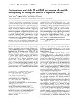
Figure 1. Current algorithms for the use of HCV virological tools in the treatment of chronic hepatitis C, according to the HCV
genotype: genotype 1 (A), genotypes 2 and 3 (B), and genotypes 4, 5 and 6 (C).
Int. J. Med. Sci. 2006, 3
39
HCV genotypes 2 and 3
Patients infected with HCV genotypes 2 or 3 have a
70%-80% likelihood of an SVR with a low dose of ribavirin
and only 24 weeks of treatment [19, 23, 28]. Thus, in the
absence of contra-indications, these patients should be
treated regardless of the severity of their liver disease and
they do not need a liver biopsy of noninvasive assessment
of liver fibrosis (Figure 1B). The recommended dose of
pegylated IFN alfa-2a or alfa-2b is the same as for HCV
genotype 1, i.e. 180 μg/week and 1.5 μg/kg/week,
respectively. The fixed recommended dose of ribavirin is
800 mg per day (Figure 1B) [18]. It is possible that even
lower doses of ribavirin and/or shorter duration of
treatment could be sufficient to achieve an SVR in certain
subgroups of patients with genotype 2 or 3 infection, such
as those with a low baseline viral load and no extensive
fibrosis or cirrhosis, as suggested by recent preliminary
data [29]. One should be careful in patients who combine
several baseline parameters of non-response, such as
extensive fibrosis, an old age and a male gender, who
might need 48 weeks of therapy to clear infection.
No monitoring of HCV RNA level changes during
therapy is recommended in the patients with genotype 2
or 3 infection, because the vast majority of them become
HCV RNA-negative early during treatment. Like in HCV
genotype 1-infected patients, the virological response
must be assessed by means of a sensitive HCV RNA assay
at the end of therapy and 24 weeks later in order to
determine whether the virological response is sustained
(Figure 1B) [1, 18].
HCV genotypes 4, 5 and 6
In the absence of any clinical trial including a
sufficient number of patients, the likelihood of an SVR
and the optimal treatment schedule remain unknown for
the patients infected with HCV genotypes 4, 5 or 6. It is
thus recommended to treat them like those infected with
HCV genotype 1, i.e. with pegylated IFN alfa at the usual
dose, combined with a high dose of ribavirin (1000-1200
mg per day, according to body weight less or greater than
75 kg) (Figure 1C). In the absence of published data, no
stopping rules have been defined and it is recommended
to treat these patients for a total of 48 weeks. The
virological response must be assessed by means of a
sensitive HCV RNA assay (lower limit of detection of 50
IU/ml or less) at the end of therapy and 24 weeks later [1,
18].
Conflict of interest
The authors have declared that no conflict of interest
exists.
REFERENCES
1. Pawlotsky JM. Use and interpretation of virological tests for hepatitis
C. Hepatology 2002, 36 (Suppl 1): S65-73.
2. Colin C, et al. Sensitivity and specificity of third-generation hepatitis
C virus antibody detection assays: an analysis of the literature. J
Viral Hepat 2001, 8: 87-95.
3. Pawlotsky JM, et al. What strategy should be used for diagnosis of
hepatitis C virus infection in clinical laboratories? Hepatology 1998,
27: 1700-2.
4. Pawlotsky JM, et al. Serological determination of hepatitis C virus
genotype: comparison with a standardized genotyping assay. J Clin
Microbiol 1997, 35: 1734-9.
5. Pawlotsky JM. Molecular diagnosis of viral hepatitis.
Gastroenterology 2002, 122:1554-68.
6. Martell M, et al. High-throughput real-time reverse transcription-
PCR quantitation of hepatitis C virus RNA. J Clin Microbiol 1999,
37:327-32.
7. Komurian-Pradel F, et al. Quantitation of HCV RNA using real-time
PCR and fluorimetry. J Virol Methods 2001, 95: 111-9.
8. Simmonds P. Viral heterogeneity of the hepatitis C virus. J Hepatol
1999, 31 (Suppl 1): 54-60.
9. Simmonds P, et al. Consensus proposals for a unified system of
nomenclature of hepatitis C virus genotypes. Hepatology 2005, 42:
962-73.
10. Germer JJ, et al. Automated sample preparation for the Trugene
HIV-1 genotyping kit using the MagNA pure LC instrument. Diagn
Microbiol Infect Dis 2004, 49: 59-61.
11. Stuyver L, et al. Second-generation line probe assay for hepatitis C
virus genotyping. J Clin Microbiol 1996, 34: 2259-66.
12. Stuyver L, et al. Hepatitis C virus genotyping by means of 5'-
UR/core line probe assays and molecular analysis of untypeable
samples. Virus Res 1995, 38: 137-57.
13. Zheng X, et al. Direct comparison of hepatitis C virus genotypes
tested by INNO-LiPA HCV II and TRUGENE HCV genotyping
methods. J Clin Virol 2003, 28: 214-6.
14. Lavillette D, et al. Human serum facilitates hepatitis C virus
infection, and neutralizing responses inversely correlate with viral
replication kinetics at the acute phase of hepatitis C virus infection. J
Virol 2005, 79: 6023-34.
15. [No authors listed]. EASL International Consensus Conference on
Hepatitis C. Paris, 26-28, February 1999, Consensus Statement.
European Association for the Study of the Liver. J Hepatol 1999, 30:
956-61.
16. Lok AS, et al. Antibody response to core, envelope and nonstructural
hepatitis C virus antigens: comparison of immunocompetent and
immunosuppressed patients. Hepatology 1993, 18: 497-502.
17. Thio CL, et al. Screening for hepatitis C virus in human
immunodeficiency virus-infected individuals. J Clin Microbiol 2000,
38: 575-7.
18. [No authors listed]. NIH Consensus Statement on Management of
Hepatitis C: 2002. NIH Consens State Sci Statements 2002, 19: 1-46.
19. Hadziyannis SJ, et al. Peginterferon-alpha2a and ribavirin
combination therapy in chronic hepatitis C: a randomized study of
treatment duration and ribavirin dose. Ann Intern Med 2004, 140:
346-55.
20. Castera L, et al. Prospective comparison of transient elastography,
Fibrotest, APRI, and liver biopsy for the assessment of fibrosis in
chronic hepatitis C. Gastroenterology 2005, 128: 343-50.
21. Poynard T, et al. FibroTest-FibroSURE: towards a universal
biomarker of liver fibrosis? Expert Rev Mol Diagn 2005, 5: 15-21.
22. Davis GL, et al. Early virologic response to treatment with
peginterferon alfa-2b plus ribavirin in patients with chronic hepatitis
C. Hepatology 2003, 38: 645-52.
23. Fried MW, et al. Peginterferon alfa-2a plus ribavirin for chronic
hepatitis C virus infection. N Engl J Med 2002, 347: 975-82.
24. Zeuzem S, et al. Efficacy of 24 weeks treatment with peginterferon
alfa-2b plus ribavirin in patients with chronic hepatitis C infected
with genotype 1 and low pretreatment viremia. J Hepatol 2006, 44:
97-103.
25. Chung RT, et al. Peginterferon Alfa-2a plus ribavirin versus
interferon alfa-2a plus ribavirin for chronic hepatitis C in HIV-
coinfected persons. N Engl J Med 2004, 351: 451-9.
26. Pawlotsky JM. Treating hepatitis C in "difficult-to-treat" patients. N
Engl J Med 2004, 351: 422-3.
27. Torriani FJ, et al. Peginterferon Alfa-2a plus ribavirin for chronic
hepatitis C virus infection in HIV-infected patients. N Engl J Med
2004, 351: 438-50.
28. Manns MP, et al. Peginterferon alfa-2b plus ribavirin compared with
interferon alfa-2b plus ribavirin for initial treatment of chronic
hepatitis C: a randomised trial. Lancet 2001, 358: 958-65.
29. Dalgard O, et al. Treatment with pegylated interferon and ribavarin
in HCV infection with genotype 2 or 3 for 14 weeks: a pilot study.
Hepatology 2004, 40: 1260-5.