Báo cáo hóa học: " Subcellular forms and biochemical events triggered in human cells by HCV polyprotein expression from a viral vector" doc
Bạn đang xem bản rút gọn của tài liệu. Xem và tải ngay bản đầy đủ của tài liệu tại đây (1.69 MB, 20 trang )
BioMed Central
Page 1 of 20
(page number not for citation purposes)
Virology Journal
Open Access
Research
Subcellular forms and biochemical events triggered in human cells
by HCV polyprotein expression from a viral vector
Andrée M Vandermeeren
1
, Carmen Elena Gómez
1
, Cristina Patiño
2
,
Elena Domingo-Gil
1
, Susana Guerra
1
, Jose Manuel González
1
and
Mariano Esteban*
1
Address:
1
Department of Molecular and Cellular Biology, Centro Nacional de Biotecnología, CSIC, Campus Universidad Autónoma, E-28049,
Madrid, Spain and
2
Electron Microscopy Service, Centro Nacional de Biotecnología, CSIC, Campus Universidad Autónoma, E-28049, Madrid,
Spain
Email: Andrée M Vandermeeren - ; Carmen Elena Gómez - ;
Cristina Patiño - ; Elena Domingo-Gil - ; Susana Guerra - ;
Jose Manuel González - ; Mariano Esteban* -
* Corresponding author
Abstract
To identify the subcellular forms and biochemical events induced in human cells after HCV
polyprotein expression, we have used a robust cell culture system based on vaccinia virus (VACV)
that efficiently expresses in infected cells the structural and nonstructural proteins of HCV from
genotype 1b (VT7-HCV7.9). As determined by confocal microscopy, HCV proteins expressed from
VT7-HCV7.9 localize largely in a globular-like distribution pattern in the cytoplasm, with some
proteins co-localizing with the endoplasmic reticulum (ER) and mitochondria. As examined by
electron microscopy, HCV proteins induced formation of large electron-dense cytoplasmic
structures derived from the ER and containing HCV proteins. In the course of HCV protein
production, there is disruption of the Golgi apparatus, loss of spatial organization of the ER,
appearance of some "virus-like" structures and swelling of mitochondria. Biochemical analysis
demonstrate that HCV proteins bring about the activation of initiator and effector caspases
followed by severe apoptosis and mitochondria dysfunction, hallmarks of HCV cell injury.
Microarray analysis revealed that HCV polyprotein expression modulated transcription of genes
associated with lipid metabolism, oxidative stress, apoptosis, and cellular proliferation. Our findings
demonstrate the uniqueness of the VT7-HCV7.9 system to characterize morphological and
biochemical events related to HCV pathogenesis.
Background
Hepatitis C virus (HCV) infection is a major cause of
chronic hepatitis, liver cirrhosis and hepatocellular carci-
noma [1]. With over 170 million people chronically
infected with HCV worldwide, this disease has emerged as
a serious global health problem.
The HCV virus is the sole member of the genus hepacivi-
rus which belongs to the Flaviviridae family, represented
by six major genotypes. The viral genome is a positive
polarity single-stranded RNA molecule of approximately
9.5 kb in length that has a unique open-reading frame,
coding for a single polyprotein. The length of the polypro-
Published: 15 September 2008
Virology Journal 2008, 5:102 doi:10.1186/1743-422X-5-102
Received: 21 July 2008
Accepted: 15 September 2008
This article is available from: />© 2008 Vandermeeren et al; licensee BioMed Central Ltd.
This is an Open Access article distributed under the terms of the Creative Commons Attribution License ( />),
which permits unrestricted use, distribution, and reproduction in any medium, provided the original work is properly cited.
Virology Journal 2008, 5:102 />Page 2 of 20
(page number not for citation purposes)
tein-encoding region varies according to the isolate and
genotype of the virus from 3,008 to 3,037 amino acids.
After virus entry and uncoating, the viral genome serves as
template for the translation of the single polyprotein
which is processed by cellular and viral proteases to yield
the mature structural (Core-E1-E2-p7) and nonstructural
proteins (NS2-NS3-NS4A-NS4B-N5A-NS5B) [2,3].
Despite the identification of HCV as the most common
etiologic agent of posttransfusion and sporadic non-A,
non-B hepatitis, its replication cycle and pathogenesis are
incompletely understood. Improvement has been made
using heterologous expression systems, functional full-
length cDNA clones, and subgenomic replicons [4-6]. The
recent establishment of a cell culture system for HCV
propagation is a major progress to analyse the complete
viral life cycle and HCV virus-host interactions [7-9].
The impact of HCV polyprotein expression in human cells
has been hampered by limitations of different cell systems
to express the entire HCV polyprotein in high yields and
in all cells. Vaccinia virus (VACV), a prototype member of
the poxvirus family, has proven to be a useful vector for
faithful expression of many proteins in cells [10,11]. We
have previously described a novel poxvirus-based delivery
system that is inducible and expresses the structural and
nonstructural (except C-terminal part of NS5B) proteins
of HCV ORF from genotype 1b [12]. In this model, we
observed that HCV proteins control cellular translation
through eIF-2α-S51 phosphorylation, with involvement
of the double-stranded RNA-dependent protein kinase
PKR. Moreover, in VT7-HCV7.9 infected cells HCV pro-
teins bring about an apoptotic response through the acti-
vation of the RNase L pathway [12].
As it has been considered that the viral cytopathic effect
might be involved in the liver-cell injuries [1,2,13], here
we have analyzed in detail the subcellular forms and bio-
chemical changes occurring in human cells (HeLa and
hepatic HepG2) following expression of the HCV poly-
protein from VACV recombinant. We found that the pro-
duction of HCV proteins in the host cell from 4 to 48 h
induced severe cellular damage with modifications in cell
organelles, formation of large cytoplasmic membrane
structures and activation of death pathways, hallmarks of
HCV cell injury. In addition, we analyzed by microarray
technology the gene expression profile of HeLa cells
infected with VT7-HCV7.9 recombinant and identified
genes that were differentially regulated by HCV proteins
and are related with HCV pathogenesis. The morphologi-
cal and biochemical changes triggered in human cells by
HCV polyprotein expression highlight the use of the pox-
virus-based system as a suitable model in the study of the
biology of HCV infection and morphogenesis, host-cell
interactions and drug-treatment.
Results
HCV proteins induced disruption of the Golgi apparatus
and co-localized with ER and mitochondria markers
We have previously described that the DNA fragment of
HCV ORF from genotype 1b included in the VT7-HCV
7.9
recombinant is efficiently transcribed and translated upon
induction with IPTG into a viral polyprotein precursor
that is correctly processed into mature structural and non-
structural (except the C-terminal part of NS5B) HCV pro-
teins [12].
To identify the cytoplasmic compartment(s) in which
viral HCV proteins accumulated during infection of HeLa
cells with VT7-HCV
7.9
, we performed immunofluores-
cence analysis using serum from an HCV-infected patient
to recognize HCV proteins and antibodies specific for the
Golgi apparatus (anti-gigantin), the endoplasmic reticu-
lum (anti-calnexin) or the mitochondria (mitotracher)
(Fig. 1). Inducible expression of HCV proteins caused
severe disruption of the Golgi apparatus as revealed by
labelling this organelle with a specific antibody (Fig. 1A).
This effect was not observed in cells infected with VT7-
HCV
7.9
in the absence of IPTG. There is no co-localization
of HVC proteins with the disrupted Golgi, whereas in the
labelling of the endoplasmic reticulum, a clear co-locali-
zation between HCV proteins expressed from VT7-HCV
7.9
and ER proteins was observed (Fig. 1B). With an in vivo
mitochondrial marker (Fig. 1C), we detected partial co-
localization between HCV proteins expressed from VT7-
HCV
7.9
and mitochondria organelles. Moreover, the mito-
chondria appeared more rounded in cells infected with
VT7-HCV
7.9
+ IPTG, in comparison with uninfected cells
or with cells infected in the absence of IPTG.
HCV polyprotein expression in human HeLa and HepG2
cells induces severe morphological alterations and
production of electron dense structures in the cytoplasm
surrounded by membranes
To gain more detail information on the subcellular
changes induced by HCV polyprotein expression, we per-
formed transmission electron microscopy (EM) analysis.
HeLa cells were infected with VT7-HCV
7.9
in the presence
or absence of IPTG, and at 16 h p.i, infected and unin-
fected cells were collected and ultrathin sections visual-
ized by EM at low and high magnification. While in cells
infected with VT7-HCV
7.9
, in the absence of IPTG there are
high number of immature (IVs) and intracellular mature
(IMVs) forms of VACV virus, in cells infected with VT7-
HCV
7.9
in the presence of IPTG fewer IVs and IMVs were
observed, corroborating our previous finding that the
expression of HCV proteins blocked VACV morphogene-
sis [12]. In addition, several morphological alterations
were detected in cells expressing the HCV polyprotein
when compared with uninfected cells (Fig. 2A) or with
cells only expressing VACV proteins (Fig. 2B). The first
Virology Journal 2008, 5:102 />Page 3 of 20
(page number not for citation purposes)
alteration seen was the loss of spatial organization of the
ER, with vesicles embedded in a membrane matrix of cir-
cular or tightly undulating membranes, forming electron
dense structures indicated as EDS (Fig. 2C and 2D). These
cytoplasmic structures resemble those structures called
"membraneous webs" that have been visualized in
human hepatoma Huh7 cells expressing a subgenomic
HCV replicon [5,14,15]. Other alterations observed were
the presence of vacuoles (indicated as V) often surround-
ing the compact structures, as well as the presence of swol-
len mitochondria (indicated as m) (Fig. 2D and 2E).
Higher magnification of the electron dense structures in
cells expressing the HCV polyprotein revealed membranes
and tubular structures (indicated as TS) as part of the EDS
(Fig. 2E).
Since hepatocytes are the main targets of HCV virus, we
next analyzed if expression of HCV polyprotein from the
VT7-HCV
7.9
infected cells also affected the ultra-structure
of hepatic cells. Thus, monolayers of a human hepatoblast
cell line (HepG2) were infected with VT7-HCV
7.9
under
the same conditions as for HeLa cells and processed at 16
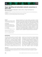
Compartmentalization of HCV proteins produced in HeLa cells infected with VT7-HCV
7.9
Figure 1
Compartmentalization of HCV proteins produced in HeLa cells infected with VT7-HCV
7.9
. Subconfluent HeLa
cells uninfected or infected at 5 PFU/cell with the recombinant VT7-HCV7.9 in the presence (+) or absence (-) of the inducer
IPTG, were fixed at 24 h p.i, labelled with the corresponding primary antibody followed by the appropriate fluorescent second-
ary antibody and visualized by confocal microscopy. The antibodies or reagents used were Hα HCV to detect HCV proteins;
Topro-3 to detect DNA; Rα Giantin to detect the Golgi complex (A); Rα Calnexine to detect the endoplasmatic reticulum
(B) and Mitotracker Deep Red 633 to detect mitochondria (C). The co-localization is shown with a higher resolution in the
white square.
Virology Journal 2008, 5:102 />Page 4 of 20
(page number not for citation purposes)
Figure 2 (see legend on next page)
Virology Journal 2008, 5:102 />Page 5 of 20
(page number not for citation purposes)
h p.i for EM analysis. In this cell line, similarly as in HeLa
cells, the inducible expression of HCV proteins blocks
VACV morphogenesis and induces the same alterations
described above. In contrast to uninfected (Fig 3A) and
infected hepG2 cells in absence of IPTG (Fig. 3B), in cells
expressing HCV proteins we distinguish EDS in membra-
nous webs (Fig. 3C and 3E), formation of large vacuoles
(Fig. 3C and 3D), and also identified "virus like-particles"
structures surrounded by membranes and dispersed in
several areas of the cell cytoplasm (Fig. 3D).
Immunogold electron microscopy revealed that HCV
proteins are part of the electron dense and membranous
structures
To assure that the electron dense structures appearing in
the cytoplasm of infected cells are the result of HCV poly-
protein expression, we performed immunogold electron
microscopy analysis with antibodies against HCV struc-
tural and nonstructural proteins (Fig. 4). Thus, HeLa cells
were infected with VT7-HCV
7.9
in the presence or absence
of IPTG and at 16 h p.i, infected and uninfected cells were
processed for immunogold labelling on ultrathin sec-
tions. Due to the fixation and embedding procedures used
in immunostaining, the cell structures are less visible than
by embedding in an epoxy resin. While in cells infected
with VT7-HCV
7.9
in absence of IPTG there was no specific
labelling detected with the serum from an HCV-infected
patient (Fig. 4A), in contrast, in antibody-reacted cells
expressing HCV proteins most gold particles were concen-
trated into electron dense and membranous structures
(Fig. 4B). Discrete labelling was observed in other parts of
the cell cytoplasm. The localization of some of the non-
structural HCV proteins was determined using rabbit pol-
yclonal antibodies against NS4B or NS5A proteins. The
membranous and electron dense structures were also spe-
cifically recognized by antibodies against NS4B (Fig. 4C)
and NS5A (Fig. 4D), indicating that both proteins are part
of electron dense membrane-associated cytoplasmic com-
plexes.
Since by confocal microscopy we observed co-localization
between ER and HCV proteins in cells infected with VT7-
HCV
7.9
in the presence of IPTG (see Fig. 1B), we per-
formed immunogold labelling using a specific ER marker
(mouse anti-PDI). As seen in Fig. 4E, strong labelling of
ER was found in the membranous webs. These results sug-
gest that the membranous webs are ER-derived structures.
As the staining pattern corresponds to that obtained with
the NS4B or NS5A proteins, the immunogold electron
microscopy indicates that the ER is a site where these pro-
teins are localized.
HCV polyprotein expression results in mitochondrial
dysfunction, as revealed by release of cytochrome c, loss of
membrane potential and generation of reactive oxygen
species (ROS)
The detection by confocal microscopy of the presence of
HCV proteins in the mitochondria (see Fig. 1C) suggests
that HCV may regulate the mitochondria homeostasis. To
confirm that, we evaluated different parameters such as,
release of proapototic proteins including cytochrome c,
loss of mitochondrial membrane potential (ΔΨm) and
production of reactive oxygen species (ROS), all hall-
marks of mitochondrial dysfunction.
To determine whether HCV polyprotein expression from
the VACV recombinant activates cytochrome c release,
HeLa cells were infected with VT7-HCV
7.9
in the presence
or absence of IPTG, or treated with staurosporine (as a
positive control). The cytochrome c release was detected
by confocal microscopy. As shown in Fig. 5A, the cyto-
chrome c remained confined to the mitochondria in both
uninfected cells and VT7-HCV
7.9
infected cells in the
absence of IPTG. However, in cells infected with VT7-
HCV
7.9
in the presence of IPTG, there is a diffuse cytosolic
pattern of cytochrome c staining, similarly as in cells
treated with staurosporine, indicating that cytochrome c
was released from the mitochondria.
Next we determine if HCV polyprotein expression affected
the mitochondria membrane potential (ΔΨm). HeLa cells
were infected either with VT7-HCV
7.9
in the presence or
absence of IPTG, or treated with staurosporine. At 48 h p.i,
cells were stained in vivo with a fluorescent mitochon-
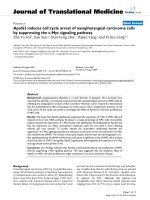
Alterations in the architecture of HeLa cells following expression of HCV proteins from VT7-HCV
7.9
seen by electron micros-copyFigure 2 (see previous page)
Alterations in the architecture of HeLa cells following expression of HCV proteins from VT7-HCV
7.9
seen by
electron microscopy. HeLa cells uninfected or infected with the recombinant VT7-HCV7.9 in the presence or absence of
the inducer IPTG, were chemically fixed at 16 h p.i and then processed for conventional embedding in an epoxy resin as
described under Materials and Methods.A: Cellular architecture of uninfected HeLa cells. B: A general view of a cell infected
with VT7-HCV7.9 in the absence of IPTG, showing the VACV forms IVs and IMVs. C and D: A general view of cells infected
with VT7-HCV7.9 in the presence of IPTG, showing few IVs, large EDS, swollen mitochondria and vacuoles. E: High magnifica-
tion of infected VT7-HCV7.9 cells in the presence of IPTG showing EDS with membranes, TS and swollen mitochondria with a
protruding membrane. Note: Nucleus (N), mitochondria (m), Golgi apparatus (G), immature virus (IV), intracellular mature
virus (IMV), tubular structures (TS), electron dense structures in membranous webs (EDS). Bar: 500 nm.
Virology Journal 2008, 5:102 />Page 6 of 20
(page number not for citation purposes)
Hepatocyte cell alterations following infection of HepG2 with VT7-HCV7.9Figure 3
Hepatocyte cell alterations following infection of HepG2 with VT7-HCV7.9. HepG2 cells uninfected or infected with
the recombinant VT7-HCV7.9 in the presence or absence of the inducer IPTG were chemically fixed at 16 h p.i and then proc-
essed for conventional embedding in an epoxy resin.A: Cellular architecture of uninfected HepG2 cells. B: A general view of a
cell infected with VT7-HCV7.9 in the absence of IPTG, showing the VACV forms IVs and IMVs.C, D and E: A general view of
a cell infected with VT7-HCV7.9 in the presence of IPTG, showing large EDS surrounded by vacuoles and the presence of
"virus-like particles" surrounded with membranes (*). Note: Vacuole (V) and electron dense structures in membranous webs
(EDS). Bar: 200 nm.
Virology Journal 2008, 5:102 />Page 7 of 20
(page number not for citation purposes)
Immunogold electron microscopy analysis of the localization of HCV proteins in VT7-HCV
7.9
infected HeLa cellsFigure 4
Immunogold electron microscopy analysis of the localization of HCV proteins in VT7-HCV
7.9
infected HeLa
cells. HeLa cells infected with VT7-HCV7.9 in the presence or absence of IPTG were chemically fixed, quickly frozen in liquid
propane and then processed at low temperature in Lowicryl K4M resin. Immunogold labelling was performed with different
antibodies. A: Cells infected with VT7-HCV7.9 in the absence of IPTG reacted with a serum from an HCV-infected patient.B:
Cells infected with VT7-HCV7.9 in the presence of IPTG reacted with a serum from an HCV-infected patient C: Cells infected
with VT7-HCV7.9 in the presence of IPTG reacted with a rabbit polyclonal anti-NS4B. D: Cells infected with VT7-HCV7.9 in
the presence of IPTG reacted with a rabbit polyclonal anti-NS5A. E: Cells infected with VT7-HCV7.9 in the presence of IPTG
reacted with a mouse monoclonal antibody anti-PDI. Note: Electron dense structures in membranous webs (EDS); mitochon-
dria (m), immature virus (IV), intracellular mature virus (IMV), nucleus (N) and Vacuole (V). Bar: 250 nm.
Virology Journal 2008, 5:102 />Page 8 of 20
(page number not for citation purposes)
HCV polyprotein expression induced dysfunction of the mitochondriaFigure 5
HCV polyprotein expression induced dysfunction of the mitochondria. A: HeLa cells uninfected or infected at 5 PFU/
cell with the recombinant VT7-HCV7.9 in the presence or absence of IPTG were labelled in vivo at 24 h p.i with Mitotracker
deep red (blue) to detect the mitochondria, with an anti-cytochrome c (red) antibody and with the serum from an HCV-
infected patient to detect HCV proteins. Cells treated with staurosporine at 0.5 μM for 16 h were used as positive control. B:
HeLa cells were either infected at 5 PFU/cell with the recombinant VT7-HCV7.9 in the presence or absence of IPTG, or
treated with staurosporine at 0.5 μM for 16 h. At 48 h p.i, the mitochrondrial membrane potential (ΔΨm) was determined
quantifying TMRE fluorescence. C: HeLa cells were either infected at 5 PFU/cell with the recombinant VT7-HCV7.9 in the
absence or presence of IPTG or treated with staurosporine at 0.5 μM for 16 h. At 48 h p.i, the uninfected and infected cells
were stained with dihydroethidium (2-HE) and subjected to flow cytometry. Note: STS: staurosporine.
Virology Journal 2008, 5:102 />Page 9 of 20
(page number not for citation purposes)
drion-specific dye, TMRE [16,17], and analysed by flow
cytometry. The loss of the ΔΨm was assessed by the ability
of the mitochondria to take up TMRE. As shown in Fig.
5B, FACS analysis demonstrated a higher proportion of
cells with decreased ΔΨm (ΔΨm Low) in HCV polypro-
tein expressing cells and in staurosporine treated cells, in
contrast with cells infected with the VT7-HCV
7.9
in the
absence of IPTG or in uninfected cells. These results indi-
cate the ability of the HCV proteins to disrupt the mito-
chondria membrane potential in HeLa cells.
As mitochondrial dysfunction is also characterized by the
generation of reactive oxygen species (ROS) [18], we
investigated whether HCV polyprotein expression trig-
gered the generation of ROS. HeLa cells were infected with
VT7-HCV
7.9
in the presence or absence of IPTG and at 48
h p.i, cells were stained with dihydroethidium (2-HE) and
subjected to flow cytometry [19]. As shown in Fig. 5C,
there is clearly production of ROS, as revealed by an
increase in ethidium staining of DNA in HeLa cells
infected with VT7-HCV
7.9
in the presence of IPTG. In con-
trast, ROS production was significantly lower (about
10%) in both uninfected cells and VT7-HCV
7.9
infected
cells in the absence of IPTG.
The above results demonstrate that HCV proteins induce
mitochondrial dysfunction evidenced by the release of
cytochrome c, mitochondrial membrane depolarization
and generation of ROS.
Expression of HCV proteins induces apoptosis through
activation of initiator and effector caspases
It has been reported in hepatic cells that expression of
structural and nonstructural proteins from HCV cDNA
[20] or from full-length RNA [21], can lead to apoptotic
cell death, which could be an important event in the
pathogenesis of chronic HCV infection in humans. We
have previously shown by an ELISA-based assay that the
inducible expression of HCV proteins from VT7-HCV
7.9
triggers apoptosis [12]. In view of the severe cellular dam-
age caused by polyprotein expression in VT7-HCV
7.9
infected cells, we wished to extend our previous observa-
tion by characterizing the apoptotic pathways in this
virus-cell system. We first performed a qualitative estima-
tion of apoptosis in HeLa cells infected with VT7-HCV
7.9
in the presence or absence of IPTG. By phase contrast
microscopy and DNA staining analysis we observed that
cells expressing HCV polyprotein developed at 24 h p.i
characteristic morphological changes of apoptosis, as
defined by cell shrinkage, granulated appearance, mem-
brane bledding and chromatin condensation (not
shown). Such morphological changes were not observed
in cells infected with VT7-HCV
7.9
in the absence of the
inducer.
To quantify the extent of apoptosis, DNA content was ana-
lyzed by flow cytometry after permeabilization and label-
ling with the DNA-specific fluorochrome propidium
iodide. As shown by flow cytometry, 78% of HeLa cells
infected with VT7-HCV
7.9
in the presence of IPTG were
apoptotic in contrast with the 26% determined when cells
were infected with VT7-HCV
7.9
in the absence of the
inducer (Fig. 6A).
Since DNA fragmentation represents a late apoptotic
event, we analyzed the activation of effector caspases as a
previous stage in the induction of apoptosis. Apoptotic
caspases are activated by a proteolytic cascade resulting in
the cleavage of critical cellular substrates, including lam-
ins and poly (ADP-ribose) polymerase (PARP). By immu-
noblot analysis using anti-poly-(ADP-ribose) polymerase
(PARP) antibody, which recognizes the full (116 kDa)
and cleaved (89 kDa) form of PARP, we determined that
the expression of HCV proteins induced the activation of
effector caspases, as revealed by the presence of the 89
kDa cleaved form in cell extracts from VT7-HCV
7.9
infected cells in the presence of IPTG. This activation was
similar to that obtained in cells expressing the apoptotic
inducer protein kinase PKR used as positive control. In
contrast, minimal PARP cleavage product was observed in
cell extracts from both uninfected cells and VT7-HCV
7.9
infected cells in the absence of IPTG (Fig. 6B, left panel).
The general caspase inhibitor zVAD-fmk blocked com-
pletely activation of caspases, as revealed by PARP cleav-
age and by ELISA (Fig. 6B).
Having established the activation of downstream effector
caspases by HCV polyprotein expression, we set out to
analyze the upstream or initiator caspases that exert regu-
latory roles, like caspase-8 and 9. Western blot analysis of
lysates from VT7-HCV
7.9
infected cells in the presence of
IPTG using an antibody that recognizes the active caspase-
8, detected a cleaved product of 43 kDa, which corre-
sponds to the active subunit of caspase-8, and a product
of 57 kDa, which corresponds to pro-caspase-8 (Fig. 6C,
left panel). The same size cleaved product was also
observed in cell lysates from VV-PKR infected cells used as
positive control. In contrast, in uninfected cells or in cells
infected with VT7-HCV
7.9
in the absence of IPTG only the
pro-caspase-8 product was observed. Caspase-8 activation
and apoptosis induction in cells infected with VT7-HCV
7.9
in the presence of IPTG was strongly inhibited by pre-
treating the cells with the specific caspase-8 inhibitor
zIETD-fmk (Fig. 6C, right panel). These results showed
that expression of HCV proteins induces caspase-8-medi-
ated apoptosis.
To define if the mitochondrial route could also be
involved in the apoptosis induced by HCV polyprotein
expression, we analyzed by Western blot the activation of
Virology Journal 2008, 5:102 />Page 10 of 20
(page number not for citation purposes)
Figure 6 (see legend on next page)
Virology Journal 2008, 5:102 />Page 11 of 20
(page number not for citation purposes)
caspase-9. Lysates from uninfected and VT7-
HCV
7.9
infected cells in the presence or absence of IPTG
were reacted with a specific antibody that detects only the
cleaved product of 37 kDa corresponding to the active
subunit of caspase-9 (Fig. 6D). The active caspase-9 was
detected in lysates from cells expressing the HCV proteins
in contrast to lysates from uninfected cells or from cells
infected with VT7-HCV
7.9
in the absence of IPTG (Fig. 6D,
left panel). The activation of caspase-9 was confirmed
after quantification by ELISA of the extent of the apoptosis
induced by HCV proteins in the presence or absence of the
specific caspase-9 inhibitor zLEHD-fmk. Severe inhibition
was obtained by pretreating cells infected with VT7-
HCV
7.9
in the presence of IPTG with zLEHD-fmk (Fig. 6D,
right panel). The above observations establish that HCV
proteins activate initiator and effector caspase-dependent
death processes, with involvement of the two caspase 8
and 9 pathways.
Identification of differentially expressed human genes in
cells expressing HCV proteins
Since identification of host genes triggered in response to
HCV proteins is important to understand the pathogenic
effects of HCV virus, we performed a microarray analysis
to profile transcripts differentially expressed in HeLa cells
infected with VT7-HCV
7.9
that inducible express HCV pro-
teins. A comparative analysis of genes regulated was done
after subtracting the values obtained from cells infected in
the absence of the inducer IPTG versus values obtained in
the presence of IPTG. Hybridization analysis revealed that
at 6 hours after induction of HCV polyprotein expression
231 genes were differentially regulated. About 71% of
these genes appear upregulated whereas the remaining
29% were downregulated. At 16 h post-induction the
number of transcripts that passed the filtering conditions
is significantly reduced. 81 genes were differentially
expressed at this time and only 43% of them appear
upregulated (see Additional file 1). The reduction
observed at 16 h p.i correlated with the cell damage
induced by HCV proteins in the infected cells, and hence
only the data from 6 h p.i will be considered. Real time
RT-PCR was used to verify the transcriptional change in
selected genes, as detected by microarray (Table 1) since
we have previously verified that microarray data corre-
lated well with RT-PCR quantification [22,23].
Most of the biochemical and morphological changes
induced by HCV proteins described in this study were
reflected in the gene expression profiling. Genes involved
in apoptosis, oxidative stress, mitochondrial functions or
membrane transport were upregulated by HCV proteins
(Table 2). Moreover, genes encoding proteins implicated
in lipid metabolism, DNA binding, cell cycle, signalling
and inflammatory response changed in expression
throughout the infection.
HCV proteins induced apoptosis in a caspase-dependent mannerFigure 6 (see previous page)
HCV proteins induced apoptosis in a caspase-dependent manner. A: Extent of apoptosis. HeLa cells were infected at
5 PFU/cell with the recombinant VT7-HCV7.9 in the presence or absence of IPTG. At 24 h p.i, uninfected and infected cells
where fixed with an EtOH 70%-PBS solution, washed and stained with propidium iodide (PI) as explained in Material and Meth-
ods. The cell cycle was measure by flow cytometry. Cells treated with staurosporine at 0.5 μM for 16 h were used as positive
control. B: Activation of effector caspases. HeLa cells were infected at 5 PFU/cell with the recombinant VT7-HCV7.9 in the
presence (+) or absence (-) of IPTG individually or in combination with a general caspase inhibitor, zVAD-fmk at 50 μM. Cell
lysates from uninfected and infected cells were collected at 48 h p.i and separated on a 12% SDS-PAGE for immunoblot analysis
using an antibody that recognizes full-length (116 kDa) and cleavage-PARP (89 kDa) (left panel) or used for the quantification of
the apoptotic levels by ELISA (right panel). C: Caspase-8 activation. HeLa cells were infected at 5 PFU/cell with the recom-
binant VT7-HCV7.9 individually or in combination with a caspase-8 inhibitor, zIEDT-fmk at 50 μM, in the presence (+) or
absence (-) of IPTG. Uninfected and infected cell lysates were collected at 48 h p.i. and separated on a 12% SDS-PAGE for
immunoblot analysis using an antibody that recognizes procaspase- (57 kDa) and active-caspase-8 (43 kDa) (left panel) or used
for the quantification of the apoptotic levels by ELISA (right panel). D: Caspase-9 activation. HeLa cells were infected at 5 PFU/
cell with the recombinant VT7-HCV7.9 individually in the presence (+) or absence (-) of IPTG or in combination with a cas-
pase-9 inhibitor, zLEHD-fmk at 50 μM. Uninfected and infected cell lysates were collected at 48 h p.i and separated on a 12%
SDS-PAGE for immunoblot analysis using an antibody that recognizes the active-caspase 9 (37 kDa) (left panel) or used for the
quantification of the apoptotic levels by ELISA (right panel). Cells infected with the inducible VV-PKR were used as positive
control.
Table 1: Confirmation of microarray data by real time RT-PCR
Gene product Fold change by:
Microarray RT-PCR
t = 6 t = 16 t = 6 t = 16
H3F3B 2.65 1.39 2.07 2.28
HIST2H4A 3.67 8.45 3.82 8.65
IL6 5.67 7.49 8.16 10.1
Virology Journal 2008, 5:102 />Page 12 of 20
(page number not for citation purposes)
Table 2: Microarray analysis revealed characteristic changes in gene expression profiling of HeLa cells during HCV protein expression
from VT7-HCV
7.9
(6 h p.i)
GENE DESCRIPTION GENBANK ACCESSION GENE SYMBOL FOLD-CHANGE
Apoptosis
RAD21 homolog (S. pombe) NM_006265
RAD21 2,93
Protein phosphatase 2 (formerly 2A), catalytic subunit, alpha isoform NM_002715
PPP2CA 2,07
Hepatocellular carcinoma-associated antigen 66 NM_018428
HCA66 1,7
Glucose regulated protein, 58 kD NM_005313
PDIA3 1,67
Insulin-like growth factor 1 receptor NM_000875
IGF1R -1,64
Sphingosine kinase type 2 isoform BC006161
SPHK2 -1,87
Mitochondrial functions
ATP synthase, H+ transporting, mitochondrial F1 complex NM_005174
ATP5C1 2,6
ATP synthase, H+ transporting, mitochondrial F1 complex, O subunit NM_001697
ATP5O 2,48
Complement component 1, q subcomponent binding protein NM_001212
C1QBP 2,27
NADH dehydrogenase (ubiquinone) 1 beta subcomplex, 9 (22 kD, B22) NM_005005
NDUFB9 2,21
Voltage-dependent anion channel 1 NM_003374
VDAC1 1,86
Surfeit 1 NM_003172
SURF1 1,69
Solute carrier family 25, member 10 NM_012140
SLC25A10 -2,41
Lipid metabolism/Oxidative stress
DnaJ (Hsp40) homolog, subfamily C, member 10 AK027647
DNAJC10 3,68
Glutathione peroxidase 4 (phospholipid hydroperoxidase) NM_002085
GPX4 2,05
Fatty acid binding protein 5 (psoriasis-associated) NM_001444
FABP5 1,81
Nuclear receptor subfamily 5, group A, member 2 NM_003822
NR5A2 1,81
Peroxiredoxin 1 NM_002574
PRDX1 1,71
StAR-related lipid transfer (START) domain containing 4 AK054566
STARD4 1,63
Cytochrome P450, family 19, subfamily A, polypeptide 1 NM_031226
CYP19A1 1,57
Glutathione S-transferase M1 NM_000561
GSTM1 -1,65
ATPase, class I, type 8B, member 4 NM_024837
ATP8B4 -1,58
24-dehydrocholesterol reductase NM_014762
DHCR24 -1,81
Peripheral myelin protein 2 NM_002677
PMP2 -1,84
Glucose-6-phosphate dehydrogenase NM_000402
G6PD -2,38
Membrane transport
Clathrin, light polypeptide (Lca) NM_007096
CLTA 3,05
Centaurin, gamma 2 NM_014914
CENTG2 2,1
Adaptor-related protein complex 3, sigma 1 subunit NM_001284
AP3S1 1,76
Coatomer protein complex, subunit beta NM_016451
COPB1 1,73
USO1 homolog, vesicle docking protein (yeast) NM_003715
USO1 1,73
SEC24 related gene family, member B (S. cerevisiae) NM_006323
SEC24B 1,72
Paralemmin NM_002579
PALM 1,67
Adaptor-related protein complex 2, mu 1 subunit NM_004068
AP2M1 -1,85
Lectin, mannose-binding 2-like NM_030805
LMAN2L -1,89
Reticulon 4 AF148537
RTN4 1,64
DNAbinding/Cell cycle
Histone cluster 1, H2am NM_003514
HIST1H2AM 7,03
Histone cluster 1, H4h NM_003543
HIST1H4H 6,09
Histone cluster 2, H4a NM_003548
HIST2H4A 3,67
H2A histone family, member Z NM_002106
H2AFZ 3,58
Histone cluster 1, H4d NM_003539
HIST1H4D 2,68
H3 histone, family 3B (H3.3B) AF218029
H3F3B 2,65
CDC28 protein kinase regulatory subunit 2 NM_001827
CKS2 2,21
Karyopherin alpha 2 (RAG cohort 1, importin alpha 1) NM_002266
KPNA2 1,78
Nuclear receptor subfamily 5, group A, member 2 NM_003822
NR5A2 1,81
Inflammatory response/Signalling
Interleukin 6 (interferon, beta 2) NM_000600
IL6 5,67
Chloride intracellular channel 1 NM_001288
CLIC1 2,81
Neuroepithelial cell transforming gene 1 BC010285
NET1 2,28
Interleukin-1 receptor-associated kinase 1 NM_001569
IRAK1 2,23
Virology Journal 2008, 5:102 />Page 13 of 20
(page number not for citation purposes)
Genes implicated in apoptosis such as RAD21, PPP2CA,
HCA66 or PDIA3 were upregulated whereas antiapoptotic
genes IGF1R or SPHK2 were downregulated by HCV pro-
teins. Interestingly, it has been described that HCA66 is
able to modulate selectively Apaf-1 dependent apoptosis
increasing downstream caspase activity following cyto-
chrome c release from the mitochondria [24], an event
observed during the inducible expression of HCV proteins
in our virus-cell system. Within the group of genes related
with mitochondrial functions, the C1QBP and SLC25A10
transcripts have been correlated with HCV infection.
C1QBP gene appears upregulated in liver biopsies from
acutely HCV-infected chimpanzees whereas downregula-
tion of SLC25A10 alters mitochondrial and cellular status
resulting in altered susceptibility of hepatic cells to apop-
tosis [25].
HCV proteins also induced disturbance in the expression
of lipid metabolism and oxidative stress. Upregulation of
GPX4, PRDX1 and CYP19A1 genes have been previously
detected in biopsies of HCV infected chimpanzees or in
human hepatocellular carcinoma (HCC) samples [25-
27]. In contrast, it was reported that GSTM1 null genotype
may facilitate HCV infection becoming chronic [28], and
also this gene was downregulated in liver cells expressing
entire HCV ORF [29]. Glucose-6-phosphate dehydroge-
nase (G6PD) activity was inhibited in hyperplastic liver as
well as in HCC [30].
In agreement with the alterations and formation of elec-
tron dense structures observed in infected cells expressing
the HCV polyprotein, genes such as CLTA, CENTG2 or
AP3S1, which are closely related with the membrane
dynamics, were upregulated. Moreover, ER-resident pro-
teins like DNAJC10 and Reticulon 4 (RTN4), which mod-
ulate the ER morphology under stress conditions, also
appear activated in HCC samples [31,32]. Gene encoding
DNA binding proteins such as HIST1HA2M, HIST1H4H,
HIST2H4A, H2AFZ and HIST1H4D, or cell cycle tran-
scripts (CKS2 or KPNA2), were consistently upregulated.
Specific increases in histones and cyclin genes were mark-
ers of proliferative changes detected in the liver of HCV
infected chimpanzees [25,33].
Other genes that have been associated with HCV infection
and were differentially expressed in our system are CLIC1,
NET1, IRAK1, DDX5, TPRKB, TCP1, OLFM1, LDOC1 and
HTRA3. CLIC1 gene was upregulated in liver biopsies
from infected chimpanzees [25], whereas DDX5 helicase
has homology with DDX3, which plays an important role
in HCV replication [34]. Relative high levels of NET1 and
IRAK1 were reported in HCC [35,36]. Genes encoding for
proteins TPRKB, T-Complex 1 (TCP1), Olftactomedin 1
(OLFM1), LDOC1 and HTRA3 have been implicated pos-
itive or negatively in cancer progression. TPRKB protein
acts as a potential inhibitor of the binding of p53-related
protein kinase PRPK to p53 [37], whereas T-Complex 1
and Olftactomedin 1 promote proliferation of cancer cells
[38,39]. On the other hand, it has been suggested that the
downregulation of LDOC1 and HTRA3 genes may play an
important role in the development and/or progression of
some cancers [40,41].
Overall, the association of the gene expression profile
obtained after induction of HCV proteins in VT7-HCV
7.9
infected HeLa cells with genomic changes in HCV patho-
genesis highlights the biological significance of the mor-
phological and biochemical events identified in this
study.
Discussion
Various in vitro model systems have been developed to
study the role of HCV polyprotein expression on host cell
responses [4,6,42-45]. However, only recently was
described a system that allows the growth of HCV in cul-
tured cells [7,9]. Although these systems produced infec-
tious HCV, the virus yields are low, not all cells become
infected and the virus growth is only observed in certain
cell lines. In this study we used the poxvirus-based system
because it allowed the regulated expression of the nearly
entire HCV polyprotein (except the C-terminal part of
NS5B) in a wide range of cell types that efficiently support
the VACV infection [46]. Confocal (CM) and electron
CDC42 small effector 1 NM_020239 CDC42SE1 1,73
Others
DEAD (Asp-Glu-Ala-Asp) box polypeptide 5 NM_004396
DDX5 2,91
TP53RK binding protein NM_016058
TPRKB 2,51
T-complex 1 NM_030752
TCP1 2,43
Eukaryotic translation initiation factor 4E NM_001968
EIF4E 2,23
Olfactomedin 1 NM_014279
OLFM1 2,03
Leucine zipper, down-regulated in cancer 1 NM_012317
LDOC1 -1,66
HtrA serine peptidase 3 AY040094
HTRA3 -1,84
Table 2: Microarray analysis revealed characteristic changes in gene expression profiling of HeLa cells during HCV protein expression
from VT7-HCV
7.9
(6 h p.i) (Continued)
Virology Journal 2008, 5:102 />Page 14 of 20
(page number not for citation purposes)
microscopy (EM) were used to determine the subcellular
localization of HCV proteins and the intracellular changes
that occurred during the course of infection.
Comparable to previous analysis of HCV proteins
expressed in culture cells [47,48], the HCV polyprotein
expressed from the VT7-HCV
7.9
recombinant virus in the
presence of the inducer IPTG, was localized largely in the
cytoplasm, with a reticular/punctuate distribution that
was more intense in the perinuclear area. In the course of
infection there is disruption of the Golgi apparatus and
co-localization between ER markers and HCV proteins.
Partial co-localization between HCV and mitochondrial
proteins was also detected. EM analysis showed the induc-
tion of membrane alterations similar to those found by
other groups in cell-culture systems [15,48] or in human
and primate liver biopsies [49-51]. The main structures
observed in infected HeLa and hepatic HepG2 cells were
the formation of cytoplasmic "membrane webs", similar
to those observed by Egger et al. [15]. These appear as elec-
tron dense structures (EDS) dispersed in several areas of
the cell cytoplasm. As revealed by immunofluorescence,
EM and immunoelectron microscopy (IEM) there is a
clear loss of ER organization and concentration of the
gold particles around the membranous webs. The electron
dense structures were coated with an outer membrane
connected to the ER membrane, where it has been
described that HCV envelope proteins (E1 and E2) and
nonstructural proteins are localized [48,52,53]. In
infected cells expressing the HCV polyprotein we detected
by EM the emergence of some "virus-like particles" struc-
tures. The shape of these structures seemed typical of
mature virions of flavivirus [54]. Their size of 40 nm are
similar to the virion-like structures observed in HeLa cells
transfected with the full-length sequence of the HCV
genome [6], but slightly smaller than the 55-nm virus-like
particles recovered from the circulation on an HCV-
infected host [55]. Nonetheless, they are consistent with
the size estimated for chimpanzee infectivity in a filtration
study [56] and the size of a tissue culture-derived virus like
particle [57]. We failed to detect HCV particles with
enclosed envelopes corresponding to the full viral parti-
cles, probably because of removal of the 5' and 3' terminal
regulatory regions of HCV genome in VT7-HCV
7.9,
the lack
of an entire NS5B protein and/or because the process of
envelope acquisition is slow or transient and affected by
specific cellular host protein(s) [58].
NS4B and NS5A expressed from the near full-length HCV
genome produced strong labelling concentrated in the
cytoplasm and were associated with the membranous
webs. While the significance of the observed membrane
alterations induced by HCV proteins cannot be assessed,
it has been recently proposed that HCV genome synthesis
occurs at lipid droplets-associated sites attached to the ER
in virus-infected cells [59,60] and that HCV assembly and
maturation occurs in the ER and post-ER compartments
[61]. Hence, the observations that NS4B and NS5A pro-
teins are associated with the membranous web and that
the same structure is found during HCV replication in
chimpanzee liver, make the membranous web, a good
candidate to act as the replication complex. In agreement
with previous observations [61-63], our results provide
evidence that the Golgi complex and the ER are subcellu-
lar compartments directly involved in HCV morphogene-
sis.
Other cellular alteration observed by EM in HeLa and
HepG2 cells expressing the HCV polyprotein was the pres-
ence of swelling mitochondria, a phenomenon that has
been previously described in patients with chronic HCV
[64]. Since partial co-localization between HCV proteins
and mitochondrial markers was also detected by immun-
ofluorescence in our VACV system, here we characterized
biochemically to what extent HCV polyprotein expression
alter mitochondrial homeostasis. We observed by CM that
in HeLa cells infected with VT7-HCV
7.9
in the presence of
the inducer IPTG there is release of cytochrome c from the
mitochondria. This release correlates with the disruption
of the mitochondrial membrane potential, as revealed by
the high proportion of cells with decreased ΔΨm, and by
the high levels of ROS. It has been reported that some
HCV proteins, in addition to the ER, localize in the mito-
chondria disturbing its function. The structural core pro-
tein targets the mitochondria and increases Ca
2+
dependent ROS production [65,66]. NS4A, when induci-
bly expressed in HepG2 transfected cells, is located in the
mitochondria and is implicated in the loss of ΔΨm [67],
while when expressed from an HCV RNA replicon it forms
a complex with NS3 changing the intracellular distribu-
tion of this organelle, triggering mitochondrial damage as
evidence by the collapsed ΔΨm and by the release of cyto-
chrome c into the cytoplasm [13]. Although we can not
assign the mitochondrial disturbance function to any
HCV protein expressed in our system, it seems clear the
need for the combined action of some HCV proteins. Our
results are compatible with those obtained in cell lines
expressing the entire HCV ORF where a profound effect
on cell oxidative metabolism, depression of mitochon-
drial membrane potential and increased production of
ROS were reported [68]. Functional analysis of human
liver biopsies suggest the impairment of key mitochon-
drial processes, as those described above, during advance
stages of fibrosis, evidencing the association between oxi-
dative stress and hepatic mitochondrial dysfunction with
HCV pathogenesis [69].
Several in vitro studies revealed that synthesis of HCV
structural proteins or the full-length genome have a direct
cytotoxic effect or activate an apoptotic response
Virology Journal 2008, 5:102 />Page 15 of 20
(page number not for citation purposes)
[13,21,70,71]. Furthermore, the alteration of ER mem-
branes [15] and the activation of signalling pathways
characteristic of an ER-stress condition, have been found
to be associated with the expression of HCV proteins [72-
74]. Although these data suggest that HCV may alter intra-
cellular events with possible consequences on liver patho-
genesis, the complex mechanism and the role of the viral
proteins implicated is under extensive study. Here we
showed that HCV polyprotein expression from a VACV
recombinant triggered morphological features of apopto-
sis, such as membrane blebbing and cell shrinkage, that
have been described as indicative of cytoskeleton rear-
rangement due to apoptosis [75,76]. Nuclear DNA frag-
mentation was also observed, as previously examined by
others groups using TUNEL staining assay with serum
from HCV infected patients [77]. As DNA fragmentation
represents a late apoptotic event, we investigated the acti-
vation of caspases which are documented to play an
important role in the apoptosis detected in various liver
disease [78,79]. Moreover, the importance of caspases in
hepatitis is underscored by studies with pharmacological
caspase inhibitors, which potently suppressed experimen-
tal hepatitis [80,81].
We found that expression of HCV proteins from the VT7-
HCV
7.9
recombinant increased the activity of initiator and
effector caspases and induced apoptosis in a caspase-
dependent manner; these effects were completely pre-
vented by treatment with specific caspase inhibitors. This
activation has been previously observed in cell culture sys-
tems individually expressing Core or E2 structural pro-
teins [71,82] and in the HCV RNA replicon when all HCV
proteins are produced [13].
The subcellular forms and biochemical effects triggered by
HCV proteins had a profound effect on gene profiling as
determined by microarrays. We found up and down regu-
lation in the transcription pattern of several genes associ-
ated with lipid metabolism, oxidative stress, apoptosis,
mitochondrial dysfunction and cellular proliferation.
Since modulation of these genes has been associated with
HCV pathogenesis, it suggest that the VAC system express-
ing the HCV polyprotein impact the host cell somewhat
similar as during HCV infection. Thus, the VACV based
system is a valuable model in which to investigate critical
features of HCV infection and morphogenesis, to charac-
terize virus-host cell interactions and to test the effect of
antiviral drugs in the different cell injuries associated with
liver diseases.
Methods
Cells and viruses
Cells were maintained in a humidified air 5% CO
2
atmos-
phere at 37°C. Human HeLa and monkey BSC40 cells
were grown in Dulbecco's modified Eagle's medium
(DMEM) supplemented with 10% newborn calf serum
(NCS). Human HepG2 hepatocellular carcinoma cells
(ATCC HB-8065) were maintained in DMEM supple-
mented with 10% fetal calf serum (FCS).
The recombinant VT7-HCV
7.9
, derived from the vaccinia
Western Reserve strain (VACV-WR), has been previously
described [12]. It contains 7.9 Kb of the HCV ORF from
genotype 1b inserted within the viral HA locus under the
transcriptional control of the T7 promoter, and expresses
the T7 RNA polymerase upon induction with IPTG. The
recombinant VV-PKR expressing IPTG-inducible dsRNA-
dependent protein kinase (PKR) was generated by homol-
ogous recombination of their respective pPR35-derived
plasmid with the VACV-WR strain as previously described
[83]. Viruses were grown and titrated in BSC40 cells and
purified by banding on sucrose gradients [84].
Immunofluorescence
HeLa cells cultured on coverslips were infected at 5 PFU/
cell with VT7-HCV
7.9
in the presence or absence of IPTG
(1.5 mM final concentration). At 24 h p.i, cells were
washed with PBS, fixed with 4% paraformaldehyde and
permeabilized with 2% Triton X-100 in PBS (room tem-
perature, 5 min). To detect the mitochondria, cells were
stained in vivo with Mitotracker Deep Red 633 (Molecular
Probes) at 500 nM in DMEM, before fixing the cells. After
blockade, cells were incubated for 1 h at 37°C with the
specific primary antibodies. The coverslips were then
extensively washed with PBS, followed by incubation in
the dark for 1 h at 37°C with specific secondary antibod-
ies conjugated with Alexa 488 (green), Alexa 594 (red) or
with the green fluorochrome Cy2 (purchased from Molec-
ular Probes), and with the DNA staining reagent ToPro-3
(diluted 1:200). Images were obtained by the Bio-Rad
Radiance 2100 confocal laser microscope at a resolution
of 63X, collected by Lasersharp 2000 software and proc-
essed in LaserPix.
Electron microscopy
Embedding of infected cells in EML-812
Monolayers of HeLa or HepG2 cells were infected with 5
PFU/cell of VT7-HCV
7.9
in the presence or absence of
IPTG. After 16 h, cells were fixed in situ with a mixture of
2% glutaraldehyde and 1% tannic acid in 0.4 M HEPES
buffer (pH 7.2) for 1 h at room temperature. Fixed mon-
olayers were removed from the culture dishes in the fixa-
tive and transferred to Eppendorf tubes. After
centrifugation and a wash with HEPES buffer, the cells
were stored at 4°C until use. For ultrastructure studies,
fixed cells were processed for embedding in the epoxy
resin EML-812 (TAAB Laboratories, Ltd., Berkshire, UK) as
previously described [85]. Postfixation of cells was done
with a mixture if 1% osmium tetroxide and 0.8% potas-
sium ferricyanide in distilled water for 1 h at 4°C. After
Virology Journal 2008, 5:102 />Page 16 of 20
(page number not for citation purposes)
four washes with HEPES buffer, samples were treated with
2% uranyl acetate, washed again, and dehydrated in
increasing concentrations of acetone (50, 70, 90, and
100%) for 15 min each time at 4°C. Infiltration in resin
was done at room temperature for 1 day. Polymerization
of infiltrated samples was done at 60°C for 3 days.
Ultrathin sections (40 to 60 nm thick) of the samples were
stained with saturated uranyl acetate and lead citrate by
standard procedures. Collections of images were done in
a JEOL 1200-EX II electron microscope operating at 100
kV.
Embedding of infected cells in Lowicryl K4M
Monolayers of HeLa cells were infected with 5 PFU/cell of
VT7-HCV
7.9
in the presence or absence of IPTG. After 16 h,
cells were fixed in situ with a mixture of 4% paraformal-
dehyde and 0.1% glutaraldehyde in PBS for 30 minutes at
4°C. Fixed cells were then removed from the dishes and
processed for low-temperature embedding in Lowicryl
K4M. After extensive washing with PBS, the cells were
incubated for 20 minutes with a solution of 0.2 M ammo-
nium chloride, to block any possible free aldehyde groups
that may remain in the preparations. Small pellets of
chemically fixed cells were cryoprotected with glycerol
and quick frozen in liquid propane. Frozen specimens
were processed by freeze-substitution for 48 h at -90°C in
a mixture of methanol and 0.5% (wt/vol) uranyl acetate.
Samples were then treated at -30°C with a mixture of
Lowicryl K4M:methanol (1:3) for 1 hour, Lowicryl
K4M:methanol (1:1) for 1 hour, Lowicryl K4M:methanol
(3:1) for 1 hour, followed by an overnight incubation in
100% Lowicryl. After replacing the resin with a fresh one,
samples were kept at -30°C for 8 hours. Finally, the sam-
ples were transferred to capsules and polymerized with
ultraviolet light for one day at -30°C, and two days at
room temperature.
Immunogold labeling of ultrathin sections
Immunogold localization on sections of infected cells was
performed by placing the sections on drops of different
solutions. After a 30 min incubation with Tris-HCl buffer
gelatine (TBG) (30 mM Tris-HCl, pH 8.0, containing 150
mM NaCl, 0.1% BSA, and 1% gelatin) to block non-spe-
cific binding of the antibodies to the samples, sections
were floated for 60 min on a drop of the specific primary
antiserum, diluted in TBG. After jet-washing with PBS,
grids were floated on 4 drops of TBG and incubated 10
min on the last drop before a 45 min incubation with the
secondary antibody, a goat anti-rabbit immunoglobulin
G conjugated with colloidal gold of 10 nm, or goat anti-
mouse IgG+igM conjugated with colloidal gold of 5 or 10
nm that was purchased from BioCell (Cardiff, UK). Wash-
ing was repeated as before, and grids were then floated on
several drops of distilled water before staining with a solu-
tion of saturated uranyl acetate for 20 min. For double-
labelling experiments, representative signals correspond-
ing to both primary antibodies were obtained after testing
different combinations of labelling steps.
Imaging and measurements
Regular thin sections were collected on formvar-coated
gold grids of 200 meshes, stained, and studied by EM.
Ultrathin sections of the samples were either stained by
standard procedures, stained with saturated uranyl acetate
in 70% ethanol (procedure that improves contrast), or
processed for immunogold labelling. Collection of
images and measurements were done with a JEOL 1200-
EX II electron microscope operating at 100 kV.
Quantification of mitochondrial membrane potential (
ΔΨ
m) and
production of reactive oxygen species (ROS)
Mitochondrial membrane potential was quantified by
flow cytometry. Infected and uninfected floating and
adhered HeLa cells were collected at 48 h p.i from the
wells, centrifuged at 2500 rpm for 15 min at 25°C,
washed once with PBS and resuspended in 1 ml of PBS
containing 0.2 μM TMRE during 30 min at 37°C, in the
dark. TMRE fluorescence was acquired through the FL-2
channel (575 nm). Bivariate flow cytometry using a FAC-
Scan was performed acquiring 10000 events per sample
with fluorescence signals at logarithmic gain analysed
with EXPO32 analysis software. The production of reac-
tive oxygen species (ROS) was monitored at 48 h p.i by
staining cells with 2-HE and analysed by FACScan. Cells
were treated as indicated above, harvested, and washed
with PBS. The pellet was resuspended in MIB buffer [86]
and incubated with 2 μM of 2-HE for 30 min at 37°C in
the dark. Analysis was carried out by flow cytometry; 2-HE
was measured in FL2 as described above. In both assays
staurosporine treated cells were used as positive control.
Measurement of apoptotic cell death
By cell cycle analysis
The different stages of cell cycle and the percentage of cells
with subG
0
DNA content were analyzed by propidium
iodide (PI) staining as previously described [87]. HeLa
cells were infected at 5 PFU/cell with VT7-HCV
7.9
, in the
presence or absence of the inducer IPTG. At 24 h p.i unin-
fected and infected cells were removed by pipetting,
washed once with cold PBS, and permeabilized with 70%
ethanol in PBS at 4°C overnight. After three washes with
PBS, the cells were incubated for 45 min at 37°C with
RNAse-A (0.1 mg/ml) and stained with PI (10 μg/ml) dur-
ing 15 min at room temperature. The percentage of cells
with hypodiploid DNA content was determined by flow
cytometry acquiring 15000 events per sample. Cells
treated with 0.5 μM of staurosporine (Sigma) for 16 h
were used as a positive control of apoptosis induction.
Virology Journal 2008, 5:102 />Page 17 of 20
(page number not for citation purposes)
By ELISA
HeLa cells were infected as described above in the pres-
ence or absence of general and specific caspase inhibitors
and harvested at 24 h p.i. The extent of apoptosis was
determined using the cell death detection enzyme-linked
immunosorbent assay (ELISA) kit (Roche) according to
the manufacturer's instructions. Duplicate samples were
measured in two independent experiments. Cells infected
with VV-PKR in presence of IPTG were used as positive
control. The specific inhibitors of caspase 8 (zIETD-fmk),
caspase 9 (zLEHD-fmk) and the general caspases inhibitor
(zVAD-fmk) were added to the cells after one hour of virus
adsorption at a final concentration of 50 μM (Calbio-
chem).
Analysis by Western blot of active caspases
To examine expression of active caspases-8 and 9, HeLa
cell monolayers were infected with 5 PFU/cell of VT7-
HCV
7.9
, in the presence or absence of the inducer IPTG.
Uninfected and infected cells were collected at 48 h p.i. in
lysis buffer (50 mM Tris-HCl pH 8.0, 0.5 M NaCl, 10%
NP40, 1% SDS). Equal amounts of protein lysates were
separated by 12% SDS-PAGE, transferred to nitrocellulose
membranes and reacted with a primary rabbit antibody
against cleaved caspase-9 or with a primary mouse anti-
body against cleaved caspase-8, followed by the respective
secondary antibody. The activation of effector caspases
was similarly assayed using a primary rabbit anti-poly-
(ADP-ribose) polymerase (PARP) antibody, which recog-
nizes the full (116 kDa) and cleaved (89 kDa) form of
PARP.
Microarray analysis
Total RNA was isolated from HeLa cells infected at 5 PFU/
cell with VT7-HCV
7.9
in the presence or absence of IPTG at
6 and 16 h p.i with Ultraspect_II RNA (Biotecx, Houston,
TX), following manufacturer's instructions. RNA was puri-
fied with Megaclear (Ambion, Foster City, CA), and the
integrity was confirmed by using an Agilent (Santa Clara,
CA) 2100 Bioanalyzer. Two independent replicates were
processed for analysis. Total RNA (1.5 μg) was amplified
with an Amino Allyl MessageAmp aRNA kit (Ambion); 54
to 88 μg of amplified RNA (aRNA) was obtained. The
mean RNA size was 1,500 nucleotides, as observed using
the Agilent 2100 Bioanalyzer. For each sample, 6 μg aRNA
was labeled with one aliquot of Cy3 or Cy5 Mono NHS
Ester (CyDye postlabeling reactive dye pack; GE Health-
care) and purified using Megaclear. Incorporation of Cy5
and Cy3 was measured using 1 μl of probe in a Nanodrop
spectrophotometer (Nanodrop Technologies). For each
hybridization, Cy5 and Cy3 probes (150 mol each) were
mixed and dried by speed vacuum and resuspended in 9
μl RNase-free water. Labeled aRNA was fragmented by
adding 1 μl 10× fragmentation buffer (Ambion), followed
by incubation (70°C for 15 min). The reaction was termi-
nated with the addition of 1 μl stop solution (Ambion) to
the mixture. Two dye-swapped hybridizations were per-
formed for each comparison; in one, the induced-infected
sample was Cy3 labeled, and the non-induced-infected
sample was Cy5 labeled; in the second, labeling was
reversed. Double labeling was used to abolish dye-specific
labeling and hybridization differences.
Slide treatment and hybridization
Slides containing 22,264 spots (21329 different oligonu-
cleotides) corresponding to Human Genome Oligo set
version 2.2 (QIAGEN, Hilden, Germany) were obtained
from the Genomic and Microarrays Laboratory (Cincin-
nati University, Cincinnati, OH). Information about
printing and the oligonucleotide set can be found on their
website
. Slides were prehybrid-
ized and hybridized as described previously [23]. Images
from Cy3 and Cy5 channels were equilibrated and cap-
tured with an Axon 4000B scanner, and spots were quan-
tified using GenePix 5.1 software. Data for replicates were
analyzed using Almazen software (Bioalma, Spain).
Briefly, background was subtracted from the signal, Log10
(signal) was plotted versus Log2 (ratio) and Lowess nor-
malization used to adjust most spots to Log Ratio 0. This
was calculated for all four replicates and a table was
obtained with mean signal, x-fold change, Log Ratio,
standard deviation of the Log Ratio, z-score and p-value
[88]. Log Ratio and x-fold change were obtained by sub-
stracting the non-induced-infected sample gene expres-
sion values from those obtained in the induced-infected
samples. In each analysis, genes with an interreplicate
mean signal of < 50 or a p-value > 0.1 were filtered out.
Quantitative real-time RT-PCR
RNA (1 μg) was reverse-transcribed (RT) using the super-
script first-strand synthesis system for reverse transcrip-
tion-PCR (RT-PCR) (Invitrogen). A 1:40 dilution of the RT
reaction mixture was used for quantitative PCR. Primers
and probe set used to amplify IL-6, H3F3B, and
HIST2H4A were purchased from Applied Biosystems. RT-
PCR reactions were performed according to Assay-on-
Demand, optimized for TaqMan Universal PCR Master-
Mix, No AmpErase UNG, as described [22]. All samples
were assayed in duplicate. Threshold cycle (Ct) values
were used to plot a standard curve in which Ct decreased
in linear proportion to the log of the template copy
number. The correlation values of standard curves were
always > 99%.
Competing interests
The authors declare that they have no competing interests.
Authors' contributions
AMV designed and performed the experiments and
drafted the manuscript. CEG designed the study, analyzed
Virology Journal 2008, 5:102 />Page 18 of 20
(page number not for citation purposes)
the data and wrote the paper. CP carried out the electron
microscopy studies. EDG performed the experiments. SG
carried out the microarrays studies. JMG participated in
the analysis of microarray data. ME conceived the study,
and participated in its design, coordination and writing.
All authors read and approved the final manuscript.
Acknowledgements
We thank Sylvia Gutierrez for help in confocal microscopy and flow citom-
etry analysis, Carlos Enríquez and Rocío Arranz for electron microscopy
support, Luis A. López Fernández for microarray performance, Victoria
Jiménez for excellent technical assistance, Dr Illka Julkunen for NS4B and
NS5A antibodies and Dr Rafel Fernández for the HCV antibody positive
human serum. This investigation was supported by grants from the Spanish
Ministry of Education and Science (BIO2002-03246), the EU (QLK2-CT-
2002-00954) and Fundación Botín.
References
1. Hoofnagle JH: Course and outcome of hepatitis C. Hepatology
2002, 36:S21-29.
2. Penin F, Dubuisson J, Rey FA, Moradpour D, Pawlotsky JM: Struc-
tural biology of hepatitis C virus. Hepatology 2004, 39:5-19.
3. von Hahn T, Rice CM: Hepatitis C virus entry. J Biol Chem 2008,
283:3689-3693.
4. Lohmann V, Korner F, Koch J, Herian U, Theilmann L, Bartenschlager
R: Replication of subgenomic hepatitis C virus RNAs in a
hepatoma cell line. Science 1999, 285:110-113.
5. Gosert R, Egger D, Lohmann V, Bartenschlager R, Blum HE, Bienz K,
Moradpour D: Identification of the hepatitis C virus RNA rep-
lication complex in Huh-7 cells harboring subgenomic repli-
cons. J Virol 2003, 77:5487-5492.
6. Mizuno M, Yamada G, Tanaka T, Shimotohno K, Takatani M, Tsuji T:
Virion-like structures in HeLa G cells transfected with the
full-length sequence of the hepatitis C virus genome. Gastro-
enterology 1995, 109:1933-1940.
7. Lindenbach BD, Evans MJ, Syder AJ, Wolk B, Tellinghuisen TL, Liu CC,
Maruyama T, Hynes RO, Burton DR, McKeating JA, Rice CM: Com-
plete replication of hepatitis C virus in cell culture. Science
2005, 309:623-626.
8. Wakita T, Pietschmann T, Kato T, Date T, Miyamoto M, Zhao Z,
Murthy K, Habermann A, Krausslich HG, Mizokami M, Bartenschlager
R, Liang TJ: Production of infectious hepatitis C virus in tissue
culture from a cloned viral genome. Nat Med 2005, 11:791-796.
9. Zhong J, Gastaminza P, Cheng G, Kapadia S, Kato T, Burton DR, Wie-
land SF, Uprichard SL, Wakita T, Chisari FV: Robust hepatitis C
virus infection in vitro. Proc Natl Acad Sci USA 2005,
102:9294-9299.
10. Moss B: Vaccinia virus: a tool for research and vaccine devel-
opment. Science 1991, 252:1662-1667.
11. Moss B: Genetically engineered poxviruses for recombinant
gene expression, vaccination, and safety. Proc Natl Acad Sci USA
1996, 93:11341-11348.
12. Gomez CE, Vandermeeren AM, Garcia MA, Domingo-Gil E, Esteban
M: Involvement of PKR and RNase L in translational control
and induction of apoptosis after Hepatitis C polyprotein
expression from a vaccinia virus recombinant. Virol J 2005,
2:81.
13. Nomura-Takigawa Y, Nagano-Fujii M, Deng L, Kitazawa S, Ishido S,
Sada K, Hotta H: Non-structural protein 4A of Hepatitis C
virus accumulates on mitochondria and renders the cells
prone to undergoing mitochondria-mediated apoptosis. J
Gen Virol 2006, 87:1935-1945.
14. Moradpour D, Brass V, Bieck E, Friebe P, Gosert R, Blum HE, Barten-
schlager R, Penin F, Lohmann V: Membrane association of the
RNA-dependent RNA polymerase is essential for hepatitis C
virus RNA replication. J Virol 2004, 78:13278-13284.
15. Egger D, Wolk B, Gosert R, Bianchi L, Blum HE, Moradpour D, Bienz
K: Expression of hepatitis C virus proteins induces distinct
membrane alterations including a candidate viral replication
complex. J Virol 2002, 76:5974-5984.
16. Antonsson B: Mitochondria and the Bcl-2 family proteins in
apoptosis signaling pathways. Mol Cell Biochem 2004,
256–257:141-155.
17. Kinnally KW, Antonsson B: A tale of two mitochondrial chan-
nels, MAC and PTP, in apoptosis. Apoptosis 2007, 12:857-868.
18. Pahl HL: Signal transduction from the endoplasmic reticulum
to the cell nucleus. Physiol Rev 1999, 79:683-701.
19. Ricci JE, Gottlieb RA, Green DR: Caspase-mediated loss of mito-
chondrial function and generation of reactive oxygen species
during apoptosis. J Cell Biol 2003, 160:65-75.
20. Kalkeri G, Khalap N, Akhter S, Garry RF, Fermin CD, Dash S: Hep-
atitis C viral proteins affect cell viability and membrane per-
meability. Exp Mol Pathol 2001, 71:194-208.
21. Kalkeri G, Khalap N, Garry RF, Fermin CD, Dash S: Hepatitis C
virus protein expression induces apoptosis in HepG2 cells.
Virology 2001, 282:26-37.
22. Guerra S, Lopez-Fernandez LA, Pascual-Montano A, Munoz M, Harsh-
man K, Esteban M: Cellular gene expression survey of vaccinia
virus infection of human HeLa cells. J Virol 2003, 77:6493-6506.
23. Guerra S, Najera JL, Gonzalez JM, Lopez-Fernandez LA, Climent N,
Gatell JM, Gallart T, Esteban M: Distinct gene expression profil-
ing after infection of immature human monocyte-derived
dendritic cells by the attenuated poxvirus vectors MVA and
NYVAC. J Virol 2007, 81:8707-8721.
24. Piddubnyak V, Rigou P, Michel L, Rain JC, Geneste O, Wolkenstein P,
Vidaud D, Hickman JA, Mauviel A, Poyet JL: Positive regulation of
apoptosis by HCA66, a new Apaf-1 interacting protein, and
its putative role in the physiopathology of NF1 microdele-
tion syndrome patients. Cell Death Differ 2007, 14:1222-1233.
25. Su AI, Pezacki JP, Wodicka L, Brideau AD, Supekova L, Thimme R,
Wieland S, Bukh J, Purcell RH, Schultz PG, Chisari FV: Genomic
analysis of the host response to hepatitis C virus infection.
Proc Natl Acad Sci USA 2002, 99:15669-15674.
26. Castagnetta LA, Agostara B, Montalto G, Polito L, Campisi I, Saetta A,
Itoh T, Yu B, Chen S, Carruba G: Local estrogen formation by
nontumoral, cirrhotic, and malignant human liver tissues
and cells. Cancer Res 2003, 63:5041-5045.
27. Tsunedomi R, Iizuka N, Hamamoto Y, Uchimura S, Miyamoto T,
Tamesa T, Okada T, Takemoto N, Takashima M, Sakamoto K,
Hamada K, Yamada-Okabe H, Oka M: Patterns of expression of
cytochrome P450 genes in progression of hepatitis C virus-
associated hepatocellular carcinoma. Int J Oncol 2005,
27:661-667.
28. Martinez C, Garcia-Martin E, Ladero JM, Herraez O, Ortega L, Tax-
onera C, Suarez A, Diaz-Rubio M, Agundez JA: GSTT1 and GSTM1
null genotypes may facilitate hepatitis C virus infection
becoming chronic. J Infect Dis 2007, 195:1320-1323.
29. Aizaki H, Harada T, Otsuka M, Seki N, Matsuda M, Li YW, Kawakami
H, Matsuura Y, Miyamura T, Suzuki T: Expression profiling of liver
cell lines expressing entire or parts of hepatitis C virus open
reading frame. Hepatology 2002, 36:1431-1438.
30. Rao KN, Elm MS, Kelly RH, Chandar N, Brady EP, Rao B, Shinozuka
H, Eagon PK: Hepatic hyperplasia and cancer in rats: meta-
bolic alterations associated with cell growth. Gastroenterology
1997, 113:
238-248.
31. Cunnea P, Fernandes AP, Capitanio A, Eken S, Spyrou G, Bjornstedt
M: Increased expression of specific thioredoxin family pro-
teins; a pilot immunohistochemical study on human hepato-
cellular carcinoma. Int J Immunopathol Pharmacol 2007, 20:17-24.
32. Teng FY, Tang BL: Cell autonomous function of nogo and retic-
ulons: The emerging story at the endoplasmic reticulum. J
Cell Physiol 2008.
33. Bigger CB, Brasky KM, Lanford RE: DNA microarray analysis of
chimpanzee liver during acute resolving hepatitis C virus
infection. J Virol 2001, 75:7059-7066.
34. Randall G, Panis M, Cooper JD, Tellinghuisen TL, Sukhodolets KE,
Pfeffer S, Landthaler M, Landgraf P, Kan S, Lindenbach BD, Chien M,
Weir DB, Russo JJ, Ju J, Brownstein MJ, Sheridan R, Sander C, Zavolan
M, Tuschl T, Rice CM: Cellular cofactors affecting hepatitis C
virus infection and replication. Proc Natl Acad Sci USA 2007,
104:12884-12889.
35. Chen L, Wang Z, Zhan X, Li DC, Zhu YY, Zhu J: Association of
NET-1 gene expression with human hepatocellular carci-
noma. Int J Surg Pathol 2007, 15:346-353.
Virology Journal 2008, 5:102 />Page 19 of 20
(page number not for citation purposes)
36. Waxman S, Wurmbach E: De-regulation of common house-
keeping genes in hepatocellular carcinoma. BMC Genomics
2007, 8:243.
37. Miyoshi A, Kito K, Aramoto T, Abe Y, Kobayashi N, Ueda N: Iden-
tification of CGI-121, a novel PRPK (p53-related protein
kinase)-binding protein. Biochem Biophys Res Commun 2003,
303:399-405.
38. Coghlin C, Carpenter B, Dundas SR, Lawrie LC, Telfer C, Murray GI:
Characterization and over-expression of chaperonin t-com-
plex proteins in colorectal cancer. J Pathol 2006, 210:351-357.
39. Koshida S, Kobayashi D, Moriai R, Tsuji N, Watanabe N: Specific
overexpression of OLFM4(GW112/HGC-1) mRNA in colon,
breast and lung cancer tissues detected using quantitative
analysis. Cancer Sci 2007, 98:315-320.
40. Nagasaki K, Manabe T, Hanzawa H, Maass N, Tsukada T, Yamaguchi
K: Identification of a novel gene, LDOC1, down-regulated in
cancer cell lines. Cancer Lett 1999, 140:227-234.
41. Bowden MA, Di Nezza-Cossens LA, Jobling T, Salamonsen LA, Nie G:
Serine proteases HTRA1 and HTRA3 are down-regulated
with increasing grades of human endometrial cancer. Gynecol
Oncol 2006, 103:253-260.
42. Chung RT, He W, Saquib A, Contreras AM, Xavier RJ, Chawla A,
Wang TC, Schmidt EV: Hepatitis C virus replication is directly
inhibited by IFN-alpha in a full-length binary expression sys-
tem. Proc Natl Acad Sci USA 2001, 98:9847-9852.
43. Ikeda M, Yi M, Li K, Lemon SM: Selectable subgenomic and
genome-length dicistronic RNAs derived from an infectious
molecular clone of the HCV-N strain of hepatitis C virus rep-
licate efficiently in cultured Huh7 cells. J Virol 2002,
76:2997-3006.
44. Kato N, Shimotohno K: Systems to culture hepatitis C virus.
Curr Top Microbiol Immunol 2000, 242:261-278.
45. Myung J, Khalap N, Kalkeri G, Garry R, Dash S: Inducible model to
study negative strand RNA synthesis and assembly of hepa-
titis C virus from a full-length cDNA clone. J Virol Methods
2001, 94:55-67.
46. Moss B: Poxviridae: the viruses and their replication. In Fields
virology 4th edition. Philadelphia: Lippincott/The Williams & Wilkins
Co; 2001:2849-2883.
47. Selby MJ, Choo QL, Berger K, Kuo G, Glazer E, Eckart M, Lee C,
Chien D, Kuo C, Houghton M: Expression, identification and
subcellular localization of the proteins encoded by the hepa-
titis C viral genome. J Gen Virol 1993, 74(Pt 6):1103-1113.
48. Rouille Y, Helle F, Delgrange D, Roingeard P, Voisset C, Blanchard E,
Belouzard S, McKeating J, Patel AH, Maertens G, Wakita T,
Wychowski C, Dubuisson J: Subcellular localization of hepatitis
C virus structural proteins in a cell culture system that effi-
ciently replicates the virus. J Virol 2006, 80:2832-2841.
49. De Vos R, Verslype C, Depla E, Fevery J, Van Damme B, Desmet V,
Roskams T: Ultrastructural visualization of hepatitis C virus
components in human and primate liver biopsies. J Hepatol
2002, 37:370-379.
50. Falcón V, Acosta-Rivero N, Chinea G, Gavilondo J, de la Rosa M-C,
Menéndez I, Dueñas-Carrera S, Viña A, García W, Gra B, Noa M, Rey-
tor E, Barceló MT, Alvarez F, Morales-Grillo J: Ultrastructural evi-
dence of HCV infection in hepatocytes of chronically HCV-
infected patients. Biochemical and Biophysical Research Communica-
tions 2003, 305:1085-1090.
51. Pfeifer U, Thomssen R, Legler K, Bottcher U, Gerlich W, Weinmann
E, Klinge O: Experimental non-A, non-B hepatitis: four types
of cytoplasmic alteration in hepatocytes of infected chim-
panzees. Virchows Arch B Cell Pathol Incl Mol Pathol 1980, 33:233-243.
52. Kim JE, Song WK, Chung KM, Back SH, Jang SK: Subcellular local-
ization of hepatitis C viral proteins in mammalian cells. Arch
Virol 1999, 144:329-343.
53. Mottola G, Cardinali G, Ceccacci A, Trozzi C, Bartholomew L, Torrisi
MR, Pedrazzini E, Bonatti S, Migliaccio G: Hepatitis C virus non-
structural proteins are localized in a modified endoplasmic
reticulum of cells expressing viral subgenomic replicons.
Virology 2002, 293:31-43.
54. Choo Q, Richman KHHJ, Berger K, Lee C, Dong C, Gallegos C, Coit
D, Medina-Selby R, Barr PJ, et al.: Genetic organization and diver-
sity of the hepatitis C virus. Proc Natl Acad Sci USA 1991,
88:2451-2455.
55. Takahashi K, Kishimoto S, Yoshizawa H, Okamoto H, Yoshikawa A,
Mishiro S: p26 protein and 33-nm particle associated with
nucleocapsid of hepatitis C virus recovered from the circula-
tion of infected hosts. Virology
1992, 191:431-434.
56. He LF, Alling D, Popkin T, Shapiro M, Alter HJ, Purcell RH: Deter-
mining the size of non-A, non-B hepatitis virus by filtration.
J Infect Dis 1987, 156:636-640.
57. Jacob JR, Burk KH, Eichberg JW, Dreesman GR, Lanford RE: Expres-
sion of infectious viral particles by primary chimpanzee
hepatocytes isolated during the acute phase of non-A, non-B
hepatitis. J Infect Dis 1990, 161:1121-1127.
58. Lim SP, Soo HM, Tan YH, Brenner S, Horstmann H, MacKenzie JM,
Ng ML, Lim SG, Hong W: Inducible system in human hepatoma
cell lines for hepatitis C virus production. Virology 2002,
303:79-99.
59. Huang H, Sun F, Owen DM, Li W, Chen Y, Gale M Jr, Ye J: Hepatitis
C virus production by human hepatocytes dependent on
assembly and secretion of very low-density lipoproteins. Proc
Natl Acad Sci USA 2007, 104:5848-5853.
60. Targett-Adams P, Boulant S, McLauchlan J: Visualization of double-
stranded RNA in cells supporting hepatitis C virus RNA rep-
lication. J Virol 2008, 82:2182-2195.
61. Gastaminza P, Cheng G, Wieland S, Zhong J, Liao W, Chisari FV: Cel-
lular determinants of hepatitis C virus assembly, matura-
tion, degradation, and secretion. J Virol 2008, 82:2120-2129.
62. Martire G, Viola A, Iodice L, Lotti LV, Gradini R, Bonatti S: Hepatitis
C virus structural proteins reside in the endoplasmic reticu-
lum as well as in the intermediate compartment/cis-Golgi
complex region of stably transfected cells. Virology 2001,
280:176-182.
63. Serafino A, Valli MB, Andreola F, Crema A, Ravagnan G, Bertolini L,
Carloni G: Suggested role of the Golgi apparatus and endo-
plasmic reticulum for crucial sites of hepatitis C virus repli-
cation in human lymphoblastoid cells infected in vitro. J Med
Virol 2003, 70:31-41.
64. Barbaro G, Di Lorenzo G, Asti A, Ribersani M, Belloni G, Grisorio B,
Filice G, Barbarini G: Hepatocellular mitochondrial alterations
in patients with chronic hepatitis C: ultrastructural and bio-
chemical findings. Am J Gastroenterol
1999, 94:2198-2205.
65. Li Y, Boehning DF, Qian T, Popov VL, Weinman SA: Hepatitis C
virus core protein increases mitochondrial ROS production
by stimulation of Ca2+ uniporter activity. Faseb J 2007,
21:2474-2485.
66. Wang T, Weinman SA: Causes and consequences of mitochon-
drial reactive oxygen species generation in hepatitis C. J Gas-
troenterol Hepatol 2006, 21(Suppl 3):S34-37.
67. Selimovic D, Hassan M: Inhibition of hepatitis C virus (HCV)
core protein- induced cell growth by non-structural protein
4A (NS4A) is mediated by mitochondrial dysregulation. Bosn
J Basic Med Sci 2008, 8:4-11.
68. Piccoli C, Scrima R, Quarato G, D'Aprile A, Ripoli M, Lecce L, Boffoli
D, Moradpour D, Capitanio N: Hepatitis C virus protein expres-
sion causes calcium-mediated mitochondrial bioenergetic
dysfunction and nitro-oxidative stress. Hepatology 2007,
46:58-65.
69. Diamond DL, Jacobs JM, Paeper B, Proll SC, Gritsenko MA, Carithers
RL Jr, Larson AM, Yeh MM, Camp DG 2nd, Smith RD, Katze MG:
Proteomic profiling of human liver biopsies: hepatitis C
virus-induced fibrosis and mitochondrial dysfunction. Hepa-
tology 2007, 46:649-657.
70. Sung VM, Shimodaira S, Doughty AL, Picchio GR, Can H, Yen TS,
Lindsay KL, Levine AM, Lai MM: Establishment of B-cell lym-
phoma cell lines persistently infected with hepatitis C virus
in vivo and in vitro: the apoptotic effects of virus infection. J
Virol 2003, 77:2134-2146.
71. Chiou HL, Hsieh YS, Hsieh MR, Chen TY: HCV E2 may induce
apoptosis of Huh-7 cells via a mitochondrial-related caspase
pathway. Biochem Biophys Res Commun 2006, 345:453-458.
72. Tardif KD, Mori K, Siddiqui A: Hepatitis C virus subgenomic rep-
licons induce endoplasmic reticulum stress activating an
intracellular signaling pathway. J Virol 2002, 76:7453-7459.
73. Pavio N, Romano PR, Graczyk TM, Feinstone SM, Taylor DR: Pro-
tein synthesis and endoplasmic reticulum stress can be mod-
ulated by the hepatitis C virus envelope protein E2 through
the eukaryotic initiation factor 2alpha kinase PERK. J Virol
2003,
77:3578-3585.
Publish with BioMed Central and every
scientist can read your work free of charge
"BioMed Central will be the most significant development for
disseminating the results of biomedical research in our lifetime."
Sir Paul Nurse, Cancer Research UK
Your research papers will be:
available free of charge to the entire biomedical community
peer reviewed and published immediately upon acceptance
cited in PubMed and archived on PubMed Central
yours — you keep the copyright
Submit your manuscript here:
/>BioMedcentral
Virology Journal 2008, 5:102 />Page 20 of 20
(page number not for citation purposes)
74. Liberman E, Fong YL, Selby MJ, Choo QL, Cousens L, Houghton M,
Yen TS: Activation of the grp78 and grp94 promoters by hep-
atitis C virus E2 envelope protein. J Virol 1999, 73:3718-3722.
75. Sebbagh M, Renvoize C, Hamelin J, Riche N, Bertoglio J, Breard J:
Caspase-3-mediated cleavage of ROCK I induces MLC phos-
phorylation and apoptotic membrane blebbing. Nat Cell Biol
2001, 3:346-352.
76. Reed JC: Mechanisms of apoptosis. Am J Pathol 2000,
157:1415-1430.
77. Calabrese F, Pontisso P, Pettenazzo E, Benvegnu L, Vario A, Chemello
L, Alberti A, Valente M: Liver cell apoptosis in chronic hepatitis
C correlates with histological but not biochemical activity or
serum HCV-RNA levels. Hepatology 2000, 31:1153-1159.
78. Bantel H, Ruck P, Gregor M, Schulze-Osthoff K: Detection of ele-
vated caspase activation and early apoptosis in liver diseases.
Eur J Cell Biol 2001, 80:230-239.
79. Bantel H, Ruck P, Schulze-Osthoff K: In situ monitoring of cas-
pase activation in hepatobiliary diseases. Cell Death Differ 2000,
7:504-505.
80. Kunstle G, Leist M, Uhlig S, Revesz L, Feifel R, MacKenzie A, Wendel
A: ICE-protease inhibitors block murine liver injury and
apoptosis caused by CD95 or by TNF-alpha. Immunol Lett 1997,
55:5-10.
81. Rouquet N, Pages JC, Molina T, Briand P, Joulin V: ICE inhibitor
YVADcmk is a potent therapeutic agent against in vivo liver
apoptosis. Curr Biol 1996, 6:1192-1195.
82. Chou AH, Tsai HF, Wu YY, Hu CY, Hwang LH, Hsu PI, Hsu PN: Hep-
atitis C virus core protein modulates TRAIL-mediated apop-
tosis by enhancing Bid cleavage and activation of
mitochondria apoptosis signaling pathway. J Immunol 2005,
174:2160-2166.
83. Lee SB, Esteban M: The interferon-induced double-stranded
RNA-activated protein kinase induces apoptosis. Virology
1994, 199:491-496.
84. Esteban M: Defective vaccinia virus particles in interferon-
treated infected cells.
Virology 1984, 133:220-227.
85. Risco C, Carrascosa JL: Visualization of viral assembly in the
infected cell. Histol Histopathol 1999, 14:905-926.
86. Domingo-Gil E, Esteban M: Role of mitochondria in apoptosis
induced by the 2–5A system and mechanisms involved. Apop-
tosis 2006, 11:725-738.
87. Martin AG, Fearnhead HO: Apocytochrome c blocks caspase-9
activation and Bax-induced apoptosis. J Biol Chem 2002,
277:50834-50841.
88. Quackenbush J: Microarray data normalization and transfor-
mation. Nat Genet 2002, 32(Suppl):496-501.