Báo cáo hóa học: " The temperature arrested intermediate of virus-cell fusion is a functional step in HIV infection" ppt
Bạn đang xem bản rút gọn của tài liệu. Xem và tải ngay bản đầy đủ của tài liệu tại đây (532.98 KB, 7 trang )
BioMed Central
Page 1 of 7
(page number not for citation purposes)
Virology Journal
Open Access
Research
The temperature arrested intermediate of virus-cell fusion is a
functional step in HIV infection
Hamani I Henderson
1
and Thomas J Hope*
2
Address:
1
University of Illinois @ Chicago, Department of Microbiology and Immunology, Chicago, IL 60612, USA and
2
Northwestern University
Department of Cell and Molecular Biology, Chicago, IL 60611, USA
Email: Hamani I Henderson - ; Thomas J Hope* -
* Corresponding author
Abstract
HIV entry occurs via membrane-mediated fusion of virus and target cells. Interactions between
gp120 and cellular co-receptors lead to both the formation of fusion pores and release of the HIV
genome into target cells. Studies using cell-cell fusion assays have demonstrated that a
temperature-arrested state (TAS) can generate a stable intermediate in fusion related events.
Other studies with MLV pseudotyped with HIV envelope also found that a temperature sensitive
intermediate could be generated as revealed by the loss of a fluorescently labeled membrane.
However, such an intermediate has never been analyzed in the context of virus infection.
Therefore, we used virus-cell infection with replication competent HIV to gain insights into virus-
cell fusion. We find that the TAS is an intermediate in the process culminating in the HIV infection
of a target cell. In the virion-cell TAS, CD4 has been engaged, the heptad repeats of gp41 are
exposed and the complex is kinetically predisposed to interact with coreceptor to complete the
fusion event leading to infection.
Introduction
The fusion process of HIV envelope (Env) is a highly con-
certed and cooperative process between viral particles and
human target cells. HIV Env mediated fusion is initiated
through gp120 interactions with cell surface CD4 [1].
These interactions lead to conformational changes in Env,
which expose binding sites to the principle cellular core-
ceptors CCR5 or CXCR4 [2]. CD4 binding also induces
conformational changes in the gp41 subunit of Env, lead-
ing to exposure of the N-terminal hydrophobic fusion
peptide and the heptad repeats [1]. The fusion peptide
then inserts into the host cell plasma membrane, which
brings the two membranes together to allow fusion.
Recently, much attention has focused on events related to
the fusion of viral and target cell membranes. These stud-
ies have provided insight into intermediate stages within
the fusion process, which has led to the development of
successful alternative drug therapies. For example, enfu-
virtide (T-20) was recently approved for clinical treatment
of HIV-1. T-20 is a peptide fusion inhibitor, which dis-
rupts fusion by interacting with the N-terminal helical
regions within gp41 to prevent six-helix bundle forma-
tion. Although enfuvirtide and other entry inhibitors uti-
lize unique mechanisms to disrupt HIV entry, the virus
can readily develop resistance to these compounds. There-
fore, much remains to be elucidated regarding the kinetics
and rate-limiting steps involved in viral fusion.
Much of the analysis of HIV fusion has been in the context
of cell-cell based fusion assays. Typically, effector cells that
express fusion proteins on their surface are coincubated
with target cells expressing the appropriate receptor and
Published: 25 May 2006
Virology Journal 2006, 3:36 doi:10.1186/1743-422X-3-36
Received: 15 April 2006
Accepted: 25 May 2006
This article is available from: />© 2006 Henderson and Hope; licensee BioMed Central Ltd.
This is an Open Access article distributed under the terms of the Creative Commons Attribution License ( />),
which permits unrestricted use, distribution, and reproduction in any medium, provided the original work is properly cited.
Virology Journal 2006, 3:36 />Page 2 of 7
(page number not for citation purposes)
coreceptors. Fusion between effector and target cells is
measured by lipid or cytoplasmic content mixing [3].
Although these assays provide valuable information
regarding fusion, it is important to fully assess all the var-
iables governing fusion of virions to their cellular targets
because of differences between virion and cellular mem-
branes.
Research has shown that the lipid composition and fluid-
ity of the HIV envelope membrane is significantly differ-
ent from that of the host cell plasma membrane [4]. The
HIV envelope membrane has an unusually high content
of cholesterol and phospholipids [4]. Other findings con-
clude that HIV preferentially selects lipid rich domains
within the host cell plasma membrane for budding from
and entry into host cells [5-7]. A number of studies also
support the notion that the specificity of the viral enve-
lope membrane plays a critical role in both entry and
infection by HIV virions [5,7]. Due to differences between
the HIV envelope membrane as well as the plasma mem-
brane of target cells, cell-cell fusion assays may not accu-
rately reflect what happens during virion-cell fusion.
Recently, Melikyan and colleagues were able to develop a
pseudoviral-cell fusion system using time-resolved imag-
ing of HIV-1 to monitor fusion of an individual virion to
a cell [3]. This assay was based on the observed loss of a
fluorescent marker located in the virion membrane. When
the virion and cell membrane merge, the viral membrane
label is free to diffuse in the cell membrane. In this assay,
fusion is scored by a loss of membrane. This approach can
provide important insights into HIV entry. However,
other studies reveal that lipid mixing can take place with-
out the completion of the fusion process. For example, for
the entry of rous sarcoma virus (RSV), lipid mixing is pH-
independent, while the completion of the fusion process
is pH-dependent [8]. Further, the formation of a fusion
pore appears to be reversible [9]. Again lipid mixing can
take place without the completion of the fusion process.
Considering the potential confounding aspects of lipid
mixing assays and differences between virion-cell fusion
and cell-cell fusion we explored the early events in HIV
entry using viral infection as the readout of successful
completion of the entry process. For this analysis we took
advantage of the temperature arrested state which has
been previously demonstrated to represent an intermedi-
ate step in the process of fusion mediated by the interac-
tion of HIV envelope and cells expressing CD4 and
coreceptor. These studies by Melikyan et. al. demonstrated
that a temperature arrested state (TAS) can be created by
pre-incubating effector cells expressing HIV envelope and
target cells expressing CD4 and coreceptor at suboptimal
temperatures before shifting to 37°C, which is permissive
for fusion [9-11]. These studies revealed a rapid increase
in the fusion kinetics of effector and target cells that were
initially maintained at suboptimal temperatures, com-
pared to those cells maintained at the biologically rele-
vant temperature of 37°C [9,11]. They found that during
cell-cell fusion TAS, CD4 had been engaged and the hep-
tad repeats in gp41 had been exposed, but coreceptor had
not yet been engaged. Our analysis reveals that an analo-
gous "temperature arrested state" can be generated for vir-
ion-cell fusion and that it is an intermediate in the process
leading to HIV infection.
Results
To determine if TAS was an intermediate step in the proc-
ess of HIV fusion leading to infection we compared the
ability of known inhibitors of the interactions required for
HIV envelope mediated fusion of virions bound to cells at
4°C followed by incubation at 23°C. For this analysis we
used Magi +/+ cells. These cells stably express CD4 and
CCR5 as well as endogenous levels of CXCR4. These cells
also contain an expression cassette for β-galactosidase
driven by an HIV LTR promoter. If the cells become
infected, the integrated provirus expresses the HIV protein
tat, which in turn activates β-galactosidase expression.
Expression can then be readily detected in a liquid assay
for β-galactosidase and read in a plate reader. Analysis was
done in the context of a 96 well plate and the cells were
infected with a 2-fold dilution series of HIV to demon-
strate that the Magi +/+ assay was in the linear phase of
infection and enzymatic detection (data not shown). To
differentiate which steps of virus-cell fusion occurred
before or after the extended 23°C (TAS) incubation, inhi-
bition by soluble CD4 (sCD4) and the CXCR4 antagonist
(AMD 3100) were utilized. Initially, target cells were incu-
bated with HIV-1
NL4.3
for 2 hours at 4°C to allow binding
and attachment of viral particles. The unbound virus was
washed away with PBS and the cells where then main-
tained at 23°C to establish TAS. Soluble CD4 (PRO 542),
which binds to gp120 and competes with the binding of
cell associated CD4, or AMD 3100 which inhibits CXCR4
binding, were added to a subset of cells during the first
hour ("before TAS") at 23°C-TAS or during the last hour
("after TAS"). Inhibitors were only present for 1 hour to
interact and then washed away. After the three hours at
23°C-TAS the cells were shifted to 37°C to promote full
fusion. 48 hours later, infection was measured by β-gal
activity. We added the inhibitors at the onset of 23°C-TAS
to provide ample opportunity to alter virus-cell fusion
early during the fusion process. Conversely, inhibitors
were added in the last hour of 23°C-TAS to examine
whether TAS allowed fusion components to proceed
towards a fusion intermediate in which the inhibitors
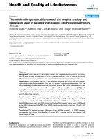
were no longer effective (figure 1). As shown in figure 1,
AMD 3100 was capable of inhibiting virus-cell fusion
both before and after 23°C-TAS. In contrast, sCD4 was
able to inhibit infection to a greater extent when present
at the beginning of the 23°C-TAS incubation period, than
Virology Journal 2006, 3:36 />Page 3 of 7
(page number not for citation purposes)
if present during the last hour of 23°C-TAS (figure 1). This
result indicates that during the 23°C-TAS incubation,
CD4 has been engaged, but interaction between HIV
envelope and CXCR4 had not yet reached an irreversible
state. This analysis also reveals that the much of the initial
binding events of HIV to the target cells at 4°C were CD4-
independent.
Next we analyzed the kinetics of coreceptor engagement
as revealed by infection becoming resistant to fusion
inhibitors that disrupt interactions between HIV envelope
and chemokine receptors. For these experiments, two sep-
arate 96 well plates of Magi +/+ cells were incubated with
virus at 4°C for 2 hours to allow virus attachment. The
virus was removed by washing with PBS. Following bind-
ing at 4°C, one 96 well plate was held at 4°C while the
other 96 well plate was shifted to 23°C-TAS. Following an
additional 2 hours at these temperatures both plates were
shifted to 37°C to promote full viral fusion. Coreceptor
antagonists were then added at various time points fol-
lowing the shift to 37°C. 48 hours later infection was
measured by β-gal activity. The kinetics of both CXCR4
tropic and CCR5 tropic virus infection were analyzed for
virus-cells preincubated at 23°C-TAS compared to those
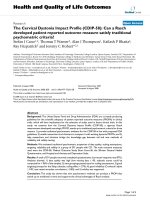
preincubated at 4°C (figure 2). For both CXCR4 tropic
and CCR5 tropic virus we found that infection became
resistant to drugs that target coreceptor engagement faster
when cells were preincubated at 23°C-TAS. The drugs
were present until the assay of β-gal activity in these exper-
iments. Resistance to the inhibitors AMD3100 and
TAK779 was observed within 7 minutes of incubation at
37°C. Conversely, resistance to the inhibitors took 30
minutes when cells were preincubated at 4°C and shifted
directly to the fusion permissive temperature of 37°C (fig-
ure 2A, 2C). We also found that by adding inhibitors 4
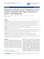
Fusion inhibition by PRO 542 and AMD 3100Figure 1
Fusion inhibition by PRO 542 and AMD 3100. Magi +/+ cells
were preincubated for 2 hours at 4°C. Unbound virus was
washed away with PBS. Fusion inhibitors AMD 3100 (a
CXCR4 antagonist) or PRO 542 (a neutralizing antibody
against gp120) were added to cells along with wild type HIV-
1
NL4.3
at the onset of the 23°C incubation period (white bars)
or after 23°C-TAS was established by two hours (black bars).
Inhibitors were allowed 1 hour for binding then washed. Fol-
lowing the 3 hour TAS incubation period, the cells were
washed with PBS and shifted to 37°C to promote fusion. For
control experiments (grey bar), TAS was established, subse-
quently shifted to 37°C, however no inhibitor was added to
block fusion. The averages of triplicate experiments are
shown (n = 4). Error bars represent the standard error of
the mean. The concentrations of inhibitors were: 4 uM of
AMD 3100 and 5 ug/mL of PRO 542.
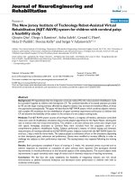
Kinetics of CXCR4 and CCR5 tropic Virus-Cell FusionFigure 2
Kinetics of CXCR4 and CCR5 tropic Virus-Cell Fusion. (A)
Magi +/+ cells were plated at 10,000 cells/well in two sepa-
rate 96 well plates. Cells were infected with wild type HIV-
1
NL4.3
or HIV-1
JRFL
and incubated at 4°C for 2 hours to allow
viral binding. Unbound virus was washed away with PBS.
Both plates were incubated for an additional 2 hours; one
plate remaining at 4°C, while the other plate was shifted to
23°C-TAS. Following the second 2-hour incubation period,
both plates were shifted directly to 37°C to promote viral
fusion. AMD 3100 (4 uM) or TAK 779 (5 ug/mL) were added
at various time points to inhibit subsequent fusion. (A, C)
Virus-cell fusion kinetics are faster when pre-incubated at
23°C-TAS (diamonds) compared to when cells are directly
shifted to 37°C following the 4°C incubation period
(squares). (B, D) CXCR4 and CCR5 fusion kinetics after 4
hours. Varying temperatures were maintained using Eppen-
dorf Centrifuge 5810 R. Relative infectivity was quantified
using a liquid β-Gal assay. OD 405 nm, optical density at 405
nm. The averages of triplicate experiments are shown (n =
4). Error bars represent the standard error of the mean.
Virology Journal 2006, 3:36 />Page 4 of 7
(page number not for citation purposes)
hours after shifting to 37°C, there was no difference in the
fusion kinetics of cells preincubated at 23°C-TAS com-
pared to those preincubated at 4°C (figure 2B, D). This
reveals that the cells preincubated at 4°C will eventually
catch up to the cells preincubated at 23°C-TAS. These
results demonstrate that TAS provides a kinetic predispo-
sition for engagement of coreceptor by virion associated
gp120.
To further define the virion-cell TAS intermediate, we car-
ried out similar experiments using the C34 peptide to
inhibit fusion. C34 is a C-helix peptide, which binds to
transiently exposed heptad repeats within gp41 [12-14].
Previous research has shown C34 to potently inhibit six-
helix bundle formation and subsequent fusion [12-14].
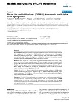
Here we find that C34 is able to block fusion after coincu-
bation of virus with target cells at TAS (figure 3, time
zero). Also, consistent with figure 2, the kinetics to
develop resistance to C34 inhibition were faster when
virus-cell complexes were maintained at 23°C-TAS before
shifting them to 37°C (figure 3a). This data suggests that
TAS allows virus-cell fusion to proceed to a point that
does not include six-helix bundle formation.
Exposure of the heptad repeats of gp41 is required for sus-
ceptibility to C34. We therefore wanted to determine
when fusion became resistant to C34 during the 23°C
incubation [9]. We therefore conducted the following
experiment where virus was allowed to bind cells at 4°C
for 2 hours in the absence of drug. Virus was removed by
washing with PBS and the cells where shifted to 23°C for
3 hours to establish TAS. In a subset of cells, we examined
the ability of C34 or sCD4 to inhibit viral fusion when
added in the first hour of 23°C-TAS ("before TAS") or in
the last hour of 23°C-TAS ("after TAS). In either case the
inhibitor was allowed 1 hour for binding and removed by
washing. After 3 hours at 23°C-TAS, cells where shifted to
37°C to promote full fusion. 48 hours later infection was
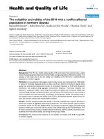
measured by β-gal activity. When C34 was added at the
onset of TAS ("before TAS"), a minimal 20% reduction in
fusion was observed, suggesting the heptad repeats of
gp41 were not fully accessible during this time. However,
a greater degree of inhibition was observed when C34 was
added in the last hour ("after TAS") of 23°C-TAS, suggest-
ing that the heptad repeats do eventually become accessi-
ble to the C34 peptide during TAS. Conversely, inhibition
by sCD4 was only achieved when sCD4 was added at the
onset of TAS and not in the last hour of TAS. This indicates
that the step of sCD4 inhibition arises before TAS while
the step of C34 inhibition arises after TAS has been estab-
lished. Further, the lack of inhibition by C34 when added
before 23°C-TAS implies that binding sites within gp41,
to which C34 is reactive, are not exposed until after TAS
has been established.
Discussion
The fusion of the HIV membrane with that of a target cell
is a complicated multistep process requiring a variety of
molecular interactions. Previous studies show that a tem-
perature sensitive intermediate can be generated for the
HIV fusion process following prolonged incubation at
temperatures ranging from 18°C to 23°C using cell-cell
based fusion assays. In the TAS identified by Cohen and
coworkers, CD4 has been engaged to a lesser extent than
coreceptor. This is revealed by sensitivity to compounds
that block interactions between the HIV envelope and
receptor/coreceptor [9,15]. Likewise, the heptad repeats
are exposed after CD4 engagement by HIV envelope [16].
The ability to generate a similar intermediate using MLV
pseudotyped with HIV envelope has recently been
reported [17]. However, in this case, the HIV envelope
lacked its normal c-terminal tail, which was removed to
allow incorporation into MLV based particles. The infec-
tivity of these pseudotyped virions was not analyzed in
this study. In the studies presented here we find that a TAS
can be generated in the context of wild-type HIV infection
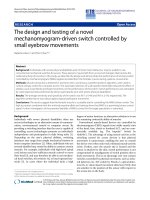
Inhibition by C34 peptideFigure 3
Inhibition by C34 peptide. (A) Fusion kinetics of CXCR4
tropic virus-cell fusion using the C34 peptide for inhibition.
Virus-cell fusion kinetics are faster when pre-incubated at
23°C-TAS (squares) compared to when cells are directly
shifted to 37°C following the 4°C incubation period (dia-
monds). (B) Fusion kinetics after 4 hours.
Virology Journal 2006, 3:36 />Page 5 of 7
(page number not for citation purposes)
of a target cell. Similar to the cell-cell based TAS, the
extended incubation of HIV with target cells at 23°C led
to a decrease in sensitivity to inhibition by soluble CD4.
We also observed an increase in sensitivity to inhibition
by the C34 peptide, which binds to the exposed heptad
repeat (figure 4). Both of these changes in sensitivity were
observed for the TAS generated in cell-cell fusion [9].
Additionally, like cell-cell fusion, coreceptor has not been
engaged to the same extent. Since the readout for fusion
in this analysis is HIV infection of target cells, it suggests
that the TAS is an intermediate in the steps leading to
functional virion fusion and subsequent infection.
Other studies presented here demonstrate that virions in
the TAS intermediate are kinetically predisposed to pass
beyond a point where they are sensitive to inhibition by
CXCR4 or CCR5 coreceptor antagonists (figure 2). The
same is true for the fusion passing beyond a point where
it is sensitive to inhibition by the C34 peptide (figure 3).
The most simple interpretation of this finding is that the
time needed for envelope to successfully engage cellular
receptors is eliminated during the 23°C incubation.
Therefore, the fusion complex can proceed directly to
complete coreceptor engagement, and go on to fuse after
shifting to 37°C. In contrast, virions incubated at 4°C for
the same period are delayed in achieving resistance to
coreceptor antagonists because they must take the time to
properly engage CD4 before proceeding to engage a core-
ceptor. Coreceptor engagement following CD4 engage-
ment has been demonstrated to take approximately 30
minutes [18,19]. After 4 hours, any kinetic advantage
imparted by the TAS is lost as virus-cell cultures become
resistant to both coreceptor antagonists or C34 peptide,
regardless of whether they were incubated at 23°C or 4°C
(figure 2, 3B). The kinetics of resistance to downstream
fusion inhibitors also demonstrates that TAS is an inter-
mediary in the process leading to fusion and infection. It
is possible that TAS represents a non-productive but
reversible intermediate. However, reversion to the func-
tional pathway would take time, resulting in the delay of
fusion of virus-cell cultures maintained at TAS relative to
control cells. Therefore, the kinetic predisposition of
virus-cell cultures to advance beyond sensitivity to down-
stream inhibitors of fusion demonstrate that the TAS
intermediate represents a discreet step in the fusion path-
way which ultimately leads to HIV infection of target cells.
Comparing our findings using virus-cell interactions to
previous studies using cell-cell based fusion assays reveals
a difference between the cell-cell TAS and virus-cell TAS at
the level of engagement of the CXCR4 coreceptor. In the
cell-cell based assays, there is a significant and detectable
engagement of the coreceptor shown by resistance to the
CXCR4 binding peptide T22 [9]. Likewise, significant
resistance to AMD3100 for blocking CXCR4 mediated
fusion after incubation at 23°C for 2 hours is also
observed. Furthermore, Mkrtchyan has shown that the
longer the incubation period, the greater the extent of
resistance [15]. In contrast, we have shown that incuba-
tion of virus and target cells at 23°C after 2–3 hours con-
fers negligible resistance to AMD3100. Conversely, our
studies with CCR5 tropic envelope in the virus-cell TAS,
mimicked previous findings using a TAS for cell-cell
fusion assays. For example, resistance to TAK779 was 20%
after incubation at 23°C (figure 2C) similar to the cell-cell
fusion studies. One possible explanation for this differ-
ence lies in the mobilities of the chemokine receptors in
the membrane. We have previously reported that CCR5 is
highly mobile in the membrane [20]. In contrast, we have
recently found that CXCR4 in much less mobile [21]. The
highly mobile CCR5 can begin to engage the CD4 bound
HIV envelope at 23°C while the less mobile CXCR4 can
not. The mobility of CXCR4 is less important in the case
of the cell-cell assays because it would be recruited to the
site of cell-cell contact within minutes of the shift to 37°C,
as has been reported for the virological synapse. It has pre-
viously been shown that receptor/co-receptor density
plays a role in the rate of HIV fusion and infection [22],
and that multiple receptor and co-receptor molecules
must engage multiple gp120 subunits in order to initiate
TAS contributes to the exposure of binding sites within gp41 necessary for inhibition by C34 peptideFigure 4
TAS contributes to the exposure of binding sites within gp41
necessary for inhibition by C34 peptide. MAGI +/+ cells were
plated at 10,000 cells/well, and coincubated with HIV-1
NL4.3
at 4°C for 2 hours. Unbound virus was washed with PBS, and
cells were shifted to 23°C-TAS for 3 hours. Fusion inhibitors
sCD4 (PRO 542) and C34 peptide were either added at the
onset of 23°C incubation (white bars) or after TAS had been
established for 2 hours (black bars). Inhibitors were allowed
1 hour for binding, then washed. Following the 3 hour TAS
incubation, the cells were washed with PBS and shifted to
37°C. The averages of triplicate experiments are shown (n =
4). Error bars represent the standard error of the mean. Rel-
ative infectivity was quantified using a liquid β-Gal assay. OD
405 nm, optical density at 405 nm. The concentrations of the
inhibitors were: sCD4 (5 ug/mL) and C34 (100 nM).
Virology Journal 2006, 3:36 />Page 6 of 7
(page number not for citation purposes)
fusion [23]. The formation of a virological synapse
recruits CXCR4 and increases the rate of these engage-
ments. The difference observed in virus-cell interactions
suggests that CXCR4 is not actively recruited to the site of
virus binding at the same rate.
Studies by Kabat's laboratory suggest that CCR5 entry is
governed by three kinetic processes. One of these proc-
esses includes the formation of 'competent complexes.'
These 'competent complexes' consist of sufficient CCR5-
gp120 associations that are capable of proceeding further
into the fusion process. The TAS intermediate reveals that
the interaction with chemokine receptor is the rate-limit-
ing step in the process of the formation of these 'compe-
tent complexes'. Potential temperature sensitive steps
might actually be differences in chemokine mobility or
affinity to the HIV envelope. It is unlikely that the temper-
ature sensitive step is associated with conformational
changes known to take place in HIV envelope because of
the differences observed between cell-cell TAS and virus-
cell TAS described above. The TAS intermediate described
here allows the HIV-1 fusion reaction to be analyzed in
the context of infectious HIV-1 particles and their respec-
tive target cells. Using suboptimal temperatures, we can
gain valuable insight into HIV-1 virus-cell fusion kinetics.
Ultimately, generating a temperature-arrested intermedi-
ate for virus-cell fusion provides a useful tool for synchro-
nizing entry and studying HIV-1 fusion microscopically.
Materials and methods
Virus-cell fusion/infection assays
In order to study virus-cell fusion, we developed a system
that allows fusion to be assayed in the context of infec-
tious virions that were preincubated with target cells. The
Magi reporter cell line for viral infection was employed
[24]. Magi +/+ cells derive from HeLa cells and stably
express CD4 and CXCR4 on the cells surface. Magi +/+
cells also contain a stably integrated copy of the β-galac-
tosidase (β-gal) gene downstream of the HIV-1 long ter-
minal repeat (LTR). Upon infection, the HIV
transactivator protein Tat activates β-gal expression.
Therefore, viral fusion leads to β-gal expression and the
level of β-gal activity can be measured from infected cells
[24]. 36 hours post infection, cells were lysed in sodium
phosphate buffer with 0.2% Triton X and assayed for β-gal
expression. β-Gal expression was quantified by monitor-
ing cleavage of the colorimetric β-gal substrate o-nitroph-
enyl-β-D-galactopyranoside (ONPG) using a 96-well
microplate reader measuring absorbance at 405 nm. The
average background values of uninfected cells were sub-
tracted from the values of infected cells. Varying tempera-
tures were maintained using the Eppendorf Centrifuge
5810 R. Using this system, we manipulated temperature
conditions during virus-cell incubation and used fusion
inhibitors to explore kinetic factors influencing HIV-1
entry.
Cell culture and virus production
Magi +/+ cells were grown in Dulbecco's modified Eagle's
growth medium (BioWhittaker, Walerville, Md.), which
contained 10% fetal bovine serum and 1% penicillin-
streptomycin-glutamine. Virus was produced by CaPO
4
transfection of 293T cells with 20 µg of HIV-1
Bru
or HIV-
1
JRFL
proviral constructs. Two days following transfection,
virus was harvested through a 0.45 µm pore sized filter.
Acknowledgements
This work was supported by National Institutes of Health Grant RO1-
AI5205 to TJH. TJH is an Elizabeth Glaser Scientist. We thank the contrib-
utors to the NIH AIDS Reagent Program for materials, especially the C34
peptide and AMD 3100. PRO 542 was obtained from Progenics
Pharmeceuticals.
References
1. Doms RW, Moore JP: HIV-1 membrane fusion: targets of
opportunity. J Cell Biol 2000, 151:F9-14.
2. Trkola A, Dragic T, Arthos J, Binley JM, Olson WC, Allaway GP,
Cheng-Mayer C, Robinson J, Maddon PJ, Moore JP: CD4-depend-
ent, antibody-sensitive interactions between HIV-1 and its
co-receptor CCR-5. Nature 1996, 384:184-7.
3. Markosyan RM, Cohen FS, Melikyan GB: Time-resolved imaging
of HIV-1 Env-mediated lipid and content mixing between a
single virion and cell membrane. Mol Biol Cell 2005, 16:5502-13.
4. Aloia RC, Tian H, Jensen FC: Lipid composition and fluidity of
the human immunodeficiency virus envelope and host cell
plasma membranes. Proc Natl Acad Sci USA 1993, 90:5181-5.
5. Guyader M, Kiyokawa E, Abrami L, Turelli P, Trono D: Role for
human immunodeficiency virus type 1 membrane choles-
terol in viral internalization. J Virol 2002, 76:10356-64.
6. Nguyen DH, Hildreth JE: Evidence for budding of human immu-
nodeficiency virus type 1 selectively from glycolipid-enriched
membrane lipid rafts. J Virol 2000, 74:3264-72.
7. Popik W, Alce TM, Au WC: Human immunodeficiency virus
type 1 uses lipid raft-colocalized CD4 and chemokine recep-
tors for productive entry into CD4(+) T cells. J Virol 2002,
76:4709-22.
8. Mothes W, Boerger AL, Narayan S, Cunningham JM, Young JA: Ret-
roviral entry mediated by receptor priming and low pH trig-
gering of an envelope glycoprotein. Cell 2000, 103:679-89.
9. Melikyan GB, Markosyan RM, Hemmati H, Delmedico MK, Lambert
DM, Cohen FS: Evidence that the transition of HIV-1 gp41 into
a six-helix bundle, not the bundle configuration, induces
membrane fusion. J Cell Biol 2000, 151:413-23.
10. Frey S, Marsh M, Gunther S, Pelchen-Matthews A, Stephens P,
Ortlepp S, Stegmann T: Temperature dependence of cell-cell
fusion induced by the envelope glycoprotein of human
immunodeficiency virus type 1. J Virol 1995, 69:1462-72.
11. Gallo SA, Clore GM, Louis JM, Bewley CA, Blumenthal R: Temper-
ature-dependent intermediates in HIV-1 envelope glycopro-
tein-mediated fusion revealed by inhibitors that target N-
and C-terminal helical regions of HIV-1 gp41. Biochemistry
2004, 43:8230-3.
12. Chan DC, Chutkowski CT, Kim PS: Evidence that a prominent
cavity in the coiled coil of HIV type 1 gp41 is an attractive
drug target. Proc Natl Acad Sci USA 1998, 95:15613-7.
13. Malashkevich VN, Chan DC, Chutkowski CT, Kim PS: Crystal
structure of the simian immunodeficiency virus (SIV) gp41
core: conserved helical interactions underlie the broad inhib-
itory activity of gp41 peptides. Proc Natl Acad Sci USA 1998,
95:9134-9.
14. Wild CT, Shugars DC, Greenwell TK, McDanal CB, Matthews TJ:
Peptides corresponding to a predictive alpha-helical domain
of human immunodeficiency virus type 1 gp41 are potent
Publish with BioMed Central and every
scientist can read your work free of charge
"BioMed Central will be the most significant development for
disseminating the results of biomedical research in our lifetime."
Sir Paul Nurse, Cancer Research UK
Your research papers will be:
available free of charge to the entire biomedical community
peer reviewed and published immediately upon acceptance
cited in PubMed and archived on PubMed Central
yours — you keep the copyright
Submit your manuscript here:
/>BioMedcentral
Virology Journal 2006, 3:36 />Page 7 of 7
(page number not for citation purposes)
inhibitors of virus infection. Proc Natl Acad Sci USA 1994,
91:9770-4.
15. Mkrtchyan SR, Markosyan RM, Eadon MT, Moore JP, Melikyan GB,
Cohen FS: Ternary complex formation of human immunode-
ficiency virus type 1 Env, CD4, and chemokine receptor cap-
tured as an intermediate of membrane fusion. J Virol 2005,
79:11161-9.
16. He Y, Vassell R, Zaitseva M, Nguyen N, Yang Z, Weng Y, Weiss CD:
Peptides trap the human immunodeficiency virus type 1
envelope glycoprotein fusion intermediate at two sites. J Virol
2003, 77:1666-71.
17. Abrahamyan LG, Mkrtchyan SR, Binley J, Lu M, Melikyan GB, Cohen
FS: The cytoplasmic tail slows the folding of human immuno-
deficiency virus type 1 Env from a late prebundle configura-
tion into the six-helix bundle. J Virol 2005, 79:106-15.
18. Gallo SA, Finnegan CM, Viard M, Raviv Y, Dimitrov A, Rawat SS, Puri
A, Durell S, Blumenthal R: The HIV Env-mediated fusion reac-
tion. Biochim Biophys Acta 2003, 1614:36-50.
19. Gallo SA, Puri A, Blumenthal R: HIV-1 gp41 six-helix bundle for-
mation occurs rapidly after the engagement of gp120 by
CXCR4 in the HIV-1 Env-mediated fusion process. Biochemis-
try 2001, 40:12231-6.
20. Steffens CM, Hope TJ: Localization of CD4 and CCR5 in living
cells. J Virol 2003, 77:4985-91.
21. Anderson A, Hope TJ: personal communication. .
22. Reeves JD, Gallo SA, Ahmad N, Miamidian JL, Harvey PE, Sharron M,
Pohlmann S, Sfakianos JN, Derdeyn CA, Blumenthal R, Hunter E,
Doms RW: Sensitivity of HIV-1 to entry inhibitors correlates
with envelope/coreceptor affinity, receptor density, and
fusion kinetics. Proc Natl Acad Sci USA 2002, 99:16249-54.
23. Kuhmann SE, Platt EJ, Kozak SL, Kabat D: Cooperation of multiple
CCR5 coreceptors is required for infections by human
immunodeficiency virus type 1. J Virol 2000, 74:7005-15.
24. Kimpton J, Emerman M: Detection of replication-competent
and pseudotyped human immunodeficiency virus with a sen-
sitive cell line on the basis of activation of an integrated beta-
galactosidase gene. J Virol 1992, 66:2232-9.