Báo cáo hóa học: " Expression and processing of the Hepatitis E virus ORF1 nonstructural polyprotein" potx
Bạn đang xem bản rút gọn của tài liệu. Xem và tải ngay bản đầy đủ của tài liệu tại đây (355.97 KB, 9 trang )
BioMed Central
Page 1 of 9
(page number not for citation purposes)
Virology Journal
Open Access
Research
Expression and processing of the Hepatitis E virus ORF1
nonstructural polyprotein
Deepak Sehgal, Saijo Thomas, Mahua Chakraborty and Shahid Jameel*
Address: Virology Group, International Center for Genetic Engineering and Biotechnology, Aruna Asaf Ali Marg, New Delhi 110 067, India
Email: Deepak Sehgal - ; Saijo Thomas - ; Mahua Chakraborty - ;
Shahid Jameel* -
* Corresponding author
Abstract
Background: The ORF1 of hepatitis E virus (HEV) encodes a nonstructural polyprotein of ~186
kDa that has putative domains for four enzymes: a methyltransferase, a papain-like cysteine
protease, a RNA helicase and a RNA dependent RNA polymerase. In the absence of a culture
system for HEV, the ORF1 expressed using bacterial and mammalian expression systems has shown
an ~186 kDa protein, but no processing of the polyprotein has been observed. Based on these
observations, it was proposed that the ORF1 polyprotein does not undergo processing into
functional units. We have studied ORF1 polyprotein expression and processing through a
baculovirus expression vector system because of the high level expression and post-translational
modification abilities of this system.
Results: The baculovirus expressed ORF1 polyprotein was processed into smaller fragments that
could be detected using antibodies directed against tags engineered at both ends. Processing of this
~192 kDa tagged ORF1 polyprotein and accumulation of lower molecular weight species took
place in a time-dependent manner. This processing was inhibited by E-64d, a cell-permeable
cysteine protease inhibitor. MALDI-TOF analysis of a 35 kDa processed fragment revealed 9
peptide sequences that matched the HEV methyltransferase (MeT), the first putative domain of the
ORF1 polyprotein. Antibodies to the MeT region also revealed an ORF1 processing pattern
identical to that observed for the N-terminal tag.
Conclusion: When expressed through baculovirus, the ORF1 polyprotein of HEV was processed
into smaller proteins that correlated with their proposed functional domains. Though the
involvement of non-cysteine protease(s) could not be be ruled out, this processing mainly
depended upon a cysteine protease.
Background
Hepatitis E virus (HEV) is the etiological agent for hepati-
tis E. It has been the cause of large epidemics as well as
many sporadic cases of acute viral hepatitis in much of the
developing world [1-5]. The viral genome is a single-
stranded 7.2-kb polyadenylated RNA of positive sense
containing three open reading frames (ORFs) [6,7]. Of
these, ORF2 encodes an 88-kDa glycoprotein that is the
major viral capsid protein [8,9]; ORF3 encodes a phos-
phoprotein [10], which is involved in cell signaling
through MAP kinase pathway [11].
Published: 26 May 2006
Virology Journal 2006, 3:38 doi:10.1186/1743-422X-3-38
Received: 25 January 2006
Accepted: 26 May 2006
This article is available from: />© 2006 Sehgal et al; licensee BioMed Central Ltd.
This is an Open Access article distributed under the terms of the Creative Commons Attribution License ( />),
which permits unrestricted use, distribution, and reproduction in any medium, provided the original work is properly cited.
Virology Journal 2006, 3:38 />Page 2 of 9
(page number not for citation purposes)
The third ORF, called ORF1 is 5109 bp long and encodes
the viral nonstructural polyprotein with a proposed
molecular mass of ~186 kDa. Based on protein sequence
homology, the ORF1 polyprotein is proposed to contain
four putative domains indicative of methyltransferase
(MeT), papain-like cysteine protease (PCP), RNA Helicase
(Hel), and RNA dependent RNA polymerase (RdRp) (Fig.
1) [12]. Of these, the MeT and RdRp enzymatic activities
have been demonstrated [13,14] while activities of the
Hel and PCP have so far not been elucidated. Attempts
have also been made to study ORF1 processing using dif-
ferent expression systems. In one study, the ~186 kDa
ORF1 polyprotein was expressed through recombinant
vaccinia virus infection of mammalian cells, but no proc-
essed products were initially observed [15]. Following
extended incubation for 24–36 hours, two processed
bands of ~107 and ~78 kDa were observed. Mutagenesis
of the proposed cysteine protease domain of ORF1 sug-
gested that the HEV protease had no role in ORF1 poly-
protein processing. The cleavage of the ~186 kDa protein
was attributed either to a vaccinia-virus encoded protease
or a cellular protease.
In another study, ORF1 processing was addressed through
in vitro transcription and translation, and expression in
either E. coli or human cells [16]. Prokaryotic expression
resulted in a ~212 kDa glutathione-S-transferase fusion
protein that exhibited strong reactivity with the antibodies
raised against the putative domains of ORF1. Since no
other smaller products were observed, ORF1 processing
did not seem to occur in the prokaryotic system. When the
expression of ORF1 was studied by carrying out in vitro
coupled transcription and translation, a polyprotein of
~186 kDa could again be immunoprecipitated with anti-
bodies against the various putative domains of ORF1, but
no smaller fragments were observed. The expression in
transiently transfected HepG2 cells also resulted in a ~186
kDa protein, but no other smaller sized fragments were
seen [16]. Transfection of an in vitro generated infectious
full-length HEV RNA into HepG2 cells has also been used
to assess ORF1 expression and processing [17]. This
resulted in the formation of processed forms of the ORF1
polyprotein that could be immunoprecipitated with vari-
ous domain-specific antibodies [17].
Though ORF1 processing was reported in at least one
study in the context of genomic RNA, it is not clear why
this was not observed in other studies. This could be due
to improper folding of the GST-ORF1 fusion protein
expressed in the prokaryotic system, and low yields of the
protein expressed in coupled in vitro transcription-transla-
tion or mammalian expression systems. To address this,
we expressed a recombinant ORF1 polyprotein tagged at
its N- and C-termini in insect cells using a baculovirus
expression system, and detected the processed fragments
using antibodies specific for the N-terminal hexa-histi-
dine and C-terminal FLAG epitopes. Using this strategy,
we show here that the ORF1 polyprotein is processed in
insect cells and that this involves both cysteine and non-
cysteine proteases. The processing of ORF1 was also con-
firmed by mass spectrometric analysis of one of the proc-
essed fragments and by western blotting with antibodies
to the methyltransferase domain.
Results
Construction of the recombinant baculovirus
The HEV-ORF1 was PCR amplified (data not shown)
using the HEV full-length cDNA as a template, so that
when expressed, the protein had an N-terminal hexa-his-
tidine tag and a C-terminal FLAG tag (Fig. 1). The ampli-
fied gene was cloned in TOPO-TA vector, in vitro
transcribed and translated to generate a polyprotein of
~192 kDa (data not shown). After confirming proper
expression of the amplified fragment, it was cloned in the
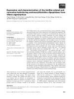
The HEV ORF1 polyproteinFigure 1
The HEV ORF1 polyprotein. A schematic illustration of the HEV ORF1 nonstructural polyprotein is shown, with the engi-
neered N- and C-terminal tags. The predicted methyltransferase (MeT), papain-like cysteine protease (PCP), helicase (Hel) and
RNA dependent RNA polymerase (RdRp) domains are shown, as is the GDD sequence that forms the RdRp active site. The
numbers on top represent amino acids of the predicted domains numbered according to the ORF1 polyprotein sequence [12].
The Y, proline-rich (Pro) and X regions with no predicted function are also shown. The tags engineered at the two ends
include the N-terminal 6XHis tag of 45 amino acids (from vector pBBHis-2b) and a FLAG epitope of 12 amino acids as
described in Materials and Methods. The entire recombinant ORF1 polyprotein engineered here is expected to be 1760 amino
acids long, with a predicted mass of 191,806 Da.
Virology Journal 2006, 3:38 />Page 3 of 9
(page number not for citation purposes)
baculovirus transfer vector pBlueBacHis-2b (Invitrogen).
Co-transfection of the recombinant plasmid and pBlue-
Bac DNA (Invitrogen), followed by selection and plaque
purification, resulted in generation of the recombinant
virus, called vORF1. For subsequent infection, this was
amplified to a titer of 10
8
pfu/ml in Sf21 cells.
ORF1 expression and processing
To study the time course of recombinant ORF1 polypro-
tein expression, vORF1 was used to infect T. ni cells. The
infected cells were harvested at various times post-infec-
tion and the lysates subjected to SDS-PAGE followed by
western blotting using anti-His or anti-FLAG antibodies
(Fig. 2). Expression of the ORF1 polyprotein was seen as
early as 24 hr post-infection (hpi) (Fig. 2A, lane1), the
time at which the polyhedrin promoter is activated. At this
time, besides the ~192 kDa fragment, other fragments
with sizes of ~98 and 47 kDa were also observed with
anti-His antibodies. Around 48 hpi, two additional bands
of ~35 and 22 kDa were seen and all of these fragments
were found to increase with time till 72 hpi (Fig. 2A).
When expression was analyzed using anti-FLAG antibod-
ies, besides the ~192 kDa polyprotein, smaller fragments
of ~122, 106, 93, 59 and 26 kDa were also observed in a
temporal manner (Fig. 2B). As a negative control, no
staining was observed with either antibody in wild type
AcMNPV (wt) infected T. ni cells (Fig. 2A and 2B, lane 6).
The expression of the ~192 kDa fragments and accumula-
tion of smaller fragments as a function of time was indic-
ative of processing of the ORF1 polyprotein. The
processing was further confirmed with antibodies against
the MeT domain, the most N-terminal predicted domain
in the polyprotein. The pattern of processing observed
with anti-MeT antibodies (Fig. 2C) was identical to that
obtained using anti-His antibodies (Fig. 2A).
Effect of cysteine protease inhibition on ORF1 processing
The ORF1 polyprotein contains a putative PCP domain.
To further validate processing of the ORF1 polyprotein
and to assess the role of cysteine protease in this, we used
the cell permeable cysteine protease inhibitor E-64d. Fol-
lowing infection of insect cells with vORF1, the cells were
treated with E-64d and the cell lysates analyzed by western
blotting with anti-His or anti-FLAG antibodies (Fig. 3A
and 3B). At 48 hr and 60 hr post-treatment E-64d was
found to inhibit ORF1 polyprotein processing as evident
from accumulation of the ~192 kDa fragment (Fig. 3A and
3B, lanes 2 and 4). Western blotting with anti-His anti-
body revealed that addition of E-64d resulted in loss of
the processed 98, 35 and 22 kDa fragments, while there
was accumulation of the 47 kDa fragment at both time
points (Fig. 3A, lanes 2 and 4). Under the same conditions
and at similar times all processed fragments were
observed in untreated cells (Fig. 3A lanes 1 and 3 respec-
tively), while none of the fragments were seen in cells
infected with the wt virus (Fig. 3A lanes 5 and 6). Equal
amounts of proteins were loaded in E-64d treated,
untreated or wt virus infected cells, as seen on Coomassie
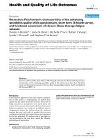
Expression and processing of the ORF1 polyproteinFigure 2
Expression and processing of the ORF1 polyprotein. T. ni cells infected with the vORF1 recombinant virus were har-
vested at various times post-infection and the lysates subjected to SDS-PAGE and western blotting with anti-His (A) or anti-
FLAG (B) antibodies. Lanes 1 to 5 show results at 24, 36, 48, 60 and 72 hr post-infection; lane 6 shows the result for wild type
AcMNPV infection after 48 hr. Panel C shows the 48 hr lysate probed with anti-MeT antibodies. In (B) the upper and lower
panels show results from 7.5% and 12% SDS-polyacrylamide gels. The estimated fragment sizes are shown based on molecular
size markers run on each gel (not shown).
Virology Journal 2006, 3:38 />Page 4 of 9
(page number not for citation purposes)
Blue stained gels (data not shown). The E-64d effect stud-
ied using anti-FLAG antibodies also showed accumula-
tion of the ~192 kDa polyprotein in inhibitor-treated cells
(Fig. 3B). Further, compared to untreated cells, it showed
disappearance of the 106, 93 and 59 kDa fragments, with
accumulation of the 122 and 26 kDa fragments (Fig. 3B,
lanes 1–4). In addition, at the 60 hr time point, partially
processed intermediates of ~130–140 kDa were also
observed in the presence of E-64d (Fig. 3B, lane 4). As ear-
lier, no background was observed in wt infected cells (Fig.
3B, lanes 5 and 6). Based on these results, various cysteine
and non-cysteine protease sites were mapped on the
ORF1 polyprotein (Fig. 4).
Purification of protein fragments and MALDI-TOF analysis
Protein fragments containing the His-tag were partially
purified by Ni-NTA affinity chromatography. After estab-
lishing their identity using anti-His antibodies, the 35-
kDa fragment was eluted from the gel and subjected to
mass spectrometric analysis. Nine tryptic peptides were
selected from the mass spectrum (Fig. 5A) and compared
for their experimentally obtained and predicted masses
(Fig. 5B). These predicted sequences matched the N- ter-
minal region of the ORF1 polyprotein spanning amino
acids 70 – 339, including the predicted MeT domain. As
shown earlier (Fig. 2C), the 35-kDa fragment also stained
with antibodies generated to the ORF1 MeT region span-
ning nucleotides 159 to 862 [16]. This antibody showed a
staining pattern similar to that observed with anti-His
antibodies (Fig. 2A).
Discussion
In all plus-strand animal RNA viruses, individual proteins
are processed from the nonstructural polyprotein through
Effect of E-64d on ORF1 polyprotein processingFigure 3
Effect of E-64d on ORF1 polyprotein processing. T. ni cells were infected with vORF1 for 12 hr at which time fresh
medium containing 200 µM E-64d was added to the cells; an equal volume of DMSO served as the control. At 48 and 60 hr fol-
lowing E-64d addition, cells were harvested and the lysates analyzed by western blotting with either anti-His (A) or anti-FLAG
(B) antibodies (lanes 1–4). Lysates from wild type AcMNPV infected cells were similarly analyzed at 48 hr after E-64d addition
(lanes 5–6). In both parts, the upper and lower panels show results obtained following separation of the proteins on 7.5% and
12% SDS-polyacrylamide gels. The estimated fragment sizes are shown based on molecular size markers run on each gel (not
shown).
Virology Journal 2006, 3:38 />Page 5 of 9
(page number not for citation purposes)
specific and limited proteolysis. Based on sequence
homology, proposed domains and replication mecha-
nism, HEV is closely related to alpha viruses with the
Rubella virus being its closest homologue [12]. Previous
studies relating to the HEV ORF1 polyprotein processing
have shown that it is not processed in mammalian cells
[15,16]. Despite the absence of processing, baculovirus
mediated expression of a 110 kDa ORF1 protein has been
shown to contain a methyltransferase activity [13]. Many
mammalian proteins have been expressed in their native
and active forms using recombinant baculoviruses [18].
Further, the baculovirus system has also been utilized to
study the expression and processing of the polyproteins of
other viruses, including the rubella viruses [19-23]. This
system also offers post-translational modifications that
are similar to those in mammalian cells, yet is capable of
expressing much higher quantities of the recombinant
protein [18]. Because of this increased signal to noise
ratio, we used baculovirus-mediated expression to study
HEV-ORF1 processing.
Unlike earlier reports, processing of the HEV nonstruc-
tural ORF1 polyprotein into smaller fragments was
detected using antibodies to the engineered N- and C- ter-
minal tags. A pattern of processing similar to that
observed with anti-His antibodies was also observed with
antibodies directed against the MeT domain. This was
expected since MeT is the N-terminal domain of ORF1,
and is closest to the His-tag in this construct. To further
check the authenticity of processing, we performed a
kinetic study of the protein expression following recom-
binant baculovirus infection. The ~192 kDa tagged poly-
protein and at least two smaller fragments of 98 and 47
kDa appeared faintly at 24 hpi. This indicated rapid, pos-
sibly cotranslational processing since the polyhedrin pro-
moter, under which ORF1 is placed, gets activated at
around 24 hpi. The polyprotein synthesis and appearance
of the processed products increased at 48 hpi and subse-
quent times in agreement with the characteristics of this
expression system. At later times, smaller N-terminal frag-
ments of 35 and 22 kDa were also found. This represents
a precursor-product relationship, indicative of polypro-
tein processing. Similar observations were made when
processing was monitored from the C-terminal end of the
polyprotein.
Since the ORF1 polyprotein has a predicted cysteine pro-
tease domain and cis-acting proteases are found within
the nonstructural polyproteins of all other positive-strand
RNA viruses [23-28], it is likely that the cysteine protease
within the ORF1 polyprotein is responsible for its process-
ing. A cell-permeable cysteine protease inhibitor, E-64d,
was also able to effectively block processing of the ORF1
polyprotein. Together with our kinetic data of rapid, pos-
Schematic illustration of the ORF1 polyproteinFigure 4
Schematic illustration of the ORF1 polyprotein. The illustration shows various predicted domains, the N- and C-termi-
nal fragments detected with anti-His and anti-FLAG antibodies, respectively, and the protease cleavage sites.
Virology Journal 2006, 3:38 />Page 6 of 9
(page number not for citation purposes)
sibly cotranslational processing of the ORF1 polyprotein,
this is suggestive of a cis-acting cysteine protease within
the HEV nonstructural polyprotein.
During a time course of E-64d inhibition of processing,
the ~192 kDa and 47 kDa fragments observed with anti-
His antibodies were found to accumulate. This suggested
that cysteine protease sites occurred at 22, 35, and 98 kDa,
while a non-cysteine protease site occurred at 47 kDa
from the N-terminus of the tagged ORF1 polyprotein.
When probed from the C-terminus with anti-FLAG anti-
bodies, the E-64d treated cells exhibited strong accumula-
tion of the ~192 kDa polyprotein, as well as fragments of
122 and 26 kDa. This meant that non-cysteine protease
sites existed at these distances from the C-terminus of the
tagged ORF1 polyprotein, while cysteine protease sites
were present around 106, 93 and 59 kDa from the C-ter-
minal end. The ~22 kDa N-terminal fragment disrupts the
MeT coding region. From the present analysis, it is not
clear whether this is due to nonspecific activity of the HEV
protease or due to a host cell cysteine protease. Similarly,
an ~26 kDa C-terminal fragment that disrupts the RdRp
region is the likely product of a non-cysteine host pro-
tease. Though our results do not unequivocally prove the
cysteine protease activity to have a viral origin, we clearly
demonstrate ORF1 polyprotein processing. As is the case
with other positive-strand RNA viruses [25-28], these
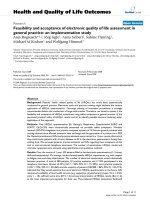
MALDI-TOF analysis of the 35 kDa N-terminal fragmentFigure 5
MALDI-TOF analysis of the 35 kDa N-terminal fragment. (A) Mass spectrum showing fragments 1–9. (B) Table shows
the experimentally observed mass, predicted mass and sequences of peptides 1–9.
Virology Journal 2006, 3:38 />Page 7 of 9
(page number not for citation purposes)
results suggest a role for viral and host cell proteases in
processing of the HEV ORF1 nonstructural polyprotein.
In order to further validate the processing, a 35 kDa frag-
ment was analyzed by tryptic digestion and mass spec-
trometry. The results showed high confidence match with
the MeT domain of ORF1 and this was confirmed by west-
ern blotting with anti-HEV MeT region antibodies.
Some earlier studies [15,16] have failed to detect ORF1
polyprotein processing. This has led Ropp et al [15] to
speculate that the proposed cysteine protease within the
HEV nonstructural polyprotein is non-functional and that
HEV is different from all other positive-strand RNA
viruses with respect to the processing of its nonstructural
polyprotein. This has important implications for the clas-
sification of HEV within the positive-strand RNA virus
group. Three lines of evidence argue against this possibil-
ity. First, using an infectious molecular clone of HEV,
Panda et al [17] were able to detect proteins smaller than
the 185 kDa ORF1 polyprotein with antisera prepared
against recombinant methyltransferase, helicase and
RdRp domains expressed in bacteria. Secondly, a 37 kDa
protein identified with anti-HEV RdRp antibodies was
observed in cells transfected with the HEV ORF1-EGFP
replicon [29]. We present the third line of evidence in this
study by demonstrating that the ORF1 polyprotein is
capable of being processed and that a cysteine protease is
partly responsible for this. We do understand that the bac-
ulovirus-mediated expression system employed in this
study is not the natural expression system for HEV. It was
used here because of our apprehension that earlier failures
to observe ORF1 processing were either due to improper
folding of the polyprotein expressed in prokaryotic sys-
tems, or due to low levels of expression in transfected
mammalian cells. The baculovirus system offered the
advantage of high expression levels and close to native
post-translational modifications and protein conforma-
tion.
A comparison of all the studies on ORF1 polyprotein
processing [15-17,29], including this one, also suggests
the interesting possibility that polyprotein processing in
the context of an infectious virus cycle [17] may require far
less protein than when ORF1 is expressed on its own
[15,16]. This may be due to subcellular compartmenta-
tion leading to high local concentrations of the protein
precursor or due to assistance from other viral and/or cel-
lular proteins, or some combination of these mecha-
nisms.
When expressed using a baculovirus system, our results
presented here show that even when expressed individu-
ally, the HEV ORF1 polyprotein undergoes processing.
This processing is primarily mediated by a cysteine pro-
tease. Additional data is needed to conclusively establish
the viral origin of this protease. To further establish this,
there would be a need to over-express the ORF1 polypro-
tein in a mammalian cell system and to use more sensitive
detection methods.
Conclusion
While the HEV nonstructural ORF1 polyprotein carries at
least four putative functional domains, its processing has
so far not been demonstrated. We reasoned this may be
due to improper folding or low expression levels of the
polyprotein in subgenomic expression systems attempted
so far. We show here expression of the ORF1 polyprotein
using a baculovirus system and demonstrate processing
using engineered tags, a domain-specific antibody and
mass spectrometric identification of a processed fragment.
A papain-like cysteine protease is predicted within the
ORF1 polyprotein. We present evidence here for the role
of a cysteine protease in ORF1 polyprotein processing; the
viral origin of this protease remains to be established.
These results have implications for the classification of
HEV among positive-sense RNA viruses.
Methods
Materials
Sf21 and T. ni cells (Invitrogen) were maintained at 28°C
in TNMFH (Gibco, BRL) and Excel 405 (JRH Biosciences)
media, respectively. Antibodies to the hexahistidine and
FLAG tags were purchased from Sigma. A rabbit serum
containing polyclonal antibodies against the methyltrans-
ferase region of HEV ORF1 have been described earlier
[16]. The Ni-NTA resin was obtained from Qiagen (Ger-
many). All common molecular biology and cell culture
grade reagents were from Sigma, unless specified other-
wise.
Construction of the ORF1 recombinant baculovirus
The ~5 kb ORF1 was PCR amplified using Gene Amp XL
PCR kit (Perkin Elmer, Applied Biosystems) according to
the suppliers guidelines. Besides other components, the
reaction mix included 20 pmoles of each primer and 1
mM Mg(OAc)
2
. The amplification primers were designed
based on alignments of the 5' and 3' ends of ORF1 in the
HEV genomic sequence (GenBank Accession Number
AF459438
) [30]. The primers used for the amplification
were EcoRI-ORF1-5', TACGGAATTC
ATGGAGGCCCAT-
CAGTTTATCAAG and Hind III-ORF1-3', CCAAAGCTT
T-
GATTTCACCCGACACAAGATTGA, containing the
underlined restriction sites. The PCR amplified fragment
was initially cloned in the TOPO-TA vector (Invitrogen).
To position a FLAG tag at the 3'end of ORF1, the FLAG
epitope was first reconstructed by annealing the oligonu-
cleotides AGCTTAACTACAAGGACGACGACGATAAG-
TAACTCGAG and
TCGACTCGAGTTACTTATCGTCGTCGTCCTTGTAGTC-
Virology Journal 2006, 3:38 />Page 8 of 9
(page number not for citation purposes)
CATA. The annealed product was ligated with the vector
pBBHis-2b (Invitrogen) at its HindIII and SalI sites. The
PCR-amplified ORF1 fragment with EcoRI and HindIII
ends was then cloned into this modified vector. The
recombinant vector, pBB-ORF1 so generated, contained
ORF1 flanked by hexahistidine and FLAG tags at its 5' and
3' ends respectively in a continuous reading frame (Fig. 1).
The insert was sequenced to confirm the junction
sequences and the translation frame before using this
transfer vector for generating the recombinant baculovi-
rus. The procedure used to construct the recombinant
ORF1 baculovirus, vORF1 was essentially the same as sug-
gested for the Bac-N-Blue DNA Transfection kit (Invitro-
gen). Essentially, 4 µg of recombinant plasmid (pBB-
ORF1) was incubated with 0.5 µg of Bac-N-blue- DNA
and Celfectin reagent (Invitrogen) at room temperature
for 20 min for the formation of the DNA-liposome com-
plex. This mixture was overlayed on Sf21 cells in 60 mm
dishes in serum-free medium and was incubated for 4 hrs
at 27°C. Following transfection, 1 ml of complete TNM-
FH medium was added and incubated further at 27°C for
72 h. Recombinant virus was harvested by collecting the
medium and subsequently used for two rounds of plaque
purification followed by the recombinant virus amplifica-
tion as described earlier [31]. This stock of virus called
vORF1 was used for infection of T. ni cells to express ORF1
for studying its processing.
Virus infection and analysis
To study ORF1 expression and processing, 1 × 10
6
T. ni
cells were infected with 10 moi of vORF1 for 1 hour, fol-
lowing which the virus was replaced with Excel 405
medium. For a time-course, the infected cells were har-
vested at 24, 48, 60 and 72 hours post-infection (hpi).
Cell lysates were prepared in SDS gel loading buffer,
lysates equivalent to 30 µg of total proteins were separated
by SDS-polyacrylamide gel electrophoresis (PAGE) and
transferred onto a nitrocellulose membrane. Western
blotting was performed with anti-His-AP conjugate
(Sigma) that was detected using NBT and BCIP substrates
(Gibco, BRL), or with anti-FLAG or anti-MeT antibodies.
These blots were incubated with a secondary anti-rabbit or
anti-mouse IgG HRP conjugate (Santa Cruz), respectively
and developed using diaminobenzidine. To test for effect
of the cysteine protease inhibitor E-64d, T. ni cells were
infected with vORF1 for 12 hours, after which time the
virus was removed. Fresh medium containing either E-
64d dissolved in DMSO at a concentration of 200 µM or
DMSO alone was added to the cells. Cells infected with
wild type AcMNPV were treated similarly. The cells were
allowed to grow for 48 and 60 hours post-treatment, then
harvested and the lysates subjected to SDS-PAGE, fol-
lowed by western blotting with anti-His or anti-FLAG
antibodies as described above.
Purification of His-tagged ORF1 fragments
T. ni cells in T75 flasks were infected with vORF1 at 10
moi and allowed to grow up to 48 hpi. The cells were then
centrifuged, washed with PBS and stored at -80°C till fur-
ther use. About 6 gm of vORF1-infected cells were sus-
pended in 12 ml of a lysis buffer containing 50 mM
sodium phosphate, pH 8.0 and 300 mM NaCl. The cells
were lysed by sonication on ice, the lysates centrifuged at
14,000 rpm in a SA600 rotor (Sorvall) for 45 min at 4°C.
The supernatant was collected and imidazole was added
to a final concentration of 10 mM. The proteins present in
the lysates were then bound with 0.5 ml of Ni-NTA resin
(Qiagen, Germany) pre-equilibrated with lysis buffer, for
one hour at 4°C. After binding, the resin-lysate mixture
was poured into a column and washed with washing
buffer containing 50 mM sodium phosphate, pH 8.0, 300
mM NaCl and 20 mM Imidazole. Following this wash, the
bound proteins were eluted in 0.5 ml fractions with an
elution buffer containing 50 mM sodium phosphate, pH
8.0, 300 mM NaCl and 250 mM imidazole. The purified
proteins were separated by SDS-PAGE and confirmed by
western blotting with anti-His antibody.
Mass spectrometry and peptide fingerprinting
The Ni-NTA purified proteins were separated by SDS-
PAGE and the gel was stained using the Silver Quest stain-
ing kit (Invitrogen). A 35 kDa band confirmed on western
blot with anti-His antibody was excised and subjected to
in-gel trypsin digestion and subjected to mass spectromet-
ric analysis using a Bruker ultraflex MALDI-TOF-TOF
instrument (Bruker Daltonics, Germany). The peptide
Mass tool />mass.html was used to generate theoretical peptide profile
of HEV ORF1 after cleaving with trypsin. These data were
compared to experimentally obtained peptide masses.
The MS analysis was carried out by TCGA, New Delhi.
Competing interests
The author(s) declare that they have no competing inter-
ests.
Authors' contributions
DS and SJ conceived of the study, analyzed the results and
wrote the manuscript. DS carried out designing of prim-
ers, construction of recombinant virus and inhibition
studies; ST carried out protein purification, western blots
and analysis of the MALDI-TOF data; MC carried out clon-
ing of HEV ORF1. All authors read and approved the final
manuscript.
Acknowledgements
We thank Dr. S.K. Panda for providing antibodies against the ORF1 MeT
region. This work was partially supported by a Wellcome Trust Senior
Research Fellowship to SJ and by internal funds from ICGEB. The ICGEB is
supported by a core grant from the Department of Biotechnology, Govern-
ment of India.
Publish with BioMed Central and every
scientist can read your work free of charge
"BioMed Central will be the most significant development for
disseminating the results of biomedical research in our lifetime."
Sir Paul Nurse, Cancer Research UK
Your research papers will be:
available free of charge to the entire biomedical community
peer reviewed and published immediately upon acceptance
cited in PubMed and archived on PubMed Central
yours — you keep the copyright
Submit your manuscript here:
/>BioMedcentral
Virology Journal 2006, 3:38 />Page 9 of 9
(page number not for citation purposes)
References
1. Khuroo MS: Chronic liver disease after non-A, non-B hepati-
tis. Lancet 1980, 18:860-861.
2. Nanda SK, Yalcinkaya K, Panigrahi AK, Acharya SK, Jameel S, Panda
SK: Etiological role of hepatitis E virus in sporadic fulminant
hepatitis. J Med Virol 1994, 42:133-137.
3. Panda SK, Datta R, Kaur K, Zuckerman AJ, Nayak NC: Enterically
transmitted non-A, non-B hepatitis: recovery of virus-like
particles from an epidemic in South Delhi and transmission
studies in rhesus monkeys. Hepatology 1989, 10:466-472.
4. Ray R, Aggarwal R, Salunke PN, Mehrotra NN, Talwar GP, Naik SR:
Hepatitis E virus genome in stools of hepatitis patients dur-
ing large epidemic in north India. Lancet 1991, 28:783-784.
5. Wong KH, Liu YM, Ng PS, Young BW, Lee SS: Epidemiology of
hepatitis A and hepatitis E infection and their determinants
in adult Chinese community in Hong Kong. J Med Virol 2004,
72:538-544.
6. Panda SK, Jameel S: Hepatitis E virus: from epidemiology to
molecular biology. Vir Hep Rev 1997, 3:227-251.
7. Purdy MA, Tam AW, Huang CC, Yarbough PO, Reyes GR: Hepatitis
E virus: A non-enveloped member of the 'alpha-like' RNA
virus supergroup. Sem Virol 1993, 4:319-326.
8. Jameel S, Zarfullah M, Ozdener MH, Panda SK: Expression in ani-
mal cells and characterization of the hepatitis E virus struc-
tural proteins. J Virol 1996, 70:207-216.
9. Zarfullah M, Ozdener MH, Kumar R, Panda SK, Jameel S: Mutational
analysis of Glycosylation, membrane translocation and cell
surface expression of the hepatitis E virus ORF2 protein. J
Virol 1999, 73:4074-4082.
10. Zarfullah M, Ozdener MH, Panda SK, Jameel S: The ORF3 protein
of Hepatitis E Virus is a phosphoprotein that associates with
the cytoskeleton. J Virol 1997, 71:1-8.
11. Korkaya H, Jameel S, Gupta D, Tyagi S, Kumar R, Zafrullah M, Maz-
umdar M, Lal SK, Li Xiaofang, Sehgal D, Das SR, Sahal D: The ORF3
protein of hepatitis virus binds to Src homology 3 domains
and activates MAPK. The J Biol Chem 2001, 276:42389-42400.
12. Koonin EV, Gorbalenya AE, Purdy MA, Rozanov MN, Reyes GR, Bra-
dley DW: Computer-assisted assignment of functional
domains in the nonstructural polyprotein of hepatitis E virus:
Delineation of an additional group of positive-strand RNA
plant and animal virus. Proc Natl Acad Sci USA 1992, 89:8259-8263.
13. Magden J, Takeda N, Li T, Auvinen P, Ahola T, Miyamura T, Merits A,
Kaariainen L: Virus-specific mRNA capping enzyme encoded
by hepatitis E virus. J Virol 2001, 75:6249-6255.
14. Agarwal S, Gupta D, Panda SK: The 3' end of hepatitis E virus
(HEV) genome binds specifically to the viral RNA polymer-
ase (RdRp). Virology 2001, 282:87-101.
15. Ropp SL, Tam AW, Purdy M, Frey TK: Expression of the hepatitis
E virus ORF1. Arch Virol 2000, 145:1321-1337.
16. Ansari IH, Nanda SK, Durgapal H, Agarwal S, Mohanty SK, Gupta D,
Jameel S, Panda SK: Cloning, sequencing, and expression of the
hepatitis E virus (HEV) nonstructural open reading frame 1
(ORF1). J Med Virol 2000, 60:275-83.
17. Panda SK, Ansari IH, Durgapal H, Agrawal S, Jameel S: The in vitro
synthesized RNA from cDNA clone of hepatitis E virus is
infectious. J Virol 2000, 74:2430-2437.
18. Kost TA, Condreay JP, Jarvis DL: Baculovirus as versatile vectors
for protein expression in insect and mammalian cells. Nat Bio-
technol 2005, 23:567-75.
19. Laco GS, Beachy RN: Rice tungro bacilliform virus encodes
reverse transcriptase, DNA polymerase, and ribonuclease H
activities. Proc Natl Acad Sci USA 1994, 91:2654-8.
20. Laco GS, Kent SB, Beachy RN: Analysis of the proteolytic
processing and activation of the rice tungro bacilliform virus
reverse transcriptase. Virology 1995, 208:207-14.
21. Merits A, Rajamaki ML, Lindholm P, Runeberg-Roos P, Kekarainen T,
Puustinen P, Makelainen K, Valkonen JP, Saarma M: Proteolytic
processing of potyviral proteins and polyprotein processing
intermediates in insect and plant cells. J Gen Virol 2002,
83:1211-21.
22. Oker-Blom C, Blomster M, Osterblad M, Schmidt M, Akerman K,
Lindqvist C: Synthesis and processing of the rubella virus p110
polyprotein precursor in baculovirus-infected Spodoptera
frugiperda cells. Virus Res 1995, 35:71-9.
23. Marr LD, Wang CY, Frey TK: Expression of the rubella virus
nonstructural protein ORF and demonstration of proteolytic
processing. Virology 1994, 198:586-92.
24. Liang Y, Gillam S: Mutational analysis of the rubella virus non-
structural polyprotein and its cleavage products in virus rep-
lication and RNA synthesis. J Virol 2000, 74:5133-41.
25. Gorbalenya AE, Koonin EV, Lai MM: Putative papain-related thiol
proteases of positive-strand RNA viruses. Identification of
rubi- and aphthovirus proteases and delineation of a novel
conserved domain associated with proteases of rubi-, alpha-
and coronaviruses. FEBS Lett 1991, 288:201-5.
26. Suzuki R, Suzuki T, Ishii K, Matsuura Y, Miyamura T: Processing and
functions of Hepatitis C virus proteins. Intervirology 1999,
42:145-52.
27. Seah EL, Marshall JA, Wright PJ: Open reading frame 1 of the
Norwalk-like virus Camberwell: completion of sequence and
expression in mammalian cells. J Virol 1999, 73:10531-5.
28. Sosnovtseva SA, Sosnovtsev SV, Green KY: Mapping of the feline
calicivirus proteinase responsible for autocatalytic process-
ing of the nonstructural polyprotein and identification of a
stable proteinase-polymerase precursor protein. J Virol 1999,
73:6626-33.
29. Thakral D, Nayak B, Rehman S, Durgapal H, Panda SK: Replication
of a recombinant hepatitis E virus genome tagged with
reporter genes and generation of a short-term cell line pro-
ducing viral RNA and proteins. J Gen Virol 2005, 8:1189-200.
30. Jameel S, Zafrullah M, Chawla YK, Dilawari JB: Reevaluation of a
North India isolate of hepatitis E virus based on the full-
length genomic sequence obtained following long RT-PCR.
Virus Res 2002, 86:53-58.
31. Sehgal D, Malik PS, Jameel S: Purification and diagnostic utility of
a recombinant hepatitis E virus capsid protein expressed in
insect larvae. Protein Expr Purif 2003, 27:27-34.