Báo cáo hóa học: " Isolation and characterization of a new simian rotavirus, YK-1" docx
Bạn đang xem bản rút gọn của tài liệu. Xem và tải ngay bản đầy đủ của tài liệu tại đây (353.46 KB, 8 trang )
BioMed Central
Page 1 of 8
(page number not for citation purposes)
Virology Journal
Open Access
Research
Isolation and characterization of a new simian rotavirus, YK-1
Larry E Westerman*
1,2
, Baoming Jiang
1,2
, Harold M McClure
2
,
Lauren J Snipes-Magaldi
1
, Dixie D Griffin
1
, Gary Shin
3
, Jon R Gentsch
1
and
Roger I Glass
1,2
Address:
1
Viral Gastroenteritis Team, Respiratory and Enteric Viruses Branch, Centers for Disease Control and Prevention, Atlanta, Georgia, USA,
2
Yerkes National Primate Research Center, Emory University, Atlanta, Georgia, USA and
3
Department of Ecology and Evolutionary Biology,
University of California at Los Angeles. Los Angeles, California, USA
Email: Larry E Westerman* - ; Baoming Jiang - ; Harold M McClure - ; Lauren J Snipes-
Magaldi - ; Dixie D Griffin - ; Gary Shin - ; Jon R Gentsch - ;
Roger I Glass -
* Corresponding author
Abstract
Background: To effectively analyze the requirements for protection to rotavirus infection, a
reliable animal model that reasonably mimics infection and disease in humans is needed. A
requirement for an effective animal model is the availability of appropriate rotavirus stocks for
challenge.
Results: A new simian rotavirus, designated YK-1, was isolated from a 2-year-old immunodeficient
pigtailed macaque with chronic diarrhea. YK-1 was distinguishable by electropherotype from the
other simian rotavirus strains, SA11 and RRV. One variant of YK-1, clone 311, which was isolated
after adaptation and plaque purification in cell cultures, displayed an unusual RNA electropherotype
with an abnormally migrating gene 11 segment. Sequence analysis demonstrated a genetic
rearrangement that involved a partial duplication of the gene 11 ORF encoding NSP5. YK-1 was
identified as a Group A rotavirus belonging to subgroup 1. To further characterize the YK-1 strain,
the genes encoding VP4, VP7, and NSP4 were sequenced. Analysis of VP4 and VP7 gene fragments
suggests that this strain is a G3P[3] rotavirus and is closely related to the simian rotavirus strain
RRV. Serotype analysis also identified YK-1 as a G3 rotavirus. The NSP4 genotype of YK-1 is C, the
same genotype as RRV.
Conclusion: This newly isolated rotavirus, YK-1, is being used to establish a nonhuman primate
model for studying the infectivity, immunity, and pathogenesis of rotavirus and for evaluating
candidate rotavirus vaccines.
Background
Rotaviruses have a wide host range and can be recovered
from many animal species [1]. The ability to isolate and
maintain rotaviruses and to use them in animal model
systems has contributed to studies of the mechanisms of
pathogenesis and immunity and to the development of
vaccines. Several rotavirus isolates and animal model sys-
tems have been successfully developed, including various
murine rotavirus strains in infant and adult mice [2-4],
Published: 31 May 2006
Virology Journal 2006, 3:40 doi:10.1186/1743-422X-3-40
Received: 30 June 2005
Accepted: 31 May 2006
This article is available from: />© 2006 Westerman et al; licensee BioMed Central Ltd.
This is an Open Access article distributed under the terms of the Creative Commons Attribution License ( />),
which permits unrestricted use, distribution, and reproduction in any medium, provided the original work is properly cited.
Virology Journal 2006, 3:40 />Page 2 of 8
(page number not for citation purposes)
C11 and Ala strains in rabbits [5], and human rotaviruses
with piglets [6].
Two simian rotavirus strains, SA11 and RRV, have been
well characterized and are currently the most widely used
reference strains in laboratories throughout the world [7-
9]. The sequences of all 11 genomic segments of SA11 are
available. In limited studies, rotavirus infection and dis-
ease have been induced in nonhuman primates inocu-
lated with SA11 [10-13]. Also, some human rotavirus
vaccines are based on the RRV strain or reassortants of
RRV with human strains [14]. The use of simian strains in
human vaccines was based on a Jennerianapproach
prompted by studies indicating that animal and human
rotaviruses share a common group antigen and that exper-
imental animals immunized with human strains of rota-
virus had a significantly lower risk of disease and
infectivity when subsequently challenged with animal
rotaviruses [15].
Although the two well-characterized simian rotavirus
strains are readily available for use as a challenge virus,
they have not been used consistently in nonhuman pri-
mate models of rotavirus infection because of their
numerous passages in cell culture, which is a common
method for viral attenuation. We wanted to isolate and
characterize a new simian virus that is with low passage
number in cell culture to be used as a challenge virus in
nonhuman primates. The isolate strain, designated YK-1
and its variant clone 311, is being used to establish a non-
human primate model for studying the infectivity, immu-
nity, and pathogenesis of rotavirus and for evaluating
candidate rotavirus vaccines [16,17].
Results
Rotavirus isolation and characterization
High titers of rotavirus antigen measured by an immu-
noassay were consistently detected in the stools of an
immunodeficient pigtailed macaque, PFm-1, which was
infected naturally with rotavirus and developed severe,
chronic diarrhea. Rotavirus-like virus particles were
detected by electron microscopy in stool extracts from this
macaque (data not shown). Polyacrylamide gel electro-
phoresis of the viral RNA segments extracted from a stool
specimen from PFm-1 revealed an electrophoretic pattern
consistent with other rotaviruses, except for a lower inten-
sity of segment 11 and an additional segment migrating
slightly slower than segments 7, 8, and 9 (data not
shown). This RNA electropherotype suggested that the
stool extract was a mixture of subpopulations of rotavirus,
as reported for other human rotavirus isolates with
genome rearrangements [18,19]. The rotaviruses from
PFm-1's stool extract were adapted to grow in MA-104 and
plaque purified, which revealed two distinct viruses,
named YK-1 and clone 311, that were distinguishable by
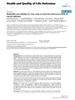
plaque size and electropherotype (Figure 1a and 1b)
[20,21]. YK-1 produced smaller plaques and had an elec-
tropherotype typical of group A rotaviruses and that was
very similar to but distinct from RRV. Variant 311 pro-
duced larger plaques and had an RNA electropherotype
identical to that for YK-1, except it had an additional seg-
ment that was migrating slightly slower than segments 7,
8, and 9 and did not have a typical migrating segment 11.
Both YK-1 and 311 showed a cytopathic effect typical of
rotavirus grown in MA-104 cells and readily grew to titers
over 10
8
ffu per ml.
Variant 311 has a rearranged segment 11
The nucleotide sequence of segment 11 from YK-1 was
first determined as a reference. It consisted of 667 nt with
a 594-bp ORF flanked by 5' and 3' UTRs of 21 and 52 nt,
respectively. Segment 11 of YK-1 had 99% similarity to
segment 11 of RRV. In the variant 311, segment 11
migrated slower than that of YK-1 and RRV, as determined
by Northern blot analysis with a probe specific for seg-
ment 11 (Figure 1b). Sequence analysis of segment 11
from variant 311 identified a rearrangement consisting of
a partial duplication of segment 11 from YK-1. The rear-
rangement occurred at nt 626 in the 3' UTR with the
duplication of the ORF starting at nt 201 and included the
entire 3' UTR (Figure 1c). The sequence of variant 311's
segment 11 has 100% identity to YK-1's segment 11 both
in the ORF and in the partial duplication.
YK-1 group, subgroup, and serotype analyses
The VP6 protein of rotavirus confers group specificity that
is divided into seven groups (A to G). The commercial
immunoassay Rotaclone utilizes a monoclonal antibody
directed against the group A VP6 antigen and identified
YK-1 as a group A rotavirus [22]. Group A rotavirus strains
have been separated into four subgroups, and YK-1 is des-
ignated subgroup 1 as determined by reactivity with a sub-
group 1 MAb 255/60 but not with subgroup 2 MAb 631/
9.
To predict the G and P serotype specificities of the YK-1
strain, the sequences of the genes encoding both VP7 and
VP4 were determined and compared with those of repre-
sentatives of established G and P serotypes. The deduced
VP7 amino acid sequence of YK-1 was closely related to
other simian G3 rotavirus strains: 89% amino acid iden-
tity with RRV and 88% amino acid identity with SA11. A
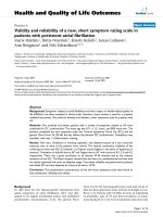
phylogenetic tree was constructed that included known
VP7 amino acid sequences of G3 and other common G
serotypes (Figure 2). The YK-1 strain clustered with other
strains of G3 serotype, including RRV, SA11, AU1, and
YO. YK-1 was also identified as a G3 serotype by reactivity
with MAbs YO-1E2 (G3) and G3-159 (G3) in an immu-
noassay utilizing MAbs reactive to VP7-specific protein.
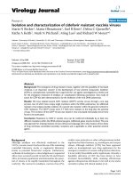
The predicted VP4 amino acid sequence of YK-1 closely
Virology Journal 2006, 3:40 />Page 3 of 8
(page number not for citation purposes)
resembled that of GRV, a newly identified P[3]G3 caprine
strain, and that of RRV P[3]G3 (Figure 3) [23].
NSP4 sequence analysis
The NSP4 gene segment of YK-1 was sequenced and its
deduced gene product was compared with those of other
known rotavirus strains. The structure of the NSP4 gene of
YK-1 was similar to those of other rotavirus strains that
were sequenced previously. The gene consists of a 528-bp
ORF that encodes a protein with a predicted size of 175
amino acids with two conserved potential N-linked glyco-
sylation sites. The deduced amino acid sequence of YK-1's
NSP4 was 98% similar to that of the simian RRV strain. At
least four genetic NSP4 groups are known, and the NSP4
of YK-1 can be classified as Group C by comparison of the
amino acid sequence (aa 131–148) of the variable portion
in the VP4 binding domain of various groups of NSP4
(Figure 4) [24,25].
Discussion
A rotavirus infection model using nonhuman primates
offers a highly relevant system to investigate the mecha-
nisms of disease and immunity to rotavirus and to deter-
mine vaccine effectiveness [16,17,26]. Since nonhuman
primates are the animals most closely related to humans,
this model may be the best predictor of infection and
immunity in humans. In order to perform such studies, it
is necessary to have a rotavirus strain that will consistently
infect nonhuman primates after oral challenge. We
describe rotavirus isolates that were obtained from a nat-
urally infected pigtailed macaque housed in a major pri-
mate research center. This monkey was
immunosuppressed and had severe chronic diarrhea pos-
sibly due to the rotavirus infection. Two isolates were
obtained from a stool of this monkey and one designated
YK-1 had an electrophoerotype typical of most group A
rotaviruses and the other, designated 311, was identical to
YK-1 except for a rearrangement in gene segment 11 that
encodes for the NSP5 protein.
The YK-1 and its variant clone 311 were fully adapted to
grow in cell culture, and both strains could produce
plaques on MA-104 cells, although the plaques from the
311 variant were larger than those from the YK-1 strain.
The significance of the difference in plaques size is not
known. Both YK-1 and 311 were identified as a group A
rotavirus, subgroup 1, genotype P[3] and serotype G3. It
is of interest that these characterizations of YK-1's G and P
types are very similar to another simian rotavirus strain,
RRV. The nucleotide sequence of the NSP4 gene from YK-
1 was also determined because of the discovery of the
NSP4 gene product as a viral enterotoxin and its implica-
tion in the virulence of rotavirus [27]. YK-1 was deter-
mined to have a group C NSP4 gene, which again was
similar to that of the RRV strain.
Comparison between the YK-1 strain and its variant 311 by (a) plaques size, (b) Northern blot analysis for rotavirus RNA segment 11, and (c) schematic diagram of the sequences of gene segment 11Figure 1
Comparison between the YK-1 strain and its variant 311 by
(a) plaques size, (b) Northern blot analysis for rotavirus RNA
segment 11, and (c) schematic diagram of the sequences of
gene segment 11. Arrows indicate segment 11 of RRV and
YK-1, or rearranged segment 11 of variant 311.
Virology Journal 2006, 3:40 />Page 4 of 8
(page number not for citation purposes)
Group A rotaviruses with atypical RNA profiles due to
genomic rearrangements have been repeatedly detected in
stools of chronically infected immunodeficient children
[18,28]. These types of rearrangements have also been
detected in rotavirus isolates from apparently immuno-
competent calves and rabbits [29-31]. We have also iso-
lated an YK-1 variant, 311, with a rearrangement in gene
segment 11. With these rotaviruses, the rearrangement
results from a partial duplication of the gene with a nor-
mal 5' UTR followed by a normal ORF and a duplication
starting at various positions after the stop codon and
extending to the 3' end and leading to a long 3' UTR. Thus,
the rearranged gene expresses a normal protein product.
Although the function of this rearrangement is unknown,
it has been proposed to play a part in the evolution of
rotaviruses and to contribute to their diversity [32]. It has
also been suggested that rearranged segments containing
a partial duplication might be more efficient templates for
double stranded RNA synthesis than are their wild-type
counterparts and thus may be preferentially selected dur-
ing viral replication [31].
Conclusion
Development of a more suitable animal model of rotavi-
rus infection requires the identification of an appropriate
challenge strain. The ideal challenge virus should be iso-
lated from the same species as that employed in the model
system because, in some systems, heterologous rotavi-
ruses tend to undergo abortive replication. We have iso-
lated a new rotavirus strain, designated YK-1, from fecal
specimens of a 2-year-old pigtailed macaque with severe
chronic diarrhea. The YK-1 strain had been used to
develop a nonhuman primate model to enhance our
understanding of the mechanisms of immunity to rotavi-
rus infection [16,17]. This report describes the characteri-
zation of this new strain and a variant of this strain and
Phylogentic tree based on amino acid sequences of the VP7-encoding genes for YK-1 and other established rotavirus strainsFigure 2
Phylogentic tree based on amino acid sequences of the VP7-encoding genes for YK-1 and other established rotavirus strains.
Virology Journal 2006, 3:40 />Page 5 of 8
(page number not for citation purposes)
compares the properties of this strain to those of the other
simian rotavirus strains, SA11 and RRV.
Materials and methods
Rotavirus isolation
The YK-1 strain of simian rotavirus was isolated from the
diarrheal stool of a 2-year-old pigtailed macaque (Macaca
nemestrina) housed at the Yerkes National Primate
Research Center, Emory University (Atlanta, GA). This
Comparision of the NSP4 deduced amino acid sequences at the variable portion in the VP4- binding domain (aa 131–148) from representative groups of rotavirus strainsFigure 4
Comparision of the NSP4 deduced amino acid sequences at the variable portion in the VP4- binding domain (aa 131–148) from
representative groups of rotavirus strains.
Phylogentic tree based on amino acid sequences of the VP4-encoding genes for YK-1 and other established rotavirus strainsFigure 3
Phylogentic tree based on amino acid sequences of the VP4-encoding genes for YK-1 and other established rotavirus strains.
Virology Journal 2006, 3:40 />Page 6 of 8
(page number not for citation purposes)
immunodeficent macaque, PFm-1, had chronic diarrhea
associated with high titers of fecal rotavirus antigen
detected by Rotaclone immunoassay (Meridian Diagnos-
tics, Cincinnati, OH). The virus was isolated by previously
described methods with modifications [20,21]. An extract
from an antigen positive stool was prepared as a 20% (wt/
vol) suspension in phosphate buffered saline (PBS, pH
7.4) and centrifuged twice at 8500 g for 10 min for clarifi-
cation. The supernatant was extracted with 1,1,2-trichlo-
rotrifluoroethane (Sigma, St. Louis, MO), and centrifuged
at 4000 × g for 5 minutes. The extract was treated with
tryspin (15 _g/ml) for 45 min at 37°C and inoculated
onto a confluent monolayer of MA104 cells (African green
monkey kidney cells) for 1 h. After being washed, the
monolayer was maintained in serum-free minimal essen-
tial medium (MEM) (Gibco, Grand Island, NY) supple-
mented with 2 _g/ml tryspin and 50 _g/ml neomycin for
3 days. A viral lysate, obtained by freeze-thawing three
times and clarification at 8500 g for 30 min, was inocu-
lated into MA104 cells and plaque purified three times.
Two distinct plaques distinguished on size were obtained
and further passed in MA104 cells.
Plaque assay
Virus stocks were activated with 15 _g/ml tryspin in MEM
for 45 min at 37°C. The activated virus was 10-fold seri-
ally diluted in MEM and 500 _l/well was inoculated onto
6-well tissue culture plates (Corning, Corning, NY) with a
confluent monolayer of MA104. After a 1 h incubation at
37°C, the inoculum was aspirated and 4 ml of a 3.5% aga-
rose (Seakem, Biowhittaker, Rockland, ME) in MEM was
overlayed on the monolayer. The agar was allowed to
solidify at room temperature (RT), after which the plates
were incubated at 37°C. Plaques were visualized by add-
ing 1 ml of MEM with 2% neutral red and 0.3% agarose 6
h prior to reading.
Purification of virus RNA
Rotavirus RNA was extracted from stools and infected cell
cultures by a modification of a previously described
method [33]. In brief, a 10% stool extract or 30% cell-cul-
ture suspension was prepared with Tris-buffered saline
supplemented with 1% sodium dodecyl sulfate (SDS),
vortexed, and incubated at RT for 10 min. Equal volumes
of virus and 1,1,2-trichlorotrifluoroethane were mixed for
1 minute, and centrifuged for at 8000 g for 10 min. The
supernatant was added to 2 volumes of 6 M guanidine iso-
thyiocyanate and incubated at 56°C for 10 min. Silica
beads were added to each sample, vortexed, and incu-
bated at RT for 10 min. The beads were washed once with
a 2:1 solution of 6 M guanidine isothyiocyanate with 50
mM Tris-HCl (pH 7.5) and then three times with 70% eth-
anol. After the final wash, the beads were air dried, incu-
bated with H
2
O for 10 minutes at 65°C, and centrifuged
for 2 min at 10,000 g. The extract was saved and stored at
-70°C until use.
Electropherotyping
Rotavirus double-stranded genomic RNA extracted from
fecal samples and cell-culture lysates were analyzed by
SDS-polyacrylamide gel electrophoresis as described pre-
viously [34].
Northern blot for segment 11
The procedures employed for Northern hybridization and
chemiluminescent detection of bound digoxigenin-
labeled probe using a commercial reagent (ECL, Amer-
sham, Piccataway, NJ) have been described [35]. Two dig-
oxigenin-labeled probes, ggcttttaaagcgctacagtgatgt and
ggtcacaaaacgggagtggggagctcc, were used to identify
genomic segment 11 of rotavirus.
Subgroup and serotype analyses
Subgroup and VP7 serotyping were determined by use of
a panel of monoclonal antibodies: 225/60 (Subgroup I),
631/9 (Subgroup II), KU-4 (G1), 5E8 (G1), S2-SG10
(G2), IC10 (G2), YO-1E2 (G3), G3-159 (G3), and ST-2G7
(G4) [36-38]. In brief, Immulon II plates (Nagle Nunc,
Rochester, NY) were coated overnight with serum from a
rabbit hyperimmunized with purified RRV rotavirus parti-
cles for positive wells and normal rabbit sera for negative
wells. After the plate was washed with wash buffer (PBS
plus 0.1% Tween 20), cell-culture lysates were added to
duplicate positive and negative wells. The plates were
incubated for 2 h at RT and washed. Specific monoclonal
antibodies were added to wells, and the plates were incu-
bated for 1 h at RT and then washed. Biotinylated goat
anti-mouse IgG (Southern Biotechnology, Birmingham,
Al) was added, incubated 30 min at RT and followed by
washing and the addition of strepavidin-horseradish per-
oxidase (Southern Biotchnology). Wells were developed
by adding tetramethylbenzidine (Sigma) and stopped
after 10 min with 1 N HCl. A sample was considered pos-
itive if the OD value of the positive coated well was >2
times and 0.100 greater than the negative coated well.
PCR amplification and sequence analysis of VP4, VP7, NSP4, and
NSP5
The PCR products of the genes coding for VP4, VP7, and
NSP4 proteins were amplified with previously described
primers, Con2/Con3 for VP4, Beg9/End9 for VP7 and
10Beg16/10End722 for NSP4 [33,39,40]. Full-length PCR
product for the YK-1 gene coding the NSP5 protein was
amplified with primers derived from the 5' and 3' ends of
the NSP5 nucleotide sequence of the SA11 strain. The
nucleotide sequence of each gene was determined from
gel-purified PCR products as previously described [41].
Phylogentic relatedness of the VP4 and VP7 genes of YK-1
was examined by comparing amino acid sequences
Virology Journal 2006, 3:40 />Page 7 of 8
(page number not for citation purposes)
between reference rotavirus strains by using the Wisconsin
Genetics Computer Group computer program [42].
Competing interests
The author(s) declare that they have no competing inter-
ests.
Authors' contributions
LEW characterized and maintained the YK-1 virus and
drafted the manuscript, BJ helped draft the manuscript,
HMM provided samples for virus isolation, LJSM isolated
YK-1, GS and DDG sequenced YK-1, JRG provided phylo-
genic analysis of YK-1, and RIG helped draft and critically
review the manuscript.
Acknowledgements
The authors thank the staff at Yerkes National Primate Research Center
for their assistance with the monkeys, Harry Greenberg, Shozo Urasawa
and Koki Taniguchi for providing monoclonal antibodies, and we thank
Claudia Chesley for editorial assistance. Supported by CRADA with
Aventis Pasteur, Lyon, France and in part by Yerkes Base Grant #RR00165.
References
1. Saif LJ, Rosen BI, Parwani AV: Animal Rotaviruses. In Viral Infec-
tions of the Gastrointestinal Tract Edited by: Kapikian AZ. New York,
NY: Marcel Dekker; 1994:279-367.
2. Burns JW, Krishnaney AA, Vo PO, Rouse RV, Anderson LJ, Green-
berg HB: Analyses of homologous rotavirus infection in the
mouse model. Virology 1995, 207:143-153.
3. McNeal MM, Belli J, Basu M, Choi AH, Ward RI: Discovery of a new
strain of murine rotavirus that is consistently shed in large
quantities after oral inoculation of adult mice. Virology 2004,
320:1-11.
4. Ward RL, McNeal MM, Sheridan JF: Development of an adult
mouse model for studies on protection against rotavirus. J
Virol 1990, 64:5070-5075.
5. Conner ME, Estes MK, Graham DY: Rabbit model of rotavirus
infection. J Virol 1988, 62:1625-1633.
6. Saif LJ, Ward LA, Yuan L, Rosen BI, To TL: The gnotobiotic piglet
as a model for studies of disease pathogenesis and immunity
to human rotaviruses. Arch Virol Suppl 1996, 12:153-161.
7. Estes MK: Rotaviruses and their replication. In Fields Virology 4th
edition. Edited by: Knipe DM, Howley PM, Griffin DE, Lamb RA, Mar-
tin MA, Roizman B, Straus SE. Philadelphia, PA: Lippincott, Williams
and Wilkins; 2001:1747-1785.
8. Malherbe HH, Strickland-Cholmley M: Simian virus SA11 and the
related O agent. Arch Gesamte Virusforsch 1967, 22:235-245.
9. Lopez S, Arias CF: Simian rotavirus SA11 strains. J Virol 1992,
66:1832.
10. Kalter SS, Heberling RL, Rodriguez AR, Lester TL: Infection of
baboons ("Papio cynocephalus") with rotavirus (SA11). Dev
Biol Stand 1983, 53:257-261.
11. Petschow BW, Litov RE, Young LJ, McGraw TP: Response of colos-
trum-deprived cynomolgus monkeys to intragastric chal-
lenge exposure with simian rotavirus strain SA11. Am J Vet Res
1992, 53:674-678.
12. Leong YK, Awang A: Experimental group A rotaviral infection
in cynomolgus monkeys raised on formula diet. Microbiol
Immunol 1990, 34:153-162.
13. Soike KF, Gary GW, Gibson S: Susceptibility of nonhuman pri-
mate species to infection by simian rotavirus SA-11. Am J Vet
Res 1980, 41:1098-1103.
14. Parashar UD, Bresee JS, Gentsch JR, Glass RI: Rotavirus. Emerg
Infect Dis 1998, 4:561-570.
15. Wyatt RG, Mebus CA, Yolken RH, Kalica AR, James HD Jr, Kapikian
AZ, Chanock RM: Rotaviral immunity in gnotobiotic calves:
heterologous resistance to human virus induced by bovine
virus. Science 1979, 203:548-550.
16. Westerman LE, McClure HM, Jiang B, Almond JW, Glass RI: Serum
IgG mediates mucosal immunity against rotavirus infection.
PNAS 2005, 102:7268-7273.
17. Westerman LE, Xu J, Jiang B, McClure HM, Glass RI: Experimental
Infection of Pigtailed Macaques with a Simian Rotavirus, YK-
1. J Med Virol 2005, 75:316-325.
18. Hundley F, McIntyre M, Clark B, Beards G, Wood D, Chrystie I, Des-
selberger U: Heterogeneity of genome rearrangements in
rotaviruses isolated from a chronically infected immunodefi-
cient child. J Virol 1987, 61:3365-3372.
19. Gault E, Schnepf N, Poncet D, Servant A, Teran S, Garbarg-Chenon
A: A human rotavirus with rearranged genes 7 and 11
encodes a modified NSP3 protein and suggests an additional
mechanism for gene rearrangement. J Virol 2001, 75:7305-14.
20. Sato K, Inaba Y, Shinozaki T, Fujii R, Matumoto M: Isolation of
human rotavirus in cell cultures: brief report. Arch Virol 1981,
69:155-160.
21. Ward RL, Knowlton DR, Pierce MJ: Efficiency of human rotavirus
propagation in cell culture. J Clin Microbiol 1984, 19:748-753.
22. Cukor G, Perron DM, Hudson R, Blacklow NR: Detection of rota-
virus in human stools by using monoclonal antibody. J Clin
Microbiol 1984,
19:888-892.
23. Lee JB, Youn SJ, Nakagomi T, Park SY, Kim TJ, Song CS, Jang HK, Kim
BS, Nakagomi O: Isolation, serologic and molecular character-
ization of the first G3 caprine rotavirus. Arch Virol 2003,
148:643-657.
24. Kapikian AZ, Hoshino Y, Chanock RM: Rotaviruses. In Fields Virol-
ogy 4th edition. Edited by: Knipe DM, Howley PM, Griffin DE, Lamb
RA, Martin MA, Roizman B, Straus SE. Philadelphia, PA: Lippincott,
Williams and Wilkins; 2001:1787-1833.
25. Kirkwood CD, Palombo EA: Genetic characterization of the
rotavirus nonstructural protein, NSP4. Virology 1997,
236:258-265.
26. McNeal MM, Sestak K, Choi AH, Basu M, Cole MJ, Ave PP, Bohm RP,
Ward RL: Development of a rotavirus-shedding model in rhe-
sus macaques, using a homologous wild-type rotavirus of a
new P genotype. J Virol 2005, 79:944-954.
27. Ball JM, Tain P, Zeng CQ, Morris AP, Estes MK: Age-dependent
diarrhea induced by a rotaviral nonstructural glycoprotein.
Science 1996, 272:101-104.
28. Eiden J, Losonsky GA, Johnson J, Yolken RH: Rotavirus RNA vari-
ation during chronic infection of immunocompromised chil-
dren. Pediatr Infect Dis 1985, 4:632-637.
29. Pocock DH: Isolation and characterization of two group A
rotaviruses with unusual genome profiles. J Gen Virol 1987,
68:653-660.
30. Thouless ME, DiGiacomo RF, Neuman DS: Isolation of two lapine
rotaviruses: characterization of their subgroup, serotype
and RNA electropherotypes. Arch Virol 1986, 89:161-170.
31. Pedley S, Hundley F, Chrystie I, McCrae MA, Desselberger U: The
genomes of rotaviruses isolated from chronically infected
immunodeficient children. J Gen Virol 1984, 65:1141-1150.
32. Desselberger U: Genome rearrangements of rotaviruses. Arch
Virol Suppl 1996, 12:37-51.
33. Gentsch JR, Glass RI, Woods P, Gouvea V, Gorziglia M, Flores J, Das
BK, Bhan MK: Identification of group A rotavirus gene 4 types
by polymerase chain reaction. J Clin Microbiol 1992,
30:1365-1373.
34. Pereira HG, Azeredo RS, Leite JP, Candeias JA, Raez ML, Linhares AC,
Gabbav YB, Trabulsi JR: Electrophoretic study of the genome of
human rotaviruses from Rio de Janeiro, Sao Paulo and
Belem, Brazil. J of Hygiene 1983, 90:117-125.
35. Palombo EA, Bishop RF: Genetic and antigenic characterization
of a serotype G6 human rotavirus isolated in Melbourne,
Australia. J Med Virol 1995, 47:348-354.
36. Woods PA, Gentsch J, Gouvea V, Mata L, Santosham M, Bai ZS, Ura-
sawa S, Glass RI: Distribution of serotypes of human rotavirus
in different populations. J Clin Microbiol 1992, 30:781-785.
37. Taniguchi K, Urasawa T, Morita Y, Greenberg HB, Urasawa S: Direct
serotyping of human rotavirus in stools by an enzyme-linked
immunosorbent assay using serotype 1-, 2-, 3-, and 4-specific
monoclonal antibodies to VP7. J Infect Dis 1987, 155:1159-1166.
38. Coulson BS, Fowler KJ, White JR, Cotton RG: Non-neutralizing
monoclonal antibodies to a trypsin-sensitive site on the
major glycoprotein of rotavirus which discriminate between
virus serotypes. Arch Virol 1987, 93:199-211.
Publish with BioMed Central and every
scientist can read your work free of charge
"BioMed Central will be the most significant development for
disseminating the results of biomedical research in our lifetime."
Sir Paul Nurse, Cancer Research UK
Your research papers will be:
available free of charge to the entire biomedical community
peer reviewed and published immediately upon acceptance
cited in PubMed and archived on PubMed Central
yours — you keep the copyright
Submit your manuscript here:
/>BioMedcentral
Virology Journal 2006, 3:40 />Page 8 of 8
(page number not for citation purposes)
39. Cunliffe NA, Woods PA, Leite JP, Das BK, Ramachandran M, Bhan
MK, Hart CA, Glass RI, Gentsch JR: Sequence analysis of NSP4
gene of human rotavirus allows classification into two main
genetic groups. J Med Virol 1997, 53:41-50.
40. Gouvea V, Glass RI, Woods P, Taniguchi K, Clark HF, Forrester B,
Fang ZY: Polymerase chain reaction amplification and typing
of rotavirus nucleic acid from stool specimens. J Clin Microbiol
1990, 28:276-282.
41. Laird AR, Gentsch JR, Nakagomi T, Nakagomi O, Glass RI: Charac-
terization of serotype G9 rotavirus strains isolated in the
United States and India from 1993 to 2001. J Clin Microbio 2003,
41:3100-3111.
42. Devereux J, Haeberli P, Smithies O: A comprehensive set of
sequence analysis programs for the VAX. Nucleic Acids Res
1984, 12:387-395.