Báo cáo hóa học: " Hepatitis C virus NS2 and NS3/4A proteins are potent inhibitors of host cell cytokine/chemokine gene expression" potx
Bạn đang xem bản rút gọn của tài liệu. Xem và tải ngay bản đầy đủ của tài liệu tại đây (1.06 MB, 13 trang )
BioMed Central
Page 1 of 13
(page number not for citation purposes)
Virology Journal
Open Access
Research
Hepatitis C virus NS2 and NS3/4A proteins are potent inhibitors of
host cell cytokine/chemokine gene expression
Pasi Kaukinen*
1
, Maarit Sillanpää
1
, Sergei Kotenko
2
, Rongtuan Lin
3
,
John Hiscott
3
, Krister Melén
1
and Ilkka Julkunen
1
Address:
1
Department of Viral Diseases and Immunology, National Public Health Institute, Helsinki, Finland,
2
Department of Biochemistry and
Molecular Biology, University of Medicine and Dentistry-New Jersey Medical School, Newark, NJ, USA and
3
Lady Davis Institute for Medical
Research, Department of Microbiology & Immunology, McGill University, Montreal, Canada
Email: Pasi Kaukinen* - ; Maarit Sillanpää - ; Sergei Kotenko - ;
Rongtuan Lin - ; John Hiscott - ; Krister Melén - ;
Ilkka Julkunen -
* Corresponding author
Abstract
Background: Hepatitis C virus (HCV) encodes several proteins that interfere with the host cell
antiviral response. Previously, the serine protease NS3/4A was shown to inhibit IFN-β gene
expression by blocking dsRNA-activated retinoic acid-inducible gene I (RIG-I) and Toll-like
receptor 3 (TLR3)-mediated signaling pathways.
Results: In the present work, we systematically studied the effect of all HCV proteins on IFN gene
expression. NS2 and NS3/4A inhibited IFN gene activation. NS3/4A inhibited the Sendai virus-
induced expression of multiple IFN (IFN-α, IFN-β and IFN-λ1/IL-29) and chemokine (CCL5,
CXCL8 and CXCL10) gene promoters. NS2 and NS3/4A, but not its proteolytically inactive form
NS3/4A-S139A, were found to inhibit promoter activity induced by RIG-I or its adaptor protein
Cardif (or IPS-1/MAVS/VISA). Both endogenous and transfected Cardif were proteolytically
cleaved by NS3/4A but not by NS2 indicating different mechanisms of inhibition of host cell
cytokine production by these HCV encoded proteases. Cardif also strongly colocalized with NS3/
4A at the mitochondrial membrane, implicating the mitochondrial membrane as the site for
proteolytic cleavage. In many experimental systems, IFN priming dramatically enhances RNA virus-
induced IFN gene expression; pretreatment of HEK293 cells with IFN-α strongly enhanced RIG-I
expression, but failed to protect Cardif from NS3/4A-mediated cleavage and failed to restore
Sendai virus-induced IFN-β gene expression.
Conclusion: HCV NS2 and NS3/4A proteins were identified as potent inhibitors of cytokine gene
expression suggesting an important role for HCV proteases in counteracting host cell antiviral
response.
Background
Hepatitis C virus (HCV) (family Flaviviridae) is an envel-
oped virus with positive-sense, single-stranded RNA
genome that causes both acute and persistent infections in
humans associated with chronic hepatitis, cirrhosis and
hepatocellular carcinoma. The HCV genome encodes for
Published: 01 September 2006
Virology Journal 2006, 3:66 doi:10.1186/1743-422X-3-66
Received: 16 June 2006
Accepted: 01 September 2006
This article is available from: />© 2006 Kaukinen et al; licensee BioMed Central Ltd.
This is an Open Access article distributed under the terms of the Creative Commons Attribution License ( />),
which permits unrestricted use, distribution, and reproduction in any medium, provided the original work is properly cited.
Virology Journal 2006, 3:66 />Page 2 of 13
(page number not for citation purposes)
a polyprotein of about 3000 amino acids, which is
cotranslationally and posttranslationally processed to
mature proteins in the ER membrane. The core and enve-
lope glycoproteins E1 and E2 form the structural proteins
of the virion. Non-structural (NS) proteins NS2, NS3,
NS4A, NS4B, NS5A and NS5B have important roles in the
polyprotein processing and HCV replication [see for
review [1]]. An alternative reading frame of the core region
encodes for F protein, whose function is presently not
known [2]. NS3 and NS4A proteins associate to form an
active enzyme possessing RNA helicase and serine pro-
tease activities. NS3/4A has an ability to interfere with
type I interferon (IFN) gene expression [3].
One of the host responses to virus infection is the produc-
tion of chemokines and antiviral cytokines such as IFN-α
and IFN-β. Virus-induced IFN production is also further
enhanced by positive feedback mechanisms via type I
IFNs [4]. The initial step for the induction of cytokine
response in RNA virus infection is the activation of cellu-
lar dsRNA receptor systems, Toll-like receptor 3 (TLR3) [5]
and DexH(D) RNA helicase, retinoic acid inducible gene-
I (RIG-I) [6]. TLR3 and RIG-I act through adaptor proteins
TRIF [7] and Cardif (also called as IPS-1/MAVS/VISA),
respectively [8-11]. TRIF and Cardif mediate the activation
of IκB kinase (IKK)α/β/γ complex and IKK-like kinases,
IKKε and TBK1 [7-10,12], which leads to activation and
nuclear translocation of NF-κB and IRF3 [13,14]. In the
nucleus IRF3, NF-κB and AP-1 (ATF-2/c-Jun) transcription
factors activate type I IFN and proinflammatory cytokine
gene expression.
The first indication for the interferon antagonistic func-
tion of HCV NS3/4A was obtained in a study showing that
NS3/4A inhibits IRF3 phosphorylation and activation [3].
Further studies demonstrated that NS3/4A disrupts both
TLR3 and RIG-I-mediated signaling pathways [15-17].
TLR3 adaptor protein, TRIF, was found to be a direct pro-
teolytic target of NS3/4A [18,19]. The RIG-I adaptor pro-
tein, Cardif, is another target for NS3/4A cleavage
[11,20,21]. NS3/4A cleaves Cardif after Cys-508 residue,
32 amino acids from the C-terminus causing the release of
Cardif from the mitochondrial outer membrane leading
to its inability to function in RIG-I signaling [11,20].
Recent studies have mainly focused on the actions of NS3/
4A in the IFN-β promoter regulation, while the role of
other HCV proteins has remained less well characterized.
We show here that NS3/4A blocks the gene expression of
several chemokine and cytokine genes by degradating
Cardif while NS2 protein inhibits gene expression
(including IFN-β) with a different mechanism. Unlike in
some other RNA virus infections, pretreatment of cells
with IFN-α does not rescue virus-induced IFN gene expres-
sion, which is due to the lack of protection of Cardif from
NS3/4A-mediated degradation. We also show that NS3/
4A colocalizes with endogenous Cardif at the mitochon-
drial membrane suggesting that the mitochondrial mem-
brane is the site of proteolytic cleavage of Cardif.
Results
HCV proteases NS2 and NS3/4A inhibit IFN-
β
promoter
activity
Recent studies have demonstrated that HCV NS3/4A pro-
tein complex interferes with IFN gene expression
[3,15,19]. Since many other HCV proteins are also capa-
ble of interfering with host cell signalling pathways, we
carried out a systematic analysis of all HCV proteins to
determine their capacity to interfere with host cell signal-
ling pathways regulating IFN gene expression. Expression
plasmids encoding 11 HCV polypeptides were transfected
into HEK293 cells together with IFN-β-Luc reporter plas-
mid; at 18 h after transfection, cells were infected with
Sendai virus for 24 h, followed by preparation of cell
lysates and measurement of luciferase activities (Fig. 1).
Sendai virus was used since it is able to activate NF-κB, IRF
and MAP kinase pathways that regulate the expression of
chemokine and antiviral cytokine genes. HCV NS3 pro-
tein inhibited Sendai virus-induced IFN-β promoter activ-
ity approximately 50%, while the expression of NS3/4A
complex reduced the promoter activity up to 85% (Fig.
1A). Strong inhibition by NS3/4A complex suggests that
the association of NS4A cofactor with NS3 is crucial for
the protein function. Viral envelope glycoprotein E2 was,
in contrast, found to activate IFN-β promoter activity (ca.
60%) while other HCV proteins did not modulate the
IFN-β promoter activity. This data indicates that serine
protease NS3/4A is a specific inhibitor of IFN-β gene
expression and other HCV proteins do not have similar
function.
Original luciferase activity data, however, revealed that
not only serine protease (NS3 and NS3/4A) but also HCV
proteins NS2 and NS4B modulate IFN-β promoter activity
(Fig. 1B). NS2 protein inhibited while NS4B protein acti-
vated the promoter 3–4-fold (Fig. 1B). Notably, NS2 pro-
tein also inhibited CCL5/RANTES and CXCL10/IP-10
promoters approx. 90% (data not shown). Both proteins
(NS2 and NS4B) regulated TK promoter (Renilla luci-
ferase) as well (Fig. 1C). Renilla luciferase activity was not
affected by NS3/4A. The data suggests that NS2 protein,
when expressed in high levels, is a general inhibitor of sev-
eral cellular promoters. The significance of these observa-
tions requires further investigation (see Discussion).
HCV NS3/4A inhibits several cytokine/chemokine
promoters
Previously, analysis of NS3/4A-mediated inhibition of
IFN gene expression has been restricted to IFN-β gene. To
further analyze whether the expression of other type I IFN
Virology Journal 2006, 3:66 />Page 3 of 13
(page number not for citation purposes)
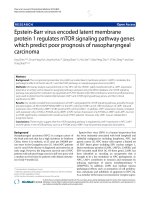
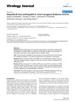
HCV NS2 and NS3/4A inhibit IFN-β gene expressionFigure 1
HCV NS2 and NS3/4A inhibit IFN-β gene expression. (A) The effect of expressed 11 HCV polypeptides on IFN-β pro-
moter activity was studied in HEK293 cells by Luc reporter driven assay. The cells were transfected in triplicates with 1.0 μg
HCV protein expression plasmids together with 0.1 μg firefly luciferase reporter under IFN-β promoter and 0.05 μg Renilla
luciferase reporter (control) plasmids. Total DNA amount was balanced with the empty plasmid (pcDNA3.1(+)-FLAG). At 18
h after transfection the cells were infected with Sendai virus (MOI 5) or mock infected for 24 h, followed by collection of cells,
preparation of cell lysates and measurement of luciferase activity. IFN-β promoter activities were normalized with Renilla luci-
ferase activities. The activity of the sample that was transfected with empty pcDNA3 plasmids was assigned to 100%. Original
values of IFN-β promoter (B) and Renilla luciferase (C) activities with HCV expression constructs are presented in the figures.
Promoter activities were measured as triplicates and expressed as the means +/- standard deviations.
0
40
80
120
160
200
pcDN
A
3
core
F protei
n
E
1
E
2
N
S
2
N
S
3
N
S
4
A
NS3/4
A
N
S
4
B
N
S
5
A
N
S
5
B
IF N -
β
p
ro m o te r a c tivit
y(
%
)
Mock
Sendai
A
0
50000
100000
150000
200000
250000
300000
pcDNA3
c
ore
F
pr
o
tei
n
E1
E2
N
S
2
NS3
N
S
4
A
NS3/4
A
N
S4
B
NS
5
A
N
S
5B
IFN-
β
promoter activity (RLU)
Moc k
Sendai
624000
B
0
400
800
1200
1600
2000
pcDNA3
core
Fprot
ei
n
E1
E2
NS
2
NS3
NS4A
NS3/4A
NS4
B
NS
5A
NS
5B
Renilla promoter activity (RLU)
Moc k
Sendai
C
6280 5440
Virology Journal 2006, 3:66 />Page 4 of 13
(page number not for citation purposes)
or IFN-like genes is also inhibited we carried out transfec-
tion analyses with IFN-β, IFN-α1, IFN-λ1/IL-29 and IFN-
λ3/IL-28B (almost identical to IFN-λ2 promoter) pro-
moter-reporter contructs together with NS3/4A-wt and
protease-inactive NS3/4A-S139A expression plasmids
(Fig. 2A). HCV NS3/4A-wt efficiently inhibited Sendai
virus-induced IFN-β, IFN-α1 and IFN-λ1/IL-29 promoter
activities while the NS3/4A-S139A did not. Thus, IFN-α
(α1), IFN-β and IFN-λ (λ1) genes are highly sensitive to
the inhibitory effect of NS3/4A and the protease activity of
NS3 is absolutely crucial for this inhibition.
The inhibitory effect of NS3/4A on other cytokine/chem-
okine gene promoters (IFN-β, CCL5/RANTES, CXCL10/
IP-10, CXCL8/IL-8, TNF-α and IFN-α4) was next studied
(Fig. 2B). NS3/4A, but not core protein, strongly inhibited
Sendai virus-induced IFN-β, CCL5/RANTES and CXCL10/
IP-10 promoters, while inhibition of CXCL8 promoter
was more moderate being only ca. 50%. The promoters of
IFN-λ3/IL-28B, TNF-α and IFN-α4 were practically not
activated in Sendai virus-infected HEK293 cells suggesting
that the transcriptional systems regulating these promot-
ers are not effectively activated by Sendai virus or certain
important components are missing in our model cell sys-
tem. Altogether, our data suggest that NS3/4A protein is
not only an effective antagonist of the IFN-β promoter but
of other cytokine/chemokine promoters as well.
Components of the RIG-I and TLR3/TLR4 pathway
activate IFN-
β
promoter in HEK293 cells
Recent studies have shown that many different signalling
pathways, including RIG-I, TLR3, RIP1 or PI3K pathways
are involved in IRF3 activation and IFN (IFN-β) gene
expression [5,6,22,23]. We analyzed whether crucial com-
ponents of these intracellular signal transduction path-
ways regulate IFN-β promoter activity in the presence or
absence of activating virus infection. The data shows that
constitutively active form of RIG-I (ΔRIG-I), Cardif, TRIF,
IKKε and TBK1 directly activated IFN-β promoter (Fig. 3;
white columns) and no further enhancement of the pro-
moter activity was seen by Sendai virus infection (Fig. 3;
black columns). The promoter activity was enhanced after
Sendai virus infection in full-length RIG-I and IRF3-
expressing cells suggesting that an additional signal
through dsRNA is needed to activate the RIG-I pathway. It
was recently shown that phosphoinositide 3-kinase
(PI3K)-Akt pathway plays a role in TLR3-mediated IRF3
activation [23]. In our experiments, PI3K or Akt expres-
sion were not able to specifically induce IFN-β promoter
activity suggesting that the expression of these molecules
by themselves cannot induce IRF3 and IFN-β promoter
activation. One may speculate that TBK1-mediated phos-
phorylation is crucial for initial IRF3 activation and the
second phosphorylation step induced by PI3K pathway is
needed for full transcriptional activity [23]. TRIF-associ-
ated RIP1 kinase was also not able to induce IFN-β pro-
moter activity. Since RIP1 mediates NF-κB activation,
RIP1 alone may not be sufficient to activate IFN gene
expression [22]. Our data are in line with other reports
showing that RIG-I [6], Cardif [8-10], TRIF [7,12], IKKε/
TBK1 [13,14] and IRF3 are the key components in IFN
gene activating pathways.
Cardif cleavage by NS3/4A but not by NS2 inhibits RIG-I
and Cardif-induced IFN-
β
promoter activity
Since we were able to reconstitute IFN-β gene expression
in HEK293 cells by overexpressing different components
of the RIG-I pathway we studied whether NS2 and NS3/
4A would interfere with RIG-I and Cardif-induced IFN-β
promoter activity. Cells were transfected with ΔRIG-I (Fig.
4A) or Cardif (Fig. 4B) expression plasmids alone or
together with NS3/4A, NS3/4A-S139A (a protease-inac-
tive mutant of NS3/4A) or NS2 expression constructs.
NS3/4A and NS2 inhibited both ΔRIG-I and Cardif-
induced IFN-β promoter activity. ΔRIG-I-induced pro-
moter activity was abolished by low amounts (0.03 μg) of
NS3/4A expression plasmids (Fig. 4A) while higher
amount (0.3 μg) of NS3/4A plasmid was needed to down-
regulate Cardif-induced activity (Fig. 4B). Protease-inac-
tive mutant NS3/4A-S139A did not inhibit the IFN-β
promoter demonstrating that the protease activity is a pre-
requisite for the action of HCV NS3/4A. Interestingly,
lower expression levels (0.03 and 0.3 μg of plasmid vs. 1
μg used in Fig. 1) of NS2 protein specifically inhibited
both ΔRIG-I and Cardif-induced IFN-β promoter activities
as well (Fig. 4A and 4B). This suggests that, in addition to
NS3/4A, NS2 is a potent inhibitor of cytokine gene expres-
sion.
The roles of RIG-I, Cardif and IKKε were studied when
cells were transfected with increasing amounts of ΔRIG-I,
Cardif or IKKε expression plasmids alone (Fig. 4C, white
columns) or together with NS3/4A expression construct
(Fig. 4C, black columns). NS3/4A was shown to abolish
ΔRIG-I and Cardif-induced IFN-β promoter activity. The
promoter activity was weakly restored with higher
amounts of Cardif expression plasmid (from 0.03 ug to
0.3 μg) indicating that Cardif is partially able to overcome
the inhibitory effect of NS3/4A. IKKε-induced activity was
not inhibited by NS3/4A suggesting that IKKε is able to
overcome the NS3/4A-mediated inhibition of IFN-β pro-
moter (Fig. 4C). All together, the data suggest that HCV
NS3/4A is likely to act only upstream from IKKε, and Car-
dif is rate limiting in this experimental setting.
Cardif has been shown to be a proteolytic target for HCV
NS3/4A [11,20]. We studied whether NS2 utilizes a simi-
lar mechanism to inhibit IFN gene expression. Cells were
transfected with Flag-Cardif and increasing amounts of
HCV NS2, NS3/4A and NS3/4A-S139A expression con-
Virology Journal 2006, 3:66 />Page 5 of 13
(page number not for citation purposes)
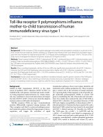
NS3/4A protein is an effective antagonist for cytokine/chemokine promotersFigure 2
NS3/4A protein is an effective antagonist for cytokine/chemokine promoters. (A) IFN-β, IFN-λ1/IL-29, IFN-λ3/IL-
28B and IFN-α1 gene promoter activities were studied in the presence of HCV core, NS3/4A-wt or NS3/4A-S139A after
Sendai virus infection. (B) Cytokine/chemokine gene promoter activities were studied in the presence of HCV core or NS3/4A
protein. The activities of IFN-β, CCL5/RANTES, CXCL8/IL-8, CXCL10/IP-10, TNF-α and IFN-α4 promoters in HCV core or
NS3/4A-expressing HEK293 cells were measured after Sendai virus infection. HEK293 cells were treated as described in the
legend for Figure 1. The activity of the sample that was transfected with empty pcDNA3 plasmids and mock infected was
assigned to value of 1.
IFN-
β
0
100
200
300
400
1 2 3 4 5
fold induction
IFN-
λ
1/IL-29
0
20
40
60
80
100
1 2 3 4 5
fold induction
IFN-
λ
3/IL-28B
0
2
4
6
1 2 3 4 5
fold in duction
IFN-
α
1
0
2
4
6
1 2 3 4 5
fold induction
SeV
-
+ + + +
CORE
+
NS3/4A-wt
+
-
NS3/4A
+
A
-S139A
0
50
100
150
200
250
1 2 3 4
fold induction
IFN-
β
B
0
50
100
150
200
250
1 2 3 4
fold induction
CCL5/RANTES
0
10
20
30
40
50
1 2 3 4
fold induction
CXCL10/IP-10
0
10
20
30
40
50
1 2 3 4
fold induction
CXCL8/IL-8
0
1
2
3
4
5
1 2 3 4
fold induction
TNF-
α
0
1
2
3
4
5
1 2 3 4
fold induction
IFN-
α
4
SeV
-
+ + +
-
+ + +
CORE
+
+
-
NS3/4A-wt
+
-
+
structs (Fig. 4D). Cardif degradation was visualized by the
appearance of CardifΔTM, which is approx. 5-kDa smaller
that the full-length Cardif (Fig. 4D, lanes 4–6). Higher
expression of NS3/4A completely destroyed full length
Cardif. Protease-inactive mutant of NS3/4A did not result
in Cardif degradation indicating that the protease activity
is crucial for the cleavage of Cardif by NS3/4A (Fig. 4D,
lanes 6–8). NS2 protein did not degrade Cardif suggesting
that inhibition of promoter activity occurs by another
mechanism apart from Cardif cleavage (Fig. 4D, lanes 1–
3). Together, these data indicate that NS3/4A and NS2
have different mechanisms to inhibit host cell cytokine
gene expression.
Virology Journal 2006, 3:66 />Page 6 of 13
(page number not for citation purposes)
Components of the RIG-I and TLR3/TLR4 pathway activate IFN-β promoter in HEK293 cellsFigure 3
Components of the RIG-I and TLR3/TLR4 pathway
activate IFN-β promoter in HEK293 cells. HEK293
cells were transfected with expression constructs (0.1 μg)
for intracellular signaling molecules as shown in the figure
and IFN-β reporter plasmid (0.1 μg). IFN-β promoter activi-
ties were measured in mock and Sendai virus-infected
HEK293 cell lysates. The activity of the control sample
(pcDNA3) was assigned to 1.
0
400
800
1200
1600
pcDNA3
NS
34A
RIG-I
R
IG-I
Cardif
TRIF
RIP1
PI3K
Akt
IKK
TBK1
IRF3
IFN-
β
promoter (fold induction)
Mock
Sendai
Δ
ε
IFN/TNF-
α
pretreatment does not rescue cells from NS3/
4A-mediated IFN-
β
promoter inhibition
Certain cytokines may mediate strong positive feedback
regulation that enhances virus-induced IFN gene expres-
sion. In many different cell types such as macrophages,
dendritic cells and epithelial cells IFN-α stimulation leads
to upregulation of TLR genes, TLR-associated adaptor
molecules, components of the RIG-I pathway as well as
IRF7 [4,24-27]. In addition to IFN-α, TNF-α pretreatment
was shown to strongly enhance chemokine and IFN gene
expression in influenza virus-infected lung epithelial cells
as compared to non-pretreated cells [28]. Based on these
findings, we studied whether IFN or TNF-α priming can
overcome the inhibitory functions of NS3/4A and rescue
Sendai virus-induced IFN-β gene expression (Fig. 5). It
was found out that cytokine pretreatments did not have
any effect on IFN-β promoter activity in HCV core or NS3/
4A-expressing cells (Fig. 5A).
We also studied whether IFN-α pretreatment affects NS3/
4A proteolytic activity and its capacity to degrade Cardif.
Immunoblotting analysis of the cell lysates showed Cardif
to be ca. 80 kDa in size (Fig. 5B). Coexpression of NS3/
4A-wt, but not that of a proteolytically inactive form of
NS3/4A-S139A, resulted in a faster migrating form of Car-
dif (approx. 5-kDa smaller) suggesting that Cardif was
proteolytically cleaved by enzymatically active NS3 pro-
tein. Longer exposure (10×) of the film showed that
endogenous Cardif was also sensitive to NS3/4A cleavage.
IFN-α priming did not protect Cardif from NS3/4A-medi-
ated proteolysis.
In primary human leukocytes and lung epithelial cells
IFN-α or TNF-α priming enhance the expression of the
components of the RIG-I pathway [24,26]. Therefore, we
analyzed whether also in HEK293 cells the expression of
RIG-I and/or its downstream components are induced by
IFNs or TNF-α. Northern blot analysis revealed that IFN-α
and to a lesser extent IFN-β induced RIG-I mRNA expres-
sion, while Cardif expression remained virtually
unchanged (Fig. 5C). Western blot analysis showed that
RIG-I protein expression was induced by IFN-α/β, while
neither IFNs nor TNF-α was able to enhance Cardif, IKKε,
IRF3 or IRF7 protein production (Fig. 5D). However,
enhanced RIG-I expression was not able to overcome
NS3/4A-mediated inhibition of IFN-β gene expression.
This is most likely due to the fact that the expression of
Cardif, the proteolytic target of NS3/4A protein complex,
is not enhanced by cytokine stimulation and it thus func-
tions as the "bottleneck" in RIG-I activated signalling
pathway. Therefore, the data demonstrate that unlike in
many viral infections, cytokine priming does not protect
cells from HCV NS3/4A-mediated inhibition of cytokine
gene expression.
HCV NS3/4A colocalizes with Cardif at mitochondrial
membrane
Recent reports have shown that Cardif localizes to the
outer mitochondrial membrane, where it is the target for
NS3/4A proteolysis [9,11,20]. HCV NS3/4A was shown to
localize into ER and/or mitochondrion-associated mem-
brane structures [20,29]. We studied whether NS3/4A or
some other HCV proteins colocalized with endogenous
Cardif, since overexpressed proteins are often mislocal-
ized in cells. Cardif showed an excellent colocalization
with MitoTracker indicating a strong mitochondrial asso-
ciation of Cardif in Huh7 cells (Fig. 6A–C). NS3/4A stain-
ing showed both a punctate pattern in the cytosol of the
cells and significant colocalization with Cardif (Fig. 6D–
F). The data is in line with another recent report [20]. It is
of interest that also HCV core protein showed partial but
significant colocalization with Cardif (Fig. 6G–I). Previ-
ously, core protein was demonstrated to form a granular
staining pattern in the cytoplasm and associate with lipid
storage vesicles and ER that may have vacuolar transport
to mitochondria as well [30-32]. NS5A protein, instead,
did not show any colocalization with Cardif or the mito-
chondria (Fig. 6j–l). Previously, NS5A protein was shown
to be an ER membrane-associated protein [33].
Discussion
Most pathogenic viruses manipulate cellular signalling
pathways for their own advantage. Several HCV proteins
interfere with important host signalling events and regu-
late e.g. cell proliferation and apoptosis. HCV uses several
different strategies to evade the antiviral response. HCV
NS3/4A inhibits IFN synthesis; core interferes with IFN
Virology Journal 2006, 3:66 />Page 7 of 13
(page number not for citation purposes)
HCV NS2 and NS3/4A inhibit RIG-I and Cardif-induced IFN promoter activityFigure 4
HCV NS2 and NS3/4A inhibit RIG-I and Cardif-induced IFN promoter activity. HEK293 cells were transfected with
ΔRIG-I (constitutively active form of RIG-I) (A) or Cardif (B) expression plasmids alone or together with NS3/4A, NS3/4A-
S139A or NS2 expression constructs (0.03 μg or 0.3 μg). IFN-β promoter activities were measured in cell lysates as described
in the legend for Figure 1. Relative IFN-β promoter activities standardized with Renilla expression. (C) IFN-β promoter was
induced by transfecting with increasing (0.03–0.3 μg) amounts of ΔRIG-I, Cardif or IKKε expression constructs either alone or
together with NS3/4A (0.1 μg) expression construct. The effect of NS3/4A on IFN-β promoter activities were measured in
HEK293 cell lysates as described in the legend for Figure 1. (D) Cells were transfected with Cardif and increasing amounts
(0.1–1.0 μg) of NS2, NS3/4A and NS3/4A-S139A expression constructs. Total cell lysates were prepared and Cardif and viral
protein expression was visualized by western blotting.
AB
0,1 ug ΔRIG-I
0,1 ug Cardif
C
NS3/4A-wt
+
-
+
-
+
(0.1 ug)
+
-
+
-
+
0.03 ug
ΔRIG-I
0.1 ug
ΔRIG-I
0.3 ug
ΔRIG-I
0.03 ug
Cardif
0.1 ug
Cardif
0.3 ug
Cardif
+
-
+
-
+
0.03 ug
IKKε
0.1 ug
IKKε
0.3 ug
IKKε
fold induction
Flag-Cardif
Flag-CardifΔTM
D
NS3/4A-S139A
NS3-wt
NS2
0,1 0,3 1,0 0,1 0,3 1,0 0,1 0,3 1,0
NS2 NS3/4A NS3/4A-S139A
1 2 3 4 5 6 7 8 9
ȝg
0
20
40
60
80
100
1 2 3 4 5 6 7
0
100
200
300
400
500
1 2 3 4 5 6 7
0
100
200
300
400
500
1 2 3 4 5 6 7
0
400
800
1200
1600
1 2 3 4 5 6 7 8
0
400
800
1200
1600
1 2 3 4 5 6 7 8
fold induction
ctrl ctrl 0.03 0.3 0.03 0.3 0.03 0.3 μg
NS3/4A
NS3/4A
S139A
NS2
ctrl ctrl 0.03 0.3 0.03 0.3 0.03 0.3 μg
NS3/4A
NS3/4A
S139A
NS2
Virology Journal 2006, 3:66 />Page 8 of 13
(page number not for citation purposes)
signalling; and core, E2 and NS5A inhibit the develop-
ment an antiviral response by inhibiting the functions of
host antiviral proteins [see for review [34]].
HCV serine protease NS3/4A has received special atten-
tion because of its capacity to inhibit IFN production. The
inhibitory mechanism began to clarify when NS3/4A was
shown to inhibit Sendai virus-induced IRF-3 activation
[3]. NS3/4A blocked IRF-3 phosphorylation and recent
studies demonstrated that NS3/4A can directly interfere
with TLR3 and RIG-I signalling pathways by cleaving the
crucial adaptor molecules, TRIF and Cardif, respectively,
thus rendering these pathways inactive [11,19,20]. Our
study was initiated in order to systematically investigate
the potential capacity of all different HCV proteins to
interfere with IFN or other cytokine/chemokine gene
expression. Interestingly, NS2 and NS3/4A inhibited and
NS4B enhanced IFN-β promoter activity. NS3/4A was,
however, demonstrated to be a more specific inhibitor for
the IFN-β promoter. When expressed in high levels NS2
and NS4B proteins regulated the control promoter activity
as well. Previously, NS2 had been found to inhibit several
cellular (e.g., TNF-α) and viral (e.g., CMV) promoters
[35]. Gene regulatory functions for NS4B have not been
previously described. The mechanism how NS2 and NS4B
regulate promoter activity is presently uncharacterized.
Both NS2 and NS4B are ER membrane proteins with mul-
tiple transmembrane domains [36,37]. NS2 is a short-
lived protein and degraded in a phosphorylation-depend-
ent manner [38]. Fast turnover of NS2 may be advanta-
geous for its functions in the inhibition of gene regulation
and apoptosis [39]. NS4B has been implicated in the for-
mation of ER-derived membranous webs that is the site
for HCV RNA replication [40]. The gene regulatory activity
of NS2 and NS4B is an interesting addition to the growing
list of their multiple functions.
NS3/4A suppressed not only IFN-β promoter but also
other IFN (IFN-α1, IFN-λ1) and chemokine gene promot-
ers (CCL5, CXCL8 and CXCL10). The inhibitory effect was
detected at the mRNA and protein expression level as well
(M. Sillanpää, unpublished observations). These data sug-
gest that HCV infection has broad-spectrum inhibitory
effects on host cell cytokine production. The disruption of
IFN production is likely to block IFN amplification loop
leading to reduced expression of both IFN genes as well as
IFN-stimulated genes (ISGs) (e.g., MHC molecules). Inhi-
bition of cytokine/chemokine and ISG expression in HCV
infection may lead to inefficient activation of adaptive
immune response and systemic immune defects [34].
Cytokine production pathway is triggered by viral dsRNA
which is produced during RNA virus replication. RIG-I
and melanoma differentiation associated gene-5 (MDA-
5)-stimulated pathway was recognized as a TLR3-inde-
pendent dsRNA-activated signalling pathway [5,6]. It
seems that TLR3 and RIG-I-induced signaling pathways
are not redundant and they are often operative in different
cell types [41]. Recently, Cardif/IPS-1/MAVS/VISA was
identified to be RIG-I-associated adaptor molecule acti-
vating IKKα/β/γ complex, IKKε and TBK-1 leading to IRF3
phosporylation and IFN gene expression [8-11]. RIG-I
and Cardif-induced IFN promoter activity was clearly
inhibited by NS3/4A. The inhibitory effect was dependent
on protease activity of NS3/4A and Cardif cleavage, since
protease-dead NS3/4A-S139A was not able to inhibit IFN
(IFN-α/β/λ) promoters and degrade Cardif. Notably, the
inhibitory effect of NS2 was not mediated by Cardif cleav-
age. Overexpression of IKKε restored IFN-β promoter
activity indicating that NS3/4A-mediated block was
upstream from IKKε. In addition, overexpression of Cardif
may also partially overcome the NS3/4A-mediated inhib-
itory effects on virus-induced IFN gene activation. These
data are in line with reports showing that Cardif is the pro-
teolytic target for NS3/4A [11,20]. The proteolysis is likely
to occur at the mitochondrial membrane where Cardif
and NS3/4A are colocalized. In the presence of NS3/4A,
the majority of Cardif became cytosolic suggesting prote-
olytic cleavage and release from the mitochondrial mem-
brane [20]. Further studies are warranted to clarify the role
of mitochondria in antiviral signalling.
Cytokine production is suppressed by many viruses such
as influenza A virus. IFN-α or TNF-α pre-treatment prior
to virus infection may restore cell machinery to induce the
IFN production. Influenza A virus infection results in a
weak cytokine response while pre-treatment prior to virus
infection dramatically enhanced host cell cytokine and
chemokine production [25,26,28]. IFN-α or TNF-α treat-
ment has been shown to enhance the expression of the
components of the TLR3 and RIG-I pathways in human
lung epithelial cells [24,28]. Enhanced expression of RIG-
I and IKKε promote dsRNA recognition and IRF3 phos-
phorylation, respectively [6,13,14]. In the present study
NS3/4A-suppressed IFN promoter activity was, however,
not restored by IFN or TNF-α pre-treatment. RIG-I expres-
sion was enhanced by the stimulation of the cells with
IFN-α/β, while the expression of Cardif or its downstream
components were not induced. Thus, even a dramatic
RIG-I expression was not sufficient to rescue the NS3/4A-
mediated block in the pathway possibly due to the fact
that Cardif expression was not enhanced, and it functions
as the bottleneck in the RIG-I pathway.
Conclusion
The present study exhibits systematic analysis of all HCV
proteins in regulating IFN-β promoter. Serine protease
NS3/4A is a crucial viral component in regulating the acti-
vation of innate immune responses. However, other viral
proteins, specifically NS2 and NS4B, have a potential to
Virology Journal 2006, 3:66 />Page 9 of 13
(page number not for citation purposes)
Cytokine priming does not protect cells from HCV NS3/4A-mediated inhibition of cytokine gene expressionFigure 5
Cytokine priming does not protect cells from HCV NS3/4A-mediated inhibition of cytokine gene expression.
(A) The effect of cytokine pre-treatment was studied in Luc-driven assay. The cells were left unprimed (non-treated) or primed
with IFN-α, IFN-β, IFN-γ, (1000 IU/ml each) or TNF-α (20 ng/ml) for 18 h followed by transfection (6 h) with IFN-β promoter/
Renilla luciferase reporter and HCV core or NS3/4A expression constructs. Transfected cells were infected with Sendai virus
(MOI 5) for 16 h, cells were collected and luciferase activity was measured as indicated in the figure. The luciferase activity of
the control sample was assigned to 1. (B) HEK293 cells were primed with IFN-α (1000 IU/ml) or left untreated for 16 h fol-
lowed by transfection with Cardif and NS3/4A-wt or NS3/4A-S139A expression plasmids. Total cell lysate was prepared and
Cardif and NS3/4A protein expression was analysed in cell lysates by immunoblotting. (C) RIG-I and Cardif mRNA was ana-
lysed in cytokine stimulated HEK293 cells. HEK293 cells were untreated (c) or stimulated with IFN-α, IFN-β, IFN-γ (1000 IU/
ml each) or TNF-α (20 ng/ml) for 6 h or 16 h. Total cellular RNA was isolated and RNA samples (10 μg/lane) were analysed by
Northern blotting with RIG-I and Cardif-specific cDNA probes. (D) HEK293 cells were untreated (c) or stimulated as above
with IFN-α, IFN-β, IFN-γ or TNF-α for 24 h. Total cell lysate was prepared and RIG-I, Cardif, IKKε, IRF3 and IRF7 protein
expression was detected by immunoblotting.
B
NS3/4A-wt - + + -
Flag-Cardif + + +
Flag-Cardif
IFN-α
pretreatment
NS3/4A -S139A
+ +
NS3-wt
0
10
20
30
40
50
pcDNA3
core
NS34A
pcDNA3
core
NS34A
pcDNA3
core
NS34A
pcDNA3
core
NS34A
pcDNA3
core
NS34A
IFN-
β
promoter (fold induction)
mock
Sendai
A
non-treated
IFN-α IFN-β IFN-γ TNF-α
NS3/4A-S139A
control
Flag-Cardif
NS3-wt
Flag-CardifΔTM
Flag-CardifΔTM
NS3/4A-S139A
Cardif
CardifΔTM
(10x exposure)
C
D
Virology Journal 2006, 3:66 />Page 10 of 13
(page number not for citation purposes)
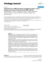
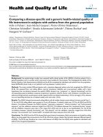
HCV NS3/4A colocalizes with Cardif at mitochondrial membraneFigure 6
HCV NS3/4A colocalizes with Cardif at mitochondrial membrane. The localization of HCV proteins and Cardif was
studied in Huh7 cells. The cells were transfected with HCV protein expression constructs (NS3/4A, core or NS5A) and 48 h
later cells were fixed and stained. The colocalization was visualised by confocal microscopy. Cells were stained for Cardif
(endogenous) (A, D, G, J), mitochondria with Mitotracker Red 580 (B), NS3/4A (E), core (H) and NS5A (K) and the signals
were merged (C, F, I, L).
A) Cardif B) MitoTracker C) Merged
D) Cardif E) HCV NS3/4A F) Merged
G) Cardif H) HCV core I) Merged
J) Cardif K) HCV NS5A L) Merged
Virology Journal 2006, 3:66 />Page 11 of 13
(page number not for citation purposes)
regulate cytokine gene expression as well. More detailed
knowledge of these proteins would help in the develop-
ment of even new antivirals that would target these com-
ponents of the virus. Although the mechanisms how NS3/
4A is suppressing cytokine response have become at least
in major part uncovered, the role of the other HCV pro-
teins in manipulating host immune responses are still rel-
atively poorly understood. This is, undoubtedly, an
important task for the future studies.
Methods
Cell culture and viruses
HEK293 cells (ATCC CLR1573) and human hepatocellu-
lar carcinoma cells (Huh7) cells were cultured in Eagle's
MEM supplemented with 0.6 ug/ml penicillin, 60 ug/ml
streptomycin, 2 mM L-glutamine and 10% heat-inacti-
vated FCS. Sendai virus (strain Cantell) was grown in 11-
day-old embryonated chicken eggs as described [42].
Cytokines
Purified human leukocyte IFN-α and IFN-γ were kindly
provided by Dr. H. Tölö from the Finnish Red Cross Blood
Transfusion Service. IFN-β was purchased from Schering-
Plough and TNF-α from R&D Biosystems.
SDS-PAGE and immunoblotting
For western blot, samples were prepared as described [25].
The blots were stained with rabbit anti-RIG-I (dilution
1:1000) [28], guinea pig anti-Cardif (1:1000), rabbit anti-
IKKε (1:250) [26], rabbit anti-IRF3 (1:200) (Santa Cruz
Biotechnology), rabbit anti-IRF7 (1:1000) [26], mouse
anti-HCV 1a NS2 (gift from Prof. C. Rice) or mouse anti-
HCV NS3 (1:400) (US Biological) antibodies in PBS con-
taining 0,05% Tween and 1% skimmed milk powder.
Anti-Cardif antibodies were prepared in guinea pigs by
immunizing the animals for 4 times (50 μg/immuniza-
tion/animal) at 4 week intervals with E. coli-expressed
GST-Cardif-C (amino acids 157–540) antigen. Peroxi-
dase-conjugated anti-mouse, anti-guinea pig and anti-rab-
bit secondary antibodies were from DAKO.
RNA isolation and Northern blot analysis
Total cellular RNA was isolated by Rneasy kit (Qiagen).
RNA samples (10 μg/lane) were size-fractionated on 1%
formaldehyde-agarose gels and transferred to nylon mem-
branes (Hybond:Amersham). Membranes were hybrid-
ised with probes for RIG-I and Cardif [26]. The probes
were labelled with [α-
32
P] dATP using a random primed
DNA labelling kit (Boehringer Mannheim).
DNA constructs
NS3/4A and F protein genes were amplified by PCR and
inserted into the BamHI site of pcDNA3.1(+)-FLAG-
tagged expression vector [43]. Primers for NS3/4A; 5'-
AAGGGGGGATCCACCATG
GCGCCCATCACGGCG-
TACGCCCAGCAG-3', 5'-GTACGGGGATCCTTATCAG-
CACTCTTCCATCTCATCGAACTCCTG-3', F gene; 5'-
AAAAAAAAGGATCCACCATG
GCACGAATCCTAAACCT-
CAAAGA-3', 5'-TTTCCCTGGGATCCTTATCACGCCGTCT-
TCCAGAACCCG-3' (initiation codon underlined).
Preparation of other HCV protein expression constructs
have been described elsewhere [32]. The mutant NS3/4A-
S139A was created using a site-directed mutagenesis kit
(Stratagene). Expression plasmids for TRIF, RIP1, PI3K
and Akt were kind gifts from Drs. K. Fitzgerald, G. Barber
and G. Sen, respectively. Expression constructs for RIG-I,
ΔRIG-I [6], IKKε, TBK1 [14], IRF3 [44] are described else-
where. The cDNA encoding Cardif was amplified from a T
cell cDNA library and cloned into pcDNA3.1(+)-FLAG.
The promoter-reporter constructs of pRANTES-Luc, pIFN-
β-Luc and pIFN-α4-Luc were described previously
[45,46]. Luc reporter constructs under CXCL10/IP-10,
CXCL8/IL-8, and TNF-α promoters were provided by Drs.
R. Ransohoff, M. Kracht and J. Economou, respectively.
The pIFN-λ1/IL-29-Luc, pIFN-λ3/IL-28B-Luc and pIFN-
α1-Luc promoter-reporter constructs have been created as
follows. The luciferase gene with SV40 mRNA polyade-
nylation signal was cloned into plasmid pcDEF3, resulting
in plasmid pEF-Luc. Promoter fragments of the human
IFN-λ1, IFN-λ3 and IFN-α1 genes were amplified with
primers 5'-GGGACGCGTTTAAACCAATGGCA-
GAAGCTCC-3' and 5'-TGCGGTACCGGCTAAATCG-
CAACTGCTTCCCCAG-3' (for IFN-λ1 promoter), 5'-
GCAACGCGTCATATTCCTGAGTCCTTCCTTGC-3' and 5'-
CCCGGTACCGTCTGTGTCACAGAGAGAAAGGGAG-3'
(for IFN-λ3 promoter), 5'-ATGACGCGTGAAATTCAG-
GAGTAATCAGATC-3' and 5'-GAGGTACCCGTAGATATT-
GCAGATACTTCTG-3' (for IFN-α1 promoter) cloned into
plasmid pEF-Luc.
Transfection and luciferase reporter assay
HEK293 cells were grown on 24-well culture plates and
transfected by using FuGene6 transfection reagent (Roche
Molecular Biochemicals). Cells were transfected with
indicated expression plasmids together with 0.1 μg firefly
luciferase reporter plasmids and 0.05 μg pRL-SV40
(Renilla luciferase) plasmids (Promega). At 18 h after
transfection the cells were mock infected or infected with
Sendai virus (MOI 5) for 24 h. The luciferase activities
were determined using the Dual-Luciferase Reporter Assay
System (Promega) and Victor multilabel reader (Wallac).
Cardif cleavage in vivo
HEK293 cells were grown on 6-well plates and treated
with IFN-α (1000 IU/ml) for 16 h. Cells were transfected
with 0.25 μg of Cardif and 1 μg of NS2, NS3/4A-wt or
NS3/4A-S139A expression plasmids followed by 24 h
incubation. Preparation of total cell lysate and immunob-
lotting were carried out as described above.
Virology Journal 2006, 3:66 />Page 12 of 13
(page number not for citation purposes)
Immunofluorescence staining
Huh7 cells were transfected with pcDNA3.1(+)-FLAG-
NS3/4A/core/NS5A plasmids for 48 h. The cells were fixed
with 3% paraformaldehyde in PBS for 15 min, permeabi-
lized with 0,1% Triton X-100 in PBS for 5 min. Staining of
mitochondria was done in 200 nM Mitotracker Red 580
(Molecular probes) for 30 min at RT. Antibody stainings
were carried out at +37°C in 0,5% BSA/PBS for 1 h. Dou-
ble-stainings were done first with guinea-pig antibody
against Cardif (dilution 1:100) and then with mouse HCV
NS3 antibody (US Biological) (dilution 1:40) or with rab-
bit antibodies against HCV core (dilution 1:50) or HCV
NS5A (dilution 1:200) [32]. The samples were treated
with IgG-FITC or IgG-Rhodamine RedX conjugate second-
ary antibodies (dilution 1:100). The samples were exam-
ined using a Leica TCS NT confocal microscope with an
100× oil immersion lens. The acquired FITC and Rhod-
amine RedX image pairs were automatically merged with
appropriate program.
Competing interests
The author(s) declare that they have no competing inter-
ests.
Authors' contributions
PK participated in the design of the study, performed most
of the experiments, analysed the results and drafted the
manuscript. MS participated in the design of the study and
carried out some experiments. SK, RL and JH provided
crucial reagents to carry out the experiments and helped to
draft the manuscript. KM participated in the design of the
study and helped with the confocal microscopy. IJ initi-
ated the study, participated in its design and coordination
and helped to draft the manuscript. All authors have read
and approved the final version of the manuscript.
Acknowledgements
We thank Hanna Valtonen, Mari Aaltonen and Johanna Lahtinen for their
expert technical assistance. This study was supported by grants from the
European Commission (grant QLK2-CT-2002-00954), Medical Research
Council of the Academy of Finland, the Sigrid Juselius foundation and the
Finnish Cancer Foundation.
References
1. Penin F, Dubuisson J, Rey FA, Moradpour D, Pawlotsky JM: Struc-
tural biology of hepatitis C virus. Hepatology 2004, 39(1):5-19.
2. Xu Z, Choi J, Yen TS, Lu W, Strohecker A, Govindarajan S, Chien D,
Selby MJ, Ou J: Synthesis of a novel hepatitis C virus protein by
ribosomal frameshift. Embo J 2001, 20(14):3840-3848.
3. Foy E, Li K, Wang C, Sumpter RJ, Ikeda M, Lemon SM, Gale MJ: Reg-
ulation of interferon regulatory factor-3 by the hepatitis C
virus serine protease. Science 2003, 300(5622):1145-1148.
4. Marie I, Durbin JE, Levy DE: Differential viral induction of dis-
tinct interferon-alpha genes by positive feedback through
interferon regulatory factor-7. Embo J 1998, 17(22):6660-6669.
5. Alexopoulou L, Holt AC, Medzhitov R, Flavell RA: Recognition of
double-stranded RNA and activation of NF-kappaB by Toll-
like receptor 3. Nature 2001, 413(6857):732-738.
6. Yoneyama M, Kikuchi M, Natsukawa T, Shinobu N, Imaizumi T, Miy-
agishi M, Taira K, Akira S, Fujita T: The RNA helicase RIG-I has
an essential function in double-stranded RNA-induced
innate antiviral responses. Nat Immunol 2004, 5(7):730-737.
7. Oshiumi H, Matsumoto M, Funami K, Akazawa T, Seya T: TICAM-1,
an adaptor molecule that participates in Toll-like receptor 3-
mediated interferon-beta induction. Nat Immunol 2003,
4(2):161-167.
8. Kawai T, Takahashi K, Sato S, Coban C, Kumar H, Kato H, Ishii KJ,
Takeuchi O, Akira S: IPS-1, an adaptor triggering RIG-I- and
Mda5-mediated type I interferon induction. Nat Immunol 2005,
6(10):981-988.
9. Seth RB, Sun L, Ea CK, Chen ZJ: Identification and characteriza-
tion of MAVS, a mitochondrial antiviral signaling protein
that activates NF-kappaB and IRF 3. Cell 2005, 122(5):669-682.
10. Xu LG, Wang YY, Han KJ, Li LY, Zhai Z, Shu HB: VISA is an
adapter protein required for virus-triggered IFN-beta signal-
ing. Mol Cell 2005, 19(6):727-740.
11. Meylan E, Curran J, Hofmann K, Moradpour D, Binder M, Barten-
schlager R, Tschopp J: Cardif is an adaptor protein in the RIG-I
antiviral pathway and is targeted by hepatitis C virus. Nature
2005, 437(7062):1167-1172.
12. Fitzgerald KA, McWhirter SM, Faia KL, Rowe DC, Latz E, Golenbock
DT, Coyle AJ, Liao SM, Maniatis T: IKKepsilon and TBK1 are
essential components of the IRF3 signaling pathway. Nat
Immunol 2003, 4(5):491-496.
13. Sharma S, tenOever BR, Grandvaux N, Zhou GP, Lin R, Hiscott J:
Triggering the interferon antiviral response through an IKK-
related pathway. Science 2003, 300(5622):1148-1151.
14. Breiman A, Grandvaux N, Lin R, Ottone C, Akira S, Yoneyama M,
Fujita T, Hiscott J, Meurs EF: Inhibition of RIG-I-dependent sign-
aling to the interferon pathway during hepatitis C virus
expression and restoration of signaling by IKKepsilon. J Virol
2005, 79(7):3969-3978.
15. Foy E, Li K, Sumpter RJ, Loo YM, Johnson CL, Wang C, Fish PM,
Yoneyama M, Fujita T, Lemon SM, Gale MJ: Control of antiviral
defenses through hepatitis C virus disruption of retinoic
acid-inducible gene-I signaling. Proc Natl Acad Sci U S A 2005,
102(8):2986-2991.
16. Sumpter RJ, Loo YM, Foy E, Li K, Yoneyama M, Fujita T, Lemon SM,
Gale MJ: Regulating intracellular antiviral defense and permis-
siveness to hepatitis C virus RNA replication through a cel-
lular RNA helicase, RIG-I. J Virol 2005, 79(5):2689-2699.
17. Ferreon JC, Ferreon AC, Li K, Lemon SM: Molecular determi-
nants of TRIF proteolysis mediated by the hepatitis C virus
NS3/4A protease. J Biol Chem 2005, 280(21):20483-20492.
18. Li K, Foy E, Ferreon JC, Nakamura M, Ferreon AC, Ikeda M, Ray SC,
Gale MJ, Lemon SM: Immune evasion by hepatitis C virus NS3/
4A protease-mediated cleavage of the Toll-like receptor 3
adaptor protein TRIF. Proc Natl Acad Sci U S A 2005,
102(8):2992-2997.
19. Li XD, Sun L, Seth RB, Pineda G, Chen ZJ: Hepatitis C virus pro-
tease NS3/4A cleaves mitochondrial antiviral signaling pro-
tein off the mitochondria to evade innate immunity. Proc Natl
Acad Sci U S A 2005, 102(49):17717-17722.
20. Loo YM, Owen DM, Li K, Erickson AK, Johnson CL, Fish PM, Carney
DS, Wang T, Ishida H, Yoneyama M, Fujita T, Saito T, Lee WM, Hage-
dorn CH, Lau DT, Weinman SA, Lemon SM, Gale MJ: Viral and
therapeutic control of IFN-{beta} promoter stimulator 1
during hepatitis C virus infection. Proc Natl Acad Sci U S A 2006.
21. Meylan E, Burns K, Hofmann K, Blancheteau V, Martinon F, Kelliher
M, Tschopp J: RIP1 is an essential mediator of Toll-like recep-
tor 3-induced NF-kappa B activation. Nat Immunol 2004,
5(5):503-507.
22. Sarkar SN, Peters KL, Elco CP, Sakamoto S, Pal S, Sen GC: Novel
roles of TLR3 tyrosine phosphorylation and PI3 kinase in
double-stranded RNA signaling. Nat Struct Mol Biol 2004,
11(11):1060-1067.
23. Yamamoto M, Sato S, Mori K, Hoshino K, Takeuchi O, Takeda K,
Akira S: Cutting edge: a novel Toll/IL-1 receptor domain-con-
taining adapter that preferentially activates the IFN-beta
promoter in the Toll-like receptor signaling. J Immunol 2002,
169(12):6668-6672.
24. Siren J, Pirhonen J, Julkunen I, Matikainen S: IFN-alpha regulates
TLR-dependent gene expression of IFN-alpha, IFN-beta, IL-
28, and IL-29. J Immunol 2005, 174(4):1932-1937.
25. Osterlund P, Veckman V, Siren J, Klucher KM, Hiscott J, Matikainen S,
Julkunen I: Gene expression and antiviral activity of alpha/beta
Publish with BioMed Central and every
scientist can read your work free of charge
"BioMed Central will be the most significant development for
disseminating the results of biomedical research in our lifetime."
Sir Paul Nurse, Cancer Research UK
Your research papers will be:
available free of charge to the entire biomedical community
peer reviewed and published immediately upon acceptance
cited in PubMed and archived on PubMed Central
yours — you keep the copyright
Submit your manuscript here:
/>BioMedcentral
Virology Journal 2006, 3:66 />Page 13 of 13
(page number not for citation purposes)
interferons and interleukin-29 in virus-infected human mye-
loid dendritic cells. J Virol 2005, 79(15):9608-9617.
26. Veckman V, Osterlund P, Fagerlund R, Melen K, Matikainen S,
Julkunen I: TNF-alpha and IFN-alpha enhance influenza-A-
virus-induced chemokine gene expression in human A549
lung epithelial cells. Virology 2006, 345(1):96-104.
27. Siren J, Imaizumi T, Pietila T, Lin R, Hiscott J, Noah DL, Krug RM,
Sarkar D, Fisher PB, Julkunen I, Matikainen S: RIG-I and mda-5 are
involved in influenza A virus-induced expression of antiviral
cytokines. Microbes Infect 2006, in press:.
28. Matikainen S, Siren J, Tissari J, Veckman V, Pirhonen J, Severa M, Sun
Q, Lin R, Meri S, Uze G, Hiscott J, Julkunen I: Tumor necrosis fac-
tor alpha enhances influenza A virus-induced expression of
antiviral cytokines by activating RIG-I gene expression. J Virol
2006, 80(7):3515-3522.
29. Wolk B, Sansonno D, Krausslich HG, Dammacco F, Rice CM, Blum
HE, Moradpour D: Subcellular localization, stability, and trans-
cleavage competence of the hepatitis C virus NS3-NS4A
complex expressed in tetracycline-regulated cell lines. J Virol
2000, 74(5):2293-2304.
30. Barba G, Harper F, Harada T, Kohara M, Goulinet S, Matsuura Y, Eder
G, Schaff Z, Chapman MJ, Miyamura T, Brechot C: Hepatitis C virus
core protein shows a cytoplasmic localization and associates
to cellular lipid storage droplets. Proc Natl Acad Sci U S A 1997,
94(4):1200-1205.
31. Moradpour D, Englert C, Wakita T, Wands JR: Characterization of
cell lines allowing tightly regulated expression of hepatitis C
virus core protein. Virology 1996, 222(1):51-63.
32. Melen K, Fagerlund R, Nyqvist M, Keskinen P, Julkunen I: Expression
of hepatitis C virus core protein inhibits interferon-induced
nuclear import of STATs. J Med Virol 2004, 73(4):536-547.
33. Brass V, Bieck E, Montserret R, Wolk B, Hellings JA, Blum HE, Penin
F, Moradpour D: An amino-terminal amphipathic alpha-helix
mediates membrane association of the hepatitis C virus non-
structural protein 5A. J Biol Chem 2002, 277(10):8130-8139.
34. Gale MJ, Foy EM: Evasion of intracellular host defence by hep-
atitis C virus. Nature 2005,
436(7053):939-945.
35. Dumoulin FL, von dem Bussche A, Li J, Khamzina L, Wands JR, Sauer-
bruch T, Spengler U: Hepatitis C virus NS2 protein inhibits
gene expression from different cellular and viral promoters
in hepatic and nonhepatic cell lines. Virology 2003,
305(2):260-266.
36. Yamaga AK, Ou JH: Membrane topology of the hepatitis C
virus NS2 protein. J Biol Chem 2002, 277(36):33228-33234.
37. Hugle T, Fehrmann F, Bieck E, Kohara M, Krausslich HG, Rice CM,
Blum HE, Moradpour D: The hepatitis C virus nonstructural
protein 4B is an integral endoplasmic reticulum membrane
protein. Virology 2001, 284(1):70-81.
38. Franck N, Le Seyec J, Guguen-Guillouzo C, Erdtmann L: Hepatitis C
virus NS2 protein is phosphorylated by the protein kinase
CK2 and targeted for degradation to the proteasome. J Virol
2005, 79(5):2700-2708.
39. Erdtmann L, Franck N, Lerat H, Le Seyec J, Gilot D, Cannie I, Gripon
P, Hibner U, Guguen-Guillouzo C: The hepatitis C virus NS2 pro-
tein is an inhibitor of CIDE-B-induced apoptosis. J Biol Chem
2003, 278(20):18256-18264.
40. Egger D, Wolk B, Gosert R, Bianchi L, Blum HE, Moradpour D, Bienz
K: Expression of hepatitis C virus proteins induces distinct
membrane alterations including a candidate viral replication
complex. J Virol 2002, 76(12):5974-5984.
41. Seth RB, Sun L, Chen ZJ: Antiviral innate immunity pathways.
Cell Res 2006, 16(2):141-147.
42. Pirhonen J, Sareneva T, Kurimoto M, Julkunen I, Matikainen S: Virus
infection activates IL-1 beta and IL-18 production in human
macrophages by a caspase-1-dependent pathway. J Immunol
1999, 162(12):7322-7329.
43. Melen K, Kinnunen L, Julkunen I: Arginine/lysine-rich structural
element is involved in interferon-induced nuclear import of
STATs. J Biol Chem 2001, 276(19):16447-16455.
44. Lin R, Heylbroeck C, Pitha PM, Hiscott J: Virus-dependent phos-
phorylation of the IRF-3 transcription factor regulates
nuclear translocation, transactivation potential, and protea-
some-mediated degradation. Mol Cell Biol 1998,
18(5):
2986-2996.
45. Lin R, Heylbroeck C, Genin P, Pitha PM, Hiscott J: Essential role of
interferon regulatory factor 3 in direct activation of
RANTES chemokine transcription. Mol Cell Biol 1999,
19(2):959-966.
46. Lin R, Genin P, Mamane Y, Hiscott J: Selective DNA binding and
association with the CREB binding protein coactivator con-
tribute to differential activation of alpha/beta interferon
genes by interferon regulatory factors 3 and 7. Mol Cell Biol
2000, 20(17):6342-6353.