Báo cáo hóa học: " Detection of seroconversion to West Nile virus, Usutu virus and Sindbis virus in UK sentinel chickens" doc
Bạn đang xem bản rút gọn của tài liệu. Xem và tải ngay bản đầy đủ của tài liệu tại đây (607.84 KB, 6 trang )
BioMed Central
Page 1 of 6
(page number not for citation purposes)
Virology Journal
Open Access
Short report
Detection of seroconversion to West Nile virus, Usutu virus and
Sindbis virus in UK sentinel chickens
Alan Buckley*
1
, Alistair Dawson
2
and Ernest A Gould
1
Address:
1
CEH Oxford, Mansfield Road, Oxford OX1 3SR, UK and
2
CEH Monks Wood, Abbots Ripton, Huntingdon, Cambridgeshire PE28 2LS,
UK
Email: Alan Buckley* - ; Alistair Dawson - ; Ernest A Gould -
* Corresponding author
Summary
We previously reported evidence of West Nile virus (WNV) circulation in UK birds, probably
introduced by migratory birds from overseas. We now demonstrate WNV-specific seroconversion
in sentinel chickens raised on an English farm. Maternal neutralizing antibodies to WNV in
hatchlings declined within three weeks. During the following months, healthy chickens developed
WNV neutralizing antibodies that were confirmed by immunoblotting and indirect
immunofluorescence tests using WNV antigens. The proportion of seropositive chickens was
higher for WNV than for Usutu virus or Sindbis virus. Attempts to isolate infectious virus or to
detect viral RNA in the sera, failed.
Background
West Nile virus (WNV) and Usutu virus (USUV) are anti-
genically closely related mosquito-borne members of the
genus Flavivirus. Sindbis virus (SINV) is an unrelated mos-
quito-borne member of the genus Alphavirus. These
ar
thropod-borne viruses (arboviruses), and many others,
are known to circulate globally as pathogens amongst
birds and mammalian species [1-4]. During their natural
life cycles, they infect ornithophilic Culex spp. mosquitoes
that replicate and transmit the viruses to birds and/or
mammals when they feed on them. Fatal encephalitic
infections of avian species have been recorded for WNV in
North America [5-7], and Israel, [8] and for USUV in Aus-
tria [9]. Nevertheless, many healthy avian species have
antibodies to these viruses, demonstrating that they are
not necessarily pathogenic for all species they infect. On
the other hand, WNV and SINV are known human patho-
gens and have been shown to be pathogenic for a very
wide range of other mammalian species both in North
America and in the Old World [10]. Previous serological
studies on sera collected from UK resident and migratory
birds demonstrated the presence of WNV-specific neutral-
izing antibodies and also small fragments of RNA with
sequence corresponding to WNV. We also previously
demonstrated the presence of WNV-reactive envelope and
non-structural protein (NS1) antibodies by western blot
analysis and by indirect immunofluorescence (IF) tests
using WNV-infected tissue culture cells as the substrate for
the IF tests. The presence of antibodies to NS1 protein
inferred that the virus had replicated in the birds since
non-structural proteins are only produced in infected cells
after virus replication, ie they would not be present in an
introduced virus. However, in view of the need for addi-
tional proof of the presence of WNV circulating amongst
birds in the UK, albeit apparently harmlessly, we have
looked for evidence of seroconversion to WNV, USUV and
SINV in sentinel chickens.
Published: 04 September 2006
Virology Journal 2006, 3:71 doi:10.1186/1743-422X-3-71
Received: 13 July 2006
Accepted: 04 September 2006
This article is available from: />© 2006 Buckley et al; licensee BioMed Central Ltd.
This is an Open Access article distributed under the terms of the Creative Commons Attribution License ( />),
which permits unrestricted use, distribution, and reproduction in any medium, provided the original work is properly cited.
Virology Journal 2006, 3:71 />Page 2 of 6
(page number not for citation purposes)
Results and discussion
Plaque reduction neutralization tests on sentinel chicken
sera
All sera were tested for the presence of virus-specific neu-
tralizing antibodies by plaque reduction neutralization
tests (PRNT
50
) against two strains of WNV, a strain iso-
lated from Israel (WN-Is) and a highly neutralization-sen-
sitive strain isolated in the Central African Republic (WN-
DAK). For these tests the sera were diluted in twofold steps
from 1/10 dilution, the minimum possible, owing to the
limited volume of serum. The World Health Organization
(WHO) standard method based on 50% plaque reduction
was employed to detect positive virus-neutralizing sera.
Following the WHO recommendations, the highest dilu-
tion of serum that produced 50% reduction of plaque
numbers (estimated 50 plaques per dish in control
dishes) was taken as the endpoint for individual sera. In
addition we also included USUV and SINV in this analysis
because it extended the range of viruses analysed and also
served as a form of internal control for virus-specificity.
The results of plaque reduction neutralization tests
(PRNT
50
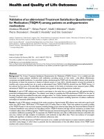
) on the individual sera are presented in Fig 1.
The inclusion of two strains of WNV maximized the data
as we have previously demonstrated differences in sensi-
tivity to neutralization of virus infectivity between differ-
ent strains of WNV [3]. As shown in Fig. 1, the sera from
6/10 and 8/10 of the four-day old chicks neutralized
WNV-DAK and WN-Is respectively, presumably reflecting
the presence of maternal antibody in the hatched chicks.
For USUV, 5/10 newly hatched chick sera contained
detectable neutralizing antibody but they were not neces-
sarily the same chicks that produced antibody against
WNV, demonstrating the specificity of the neutralization
test. However, by the time the chicks were 10 days old, the
proportion of maternally derived neutralization positive
sera against the two strains of WNV and against USUV had
dropped to 2/20, 0/20 and 2/20 respectively and at days
21 and 46 the figures remained low, ie 3/20, 0/20 and 0/
20 at day 21, but by day 46 the figures showed evidence of
increasing, ie 1/10, 3/10 and 2/10. In the case of SINV, 4/
10 four-day old chicks were positive. This figure then
dropped to 4/20 ten-day old chicks and 0/20 chicks by
day 21 and was still zero at day 46. From this time
onwards, the proportion of WNV positive sera increased
noticeably, until by October 8/8, and 7/8 of the sera were
positive for WN-DAK and WN-Is respectively. In many
cases the titres of these sera were noticeably higher than
those recorded in previous months. In contrast, the pro-
portions of seropositive chickens for USUV and SINV
remained lower than those observed for WNV, once again
demonstrating that the PRNT can discriminate between
WNV and USUV. It is important to note that the major
increases in WNV-antibody positive sera were detected in
samples collected from late July to the end of September,
Neutralization results (PRNT
50
) obtained for all sentinel chicken sera tested against each of the four viruses and grouped according to age at time of samplingFigure 1
Neutralization results (PRNT
50
) obtained for all sentinel chicken sera tested against each of the four viruses and grouped
according to age at time of sampling. All chicken sera were coded and all tests were carried out on these coded sera. The
codes were revealed only after the results had been presented. The percentages of positive sera recorded at each antibody
dilution are shown using a colour scheme; White <1/10; Green 1/10; Yellow 1/20; Orange 1/40; Red 1/80.
X
0%
20%
40%
60%
80%
100%
WN-DAK
WN-Is
USUV
SINV
WN-DAK
WN-Is
USUV
SINV
WN-DAK
WN-Is
USUV
SINV
WN-DAK
WN-Is
USUV
SINV
WN-DAK
WN-Is
USUV
SINV
WN-DAK
WN-Is
USUV
SINV
WN-DAK
WN-Is
USUV
SINV
WN-DAK
WN-Is
USUV
SINV
Antibody titres as a proportion of total
1/80
1/40
1/20
1/10
<1/10
4 Days 10 Days 3 Weeks 6 Weeks 9 Weeks 13 Weeks 14 Weeks 20 Weeks
Virology Journal 2006, 3:71 />Page 3 of 6
(page number not for citation purposes)
regardless of the date of hatching of the chicks. Moreover,
the results in Fig. 1 emphasise the importance of using a
highly neutralization sensitive strain of virus, in this case
the WN-DAK strain.
Interestingly, thirteen chicks sampled at 9 weeks post-
hatching had been kept indoors for the entire period since
hatching. Nevertheless, specific antibody responses to
WNV in particular were detected in these chicks, viz., 12/
13, and 6/13, for WNV-DAK and WNV-Is respectively and
1/13, and 3/13 for USUV and SINV respectively. However,
whilst these chicks had been kept indoors, the airflows to
their rooms came directly from the outside without isola-
tion by filtration.
Western blot analysis
Western blot analysis of sera was performed, to confirm
the presence of antibodies to viral envelope glycoprotein,
7% w/v polyethylene-glycol-precipitated virus was puri-
fied by centrifugation at 100,000 g on a continuous
sucrose gradient (15% to 60% w/w sucrose in Tris buffer
at pH 7.4). Serial fractions were collected from the sucrose
gradient and subjected to immunoblotting using a high
titre hyperimmune mouse antiserum prepared against
WNV. Whilst many fractions contained large quantities of
the recognized structural WNV proteins, the fraction col-
lected from the 60% sucrose cushion produced a very
strong and relatively clean band at 51kDa on the western
blot, corresponding to the viral envelope (E) protein of
WNV as confirmed (data not shown but equivalent
appearance to track 1 of Fig 2) using an E protein-specific
monoclonal antibody (MAb) designated MAb 528 [11].
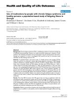
WNV-neutralization positive chicken sera were tested for
the presence of E protein-specific antibodies using the gra-
dient-purified fraction obtained from the 60% sucrose
cushion (Fig. 2). As can be seen, tracks 3 to 10 produced
increasingly intense labelling of the E protein when tested
at a 1/100 dilution. These tracks corresponded to sera
with neutralization titres of 1/10 (tracks 3 and 4), 1/20
(tracks 5 and 6), 1/40 (tracks 7 and 8), and 1/80 (tracks 9
and 10). Track 1 contained a positive control hyperim-
mune mouse antiserum against WNV and Track 2 con-
tained a negative control hyperimmune antiserum raised
against SINV (both positive and negative control sera were
diluted 1/500). Chicken sera that failed to neutralize
WNV were negative in immunoblots when tested at 1/100
dilution. The bands at 45, 42 and 36 kDa in track 9 of Fig
2 probably correspond to breakdown products of the E
protein. They were only detected by chicken sera with the
highest neutralization titres (tracks 9 and 10 of Fig 2). It is
Western blot using gradient-purified West Nile virus antigen and sera (diluted 1/100) from sentinel chickensFigure 2
Western blot using gradient-purified West Nile virus antigen and sera (diluted 1/100) from sentinel chickens. Tracks 1 and 2,
hyperimmune mouse serum prepared against WNV (positive control) or SINV (negative control) respectively; tracks 3 to 10
pairs of chicken sera that produced neutralization titres of 1/10, 1/20, 1/40, and 1/80 respectively.
51kDa ►
45kDa ►
42kDa ►
36kDa ►
1 2 3 4 5 6 7 8 9 10
Virology Journal 2006, 3:71 />Page 4 of 6
(page number not for citation purposes)
also important to note that separate immunoblots that
employed non-purified WNV-infected cell lysates as sub-
strate, and the 1/40 or 1/80 neutralization positive
chicken sera, performed exactly as published previously
[3], i.e., these neutralization-positive sera highlighted the
viral E, NS1, NS3 and NS5 proteins, confirming that WNV
must have replicated in the chickens to elicit immune
responses against the non-structural viral proteins.
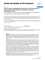
Indirect immunofluorescence tests
Indirect immunofluorescence microscopy was carried out
using a PRNT-positive chicken serum (track 9 from Fig. 2)
diluted 1/100 in PBS as described previously [3]. Vero
cells infected with WNV for 48 hours at 37°C were
washed in PBS and fixed in acetone. The diluted chicken
serum was allowed to react with the acetone-fixed infected
cells for 1 hour at 37°C before the cells were washed in
PBS. Fluorescein-conjugated anti-chicken serum diluted
1/400 in PBS was used to identify the fluorescent infected
cells. Fig. 3 illustrates the typical appearance of groups of
WNV-infected fluorescent cells produced by PRNT-posi-
tive chicken antisera. In Fig. 3, non-infected cells are not
stained by the antibody. Moreover, PRNT-negative con-
trol sera produced no fluorescence (not shown).
Attempts to isolate infectious virus from seropositive
chickens
The sera from 46-day old, and older chickens (a total of 46
sera) and five 10% brain suspensions harvested from
chickens that were seropositive in a WNV-PRNT-analysis
were inoculated directly onto monolayers of SW13 cells
which were then incubated at 37°C for 14 days. The
supernatant medium from each sample was then inocu-
lated onto fresh monolayers of SW13 cells and these were
incubated for a further 14 days. Each monolayer from the
first inoculation and subsequently each monolayer from
the second inoculation was tested for the presence of fla-
vivirus antigens using a flavivirus pan-specific mono-
clonal antibody (MAb 813), followed by fluorescein-
conjugated mouse antiglobulin, as described previously
[11]. Although some monolayers deteriorated during the
incubation period, suggesting that cytopathic effects (cpe)
were developing, we were unable to demonstrate the pres-
ence of an infectious flavivirus in any of the tested sam-
ples either by indirect immunofluorescence microscopy
using flavivirus-group-reactive MAb 813 or by RT-PCR
using flavivirus-group-reactive primers [12]. Moreover,
the mild cpe that was observed in some cultures was not
observed during subsequent passage of harvested mate-
rial, ruling out the possibility of a different cytopathic
arbovirus being isolated.
Although it was not possible to obtain sequential samples
of serum from each animal the PRNT studies with groups
of newly hatched, juvenile and young adult chickens pro-
duced evidence that these animals had been exposed
either to infectious WNV or a very closely related virus
during the summer of 2004. The supplementary positive
results obtained by immunoblotting and immunofluores-
cence microscopy also support this conclusion by demon-
strating specific immune responses against the WNV
envelope protein. Many of the newly hatched chicks had
antibodies that neutralized WNV and to a lesser extent
USUV and SINV. It is well known that maternal antibod-
ies are concentrated in the fertile egg and that the quantity
of these antibodies declines rapidly in the newly hatched
chick [13]. Our PRNT results are totally consistent with
these known observations and they demonstrate that
WNV, USUV and SINV (at least), or closely related viruses,
must have circulated on the farm in the previous year. The
decline in antibody prevalence during the first few weeks
after hatching is also consistent with the idea that WNV is
unlikely to have been circulating significantly during the
first three or four months of the year, i.e. late winter and
early spring. The detection of a significant increase in the
numbers of serologically positive chickens from July
onwards can probably be explained most appropriately as
due to this being the time immediately after the arrival of
migratory birds from Africa, Europe and the Middle East
and also being the warmest time of the year when mosqui-
toes would be relatively active and therefore capable of
transmitting arboviruses, even in England. Some chickens
seroconverted even though they had been kept indoors
for most of their lives. However, the ventilation system for
the building in which they were housed is positive and
not filtered inwards, moreover, adjoining rooms con-
tained wild birds, inferring that the chickens could have
Indirect immunofluorescence microscopy performed on chicken sera with a neutralization titre of 1/80 (diluted 1:1000 in PBS) on West Nile virus infected Vero cells (Bar 50μm)Figure 3
Indirect immunofluorescence microscopy performed on
chicken sera with a neutralization titre of 1/80 (diluted
1:1000 in PBS) on West Nile virus infected Vero cells (Bar
50μm).
Virology Journal 2006, 3:71 />Page 5 of 6
(page number not for citation purposes)
been exposed to aerosols containing virus. In addition to
virus transmission by blood transfusion and organ trans-
plantation, there is now compelling evidence that arbovi-
ruses such as WNV may be transmitted between
vertebrates using a variety of mechanisms other than
direct transmission by arthropods. These include the aer-
osol and faecal/oral routes, transmission via direct physi-
cal contact or maternal milk, and through contaminated
water. It is also clear that WNV can persist in vertebrate
hosts for months if not years without inducing obvious
clinical symptoms [5,14-21]. It seems likely that these
properties provide WNV with the tools to circulate silently
in many regions of the world and this may explain our
observations of seroconversion in sentinel chickens in the
UK. It is also important to emphasize that similar studies
using sera from sequentially bled sentinel chickens in
Italy, known to circulate WNV but with no associated dis-
ease, have been carried out and will report similar find-
ings to those reported herein (manuscript submitted for
publication).
Our observations support and extend the findings of oth-
ers that although mosquitoes are important vectors in dis-
ease transmission, other modes of transmission and
persistence may also be important in the transmission
and circulation of WNV and other arboviruses. We now
need to understand why in most cases, WNV can disperse
very successfully without causing overt disease but in
other situations it can cause significant epidemic out-
breaks involving substantial morbidity and mortality.
Materials and methods
Sentinel chickens
Three groups of chickens were hatched in early April, mid
May and mid June 2004 respectively on a farm in Cam-
bridgeshire and reared outdoors. Individual sera were col-
lected from birds at various ages from 4 days to 20 weeks.
The last samples (20 weeks) were collected at the end of
October 2004, when outdoor temperatures had dropped
sufficiently to reduce insect-biting activity in the UK to rel-
atively low levels. Groups of these animals were moni-
tored periodically for the presence, in the sera, of
neutralizing antibodies to WNV, USUV and SINV. For
obvious technical reasons, only very small quantities of
serum were obtainable from the very young chicks, limit-
ing the scope of their investigation. Another group of
chickens was hatched and reared indoors, and serum sam-
ples collected at 9 weeks of age.
Plaque reduction neutralization tests
These tests were carried out as described previously [3]
and are based on the WHO standard method. Briefly, each
heat-inactivated (56°C for 30 minutes) serum sample was
diluted serially in twofold stages. These were mixed in
equal volume with 50 plaque-forming units of either
WNV-Is, WNV-DAK, USUV or SINV. The mixtures were
incubated overnight at 4°C. Each mixture was then placed
on a monolayer of SW13 cells in 24-well Petri-plates and
incubated for 60 mins at room temperature. 1 ml of over-
lay medium (RPMI-1640 with Hepes buffer, 1% foetal
bovine serum, penicillin, streptomycin and 1% SeaPlaque
Agarose) was added to each well and allowed to set at
room temperature, then the plates were incubated at
37°C until plaques were identifiable in control wells. The
monolayers were fixed in 10% formol-saline and stained
with 0.1% naphthalene black stain. Serum neutralization
titres were estimated as the highest dilution causing at
least 50% reduction of plaque numbers. Titres less than 1/
10 were considered to be negative.
Purification of WNV
The supernatant medium collected from 10 × 175 cm
2
plastic tissue culture bottles was clarified by centrifugation
at 5000 g for 30 mins and the virus was then precipitated
from this clarified medium by the addition of 7% polyeth-
ylene glycol and 0.4 M NaCl. After stirring overnight at
4°C, the virus was sedimented by centrifugation at 5000 g
for 1 hour. The pellets were resuspended in PBS and lay-
ered onto 15–60% (w/w) sucrose gradients prepared in
Tris-EDTA buffer pH7.4. The gradients were spun at
90,000 g for 3 hours and the tube was then fractionated by
upward displacement. Each fraction was tested for the
presence of viral antigens by western blotting (see below).
The sample in the 60% sucrose fraction produced a very
distinct band of viral envelope (E) protein as deduced
using a monoclonal antibody known to bind to WNV-E
protein (see Results).
Western blotting
Gradient-purified West Nile virus antigen and sera
(diluted 1/100) from sentinel chickens were used for the
analysis. The virus proteins were separated by10% poly-
acrylamide gel electrophoresis under reducing conditions
until the dye front had run off the bottom of the gel. A
Biorad semi-dry blotter was used to transfer the protein
bands from the gel onto the Hybond-P PVDF transfer
membrane. After transfer the membrane was blocked in
5% milk powder (in TBS and 0.05% Tween 20) for 1 hour
at room temperature. The blot was then cut into identical
strips (approximately 6 mm wide) which were individu-
ally treated with a chicken serum diluted 1/100 to test for
antibodies to WNV. The strips were washed in TBS/Tween
20 three times before addition of 1:20,000 dilution of
Rabbit anti Chicken conjugated with alkaline phos-
phatase (Sigma) for 1 hour at room temperature. The
strips were washed three times in TBS/Tween 20 then once
in 0.1 M Tris pH9.6 before addition of the BCIP/NBT liq-
uid substrate system (Sigma).
Publish with BioMed Central and every
scientist can read your work free of charge
"BioMed Central will be the most significant development for
disseminating the results of biomedical research in our lifetime."
Sir Paul Nurse, Cancer Research UK
Your research papers will be:
available free of charge to the entire biomedical community
peer reviewed and published immediately upon acceptance
cited in PubMed and archived on PubMed Central
yours — you keep the copyright
Submit your manuscript here:
/>BioMedcentral
Virology Journal 2006, 3:71 />Page 6 of 6
(page number not for citation purposes)
Indirect immunofluorescence microscopy
This was performed on chicken sera (diluted 1:100 or
1:1000 in PBS). Each diluted serum was added to acetone-
fixed WNV-infected Vero cells on glass coverslips. After
incubation for 1 hour at 37°C the cells were washed in
warm PBS for 30 minutes. Rabbit anti-chicken FITC
(Sigma) diluted to 1:400 was then added and after incu-
bation for 1 hour at 37°C, the coverslips were washed in
warm PBS and water before mounting in DABCO/Glyc-
erol/PBS pH8.6, on microscope slides. Each monolayer
was examined for virus-specific immunofluorescence
under a UV light microscope.
Competing interests
The author(s) declare that they have no competing inter-
ests.
Authors' contributions
AB carried out all the immunoassays and data processing
and helped draft the manuscript, AD designed, set up and
carried out the sentinel study, EAG conceived and co-ordi-
nated the study, supervised the research and drafted the
manuscript. All authors have read and approved the sub-
mitted manuscript.
References
1. Lundstrom JO: Mosquito-borne viruses in Western Europe: A
review. Journal of Vector Ecology 1999, 24:1-39.
2. Gould EA, Higgs S, Buckley A, Gritsun TS: Potential Arbovirus
Emergence and Implications for the United Kingdom. Emerg-
ing Infectious Diseases 2006, 12:549-555.
3. Buckley A, Dawson A, Moss SR, Hinsley SA, Bellamy PE, Gould EA:
Serological evidence of West Nile virus, Usutu virus and
Sindbis virus infection of birds in the UK. Journal of General Virol-
ogy 2003, 84:2807-2817.
4. Hubalek Z, Cerny V, Mittermayer T, Kilik J, Halouzka J, Juricova Z,
Kuhn I, Bardos V: Arbovirological survey in Silica plateau area,
Roznava District, Czechoslovakia. Journal of Hygiene, Epidemiol-
ogy, Microbiology and Immunology 1986, 30:87-98.
5. Komar N, Langevin S, Hinten S, Neneth N, Edwards E, Hettler D,
Davis B, Bowen R, Bunning M: Experimental infection of North
American birds with the New York strain of West Nile virus.
Emerging Infectious Diseases 2003, 9:311-327.
6. Murgue B, Zeller H, Deubel V: The Ecology and Epidemiology of
West Nile Virus in Africa, Europe and Asia. In Current Topics in
Microbiology and Immunology Volume 267. Edited by: Mackenzie JM,
Barrett AD and Deubel V. Berlin, Springer-Verlag; 2002:195-221.
7. Stone WB, Therrien JE, Benson R, Kramer L, Kauffman EB, Eldson M,
Campbell S: Assays to detect West Nile virus in dead birds.
Emerging Infectious Diseases 2005, 11:1770-1773.
8. Malkinson M, Banet C, Weisman Y, Pokamunski S, King R, Drouet
MT, Deubel V: Introduction of West Nile virus in the Middle
East by migrating white storks. Emerging Infectious Diseases 2002,
8:392-397.
9. Weissenbock H, Kolodziejek J, Url A, Lussy H, Rebel-Bauder B, Now-
otny N: Emergence of Usutu virus, an African mosquito-
borne flavivirus of the Japanese encephalitis virus group,
Central Europe. Emerging Infectious Diseases 2002, 8:652-656.
10. McClean RG, Ubico SR, Bourne D, Komar N: West Nile Virus in
Livestock and Wildlife. In Current Topics in Microbiology and Immu-
nology Volume 267. Edited by: Mackenzie JM, Barrett AD and Deubel
V. Berlin, Springer-Verlag; 2002:272-303.
11. Gould EA, Buckley A, Cammack N, Barrett ADT, Clegg JCS, Ishak R,
Varma MGR: Examination of the immunological relationships
between flaviviruses using yellow fever virus monoclonal
antibodies.
Journal of General Virology 1985, 66:1369-1382.
12. Gaunt MW, Gould EA: Rapid subgroup identification of the fla-
viviruses using degenerate primer E-gene RT-PCR and site
specific restriction enzyme analysis. Journal of Virological Methods
2005, 128:113-127.
13. Rose ME, Orlans E, Buttress N: Immunoglobulin classes in hen's
egg; their segregation in yolk and white. European Journal of
Immunology 1974, 4:521-523.
14. Nir Y, Beemer A, Goldwasser RA: West Nile virus infection in
mice following exposure to a viral aerosol. British Journal of
Experimental Pathology 1965, 46:443-449.
15. Ravindra KV, Friefeld AG, Kalil AC, Mercer DF, Grant WJ, Botha JF,
Wrenshall LE, Stevens RB: West Nile virus-associated encepha-
litis in recipients of renal and pancreas transplants: case
series and literature review. Clinical Infectious Disease 2004,
38:1257-1260.
16. Iwamoto M, Jerrigan DB, Guasch A, Trepka MJ, Blackmore CG, Hell-
inger WC, Pham SM, Zaki S, Lanciotti RS, Lance-Parker SE, Diaz Gra-
nados CA, Winquist AG, Perlino CA, Wiersma S, Hillyer KL,
Goodman JL, Marfin AA, Chamberland ME, Petersen LR: Transmis-
sion of West Nile virus from an organ donor to four trans-
plant recipients. New England Journal of Medicine 2003,
348:2196-2203.
17. CDC: Possible West Nile virus transmission to an infant
through breast feeding-Michigan. Morbidity and Mortality Weekly
Reports 2002, 51:577-578.
18. Banet-Noach C, Simanov L, Malkinson M: Direct (non-vector)
transmission of West Nile virus in geese. Avian Pathology 2003,
32:489-494.
19. Pogodina VV, Frolova MP, Malenko GV, Fokina GI, Koreshkova GV,
Kiseleva LL, Bochkova NG, Ralph NM: Study on West Nile virus
persistence in monkeys. Arch Virol 1983, 75:71-86.
20. Tonry JH, Xiao CY, Siirin M, Chen H, Travassos da Rosa APA, Tesh
RB: Persistent shedding of West Nile virus in urine of exper-
imentally infected hamsters. American Journal of Tropical Medicine
and Hygiene 2005, 72:320-324.
21. Sbrana E, Tonry JH, Xiao SY, Travassos da Rosa APA, Higgs S, Tesh
RB: Oral transmission of West Nile virus in a hamster model.
American Journal of Tropical Medicine and Hygiene 2005, 72:325-329.