Báo cáo hóa học: " Characterization of neutralizing epitopes within the major capsid protein of human papillomavirus type 33" pot
Bạn đang xem bản rút gọn của tài liệu. Xem và tải ngay bản đầy đủ của tài liệu tại đây (1.86 MB, 11 trang )
BioMed Central
Page 1 of 11
(page number not for citation purposes)
Virology Journal
Open Access
Research
Characterization of neutralizing epitopes within the major capsid
protein of human papillomavirus type 33
Stefanie D Roth
1
, Martin Sapp
1,2,3,4
, Rolf E Streeck
1
and Hans-Christoph Selinka*
1
Address:
1
Institute for Medical Microbiology, Johannes Gutenberg-University 55101 Mainz, Germany,
2
Center for Molecular and Tumor Virology,
Louisiana State University Health Sciences Center, 1501 Kings Highway, Shreveport, LA 71130, USA,
3
Feist Weiller Cancer Center, Louisiana State
University Health Sciences Center, 1501 Kings Highway, Shreveport, LA 71130, USA and
4
Department of Microbiology and Immunology,
Louisiana State University Health Sciences Center, 1501 Kings Highway, Shreveport, LA 71130, USA
Email: Stefanie D Roth - ; Martin Sapp - ; Rolf E Streeck - ; Hans-
Christoph Selinka* -
* Corresponding author
Abstract
Background: Infections with papillomaviruses induce type-specific immune responses, mainly
directed against the major capsid protein, L1. Based on the propensity of the L1 protein to self-
assemble into virus-like particles (VLPs), type-specific vaccines have already been developed. In
order to generate vaccines that target a broader spectrum of HPV types, extended knowledge of
neutralizing epitopes is required. Despite the association of human papillomavirus type 33 (HPV33)
with cervical carcinomas, fine mapping of neutralizing conformational epitopes on HPV33 has not
been reported yet. By loop swapping between HPV33 and HPV16 capsid proteins, we have
identified amino acid sequences critical for the binding of conformation-dependent type-specific
neutralizing antibodies to surface-exposed hyper variable loops of HPV33 capsid protein L1.
Results: Reactivities of monoclonal antibodies (mAbs) H33.B6, H33.E12, H33.J3 and H16.56E with
HPV16:33 and HPV33:16 hybrid L1 VLPs revealed the complex structures of their conformational
epitopes as well as the major residues contributing to their binding sites. Whereas the epitope of
mAb H33.J3 was determined by amino acids (aa) 51–58 in the BC loop of HPV33 L1, sequences of
at least two hyper variable loops, DE (aa 132–140) and FGb (aa 282–291), were found to be
essential for binding of H33.B6. The epitope of H33.E12 was even more complex, requiring
sequences of the FGa loop (aa 260–270), in addition to loops DE and FGb.
Conclusion: These data demonstrate that neutralizing epitopes in HPV33 L1 are mainly located
on the tip of the capsomere and that several hyper variable loops contribute to form these
conformational epitopes. Knowledge of the antigenic structure of HPV is crucial for designing
hybrid particles as a basis for intertypic HPV vaccines.
Background
Human papillomavirus (HPV) infection is the obligate
first step in the development of cervical cancer [1]. How-
ever, of the more than 100 types of HPV, only 15 so-called
high risk types, most commonly types 16, 18, 31, 33, 39,
45, 52, and 58, account for at least 95% of HPV-induced
cervical cancer [2,3]. Vaccination against these high risk
types seems to be the most feasible prevention for cervical
Published: 02 October 2006
Virology Journal 2006, 3:83 doi:10.1186/1743-422X-3-83
Received: 10 August 2006
Accepted: 02 October 2006
This article is available from: />© 2006 Roth et al; licensee BioMed Central Ltd.
This is an Open Access article distributed under the terms of the Creative Commons Attribution License ( />),
which permits unrestricted use, distribution, and reproduction in any medium, provided the original work is properly cited.
Virology Journal 2006, 3:83 />Page 2 of 11
(page number not for citation purposes)
cancer. Indeed, clinical trials have shown prophylactic
HPV vaccines to be effective against HPV infection, cervi-
cal intraepithelial neoplasia (CIN), and genital warts, but
protection is type-specific and the currently developed
vaccines target only a few types [4-6]. These vaccines are
based on papillomavirus-like particles (VLPs) composed
of the major capsid protein, L1. The L1 protein self assem-
bles into VLPs when expressed at high levels in eukaryotic
or insect cells [7-10]. VLPs are composed of 360 copies of
L1 protein organized into 72 pentamers, so called cap-
someres, to form particles which are immunologically
indistinguishable from native virions. Experimentally
induced VLP antisera have been shown to be mostly type-
specific with respect to neutralization [11-13]. Minor
cross-neutralization has been observed only between
closely related HPV types, e.g. HPV6 and 11, HPV18 and
45, or HPV16 and 31 [14-16]. Structure analysis has
revealed the presence of several hyper variable loops on
the outer surface of the capsid [17]. With a few exceptions,
all HPV-neutralizing monoclonal antibodies analyzed so
far are type-specific and recognize conformational
epitopes within surface-exposed hyper variable loops of
the major capsid protein L1 [18-21]. Since capsomeres are
also potent immunogens for induction of neutralizing
antibodies, the formation of these conformational
epitopes does not necessarily require capsid assembly
[22,23]. In a few cases, cross-neutralizing monoclonal
antibodies raised against VLPs in animals that recognize
surface-exposed linear epitopes have been described
[14,16,21].
A prerequisite for generating vaccines that prevent infec-
tion with a broad spectrum of HPV types is extended
knowledge of viral determinants provoking common and
type-specific immune responses. In the present study, we
have fine mapped the binding sites of three neutralizing
monoclonal antibodies (H33.B6, H33.E12, and H33.J3)
with specificity for the human papillomavirus high risk
type 33 (HPV33) by site-directed mutagenesis of surface-
exposed amino acids in the major capsid protein L1.
Moreover, HPV16:33BC hybrid pseudovirions, formed by
HPV16 L1 proteins containing amino acids 51–58 of
HPV33 L1 and HPV16 L2, assembled into particles which
could be neutralized by both HPV33- and HPV16-specific
antibodies, confirming the functional expression of
intrinsic and ectopically expressed epitopes.
Results
Neutralization of HPV33 pseudovirus infection
Papillomavirus pseudovirions that encapsidate a marker
plasmid instead of the viral genome are widely used to
study HPV biology and infection, circumventing the diffi-
culties to obtain biochemical quantities of native virions
[12,24]. Using such HPV16 and HPV33 pseudovirions, we
first determined the neutralizing potential of various
HPV-specific antibodies (Fig. 1). Three days post infection
with HPV pseudovirions, infection was monitored by the
number of cells with green nuclear fluorescence, caused
by transmission of a GFP marker gene to the nucleus via
the HPV vector. Pseudovirus infection in the presence of
the HPV33-specific neutralizing monoclonal antibodies
(mAbs H33.B6, H33.J3, and H33.E12) was abolished
only with pseudovirions of the respective type. Moreover,
we used the recently described mAb H16.56E, generated
after immunization with HPV16 VLPs, and also observed
type-specific neutralization, demonstrating the validity of
this surrogate system for use in testing papillomavirus
neutralizing antibodies (Fig. 1A). Binding of these anti-
bodies to conformationally intact HPV VLPs bound to
Heparin-BSA-coated Elisa plates confirmed the selective
specificity of antibodies H33.J3, H33.B6 and H33.E12 for
HPV33 (Fig. 1B). Subsequent experiments were per-
formed to characterize and fine map the epitopes of these
HPV33-specific antibodies.
Characterization of hyper variable regions in HPV33 L1
For various HPV types it has been reported that type-spe-
cific monoclonal antibodies primarily reside in surface-
exposed hyper variable loops. Our experimental approach
for defining residues involved in neutralization of HPV33
by mAbs H33.B6, H33.J3 and H33.E12 was therefore
based on the exchange of type-specific loop sequences
between the closely related papillomavirus types 16 and
33. Poorly conserved regions in HPV major capsid pro-
teins L1 were identified by sequence alignment and local-
ized by RasMol, based on the coordinates of HPV16 (Pdb
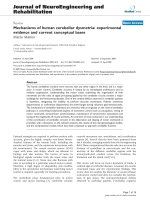
file 1DZL). As shown in Fig. 2, 30 divergent amino acids
between HPV33 and HPV16 were localized in 4 surface-
exposed hyper variable loops, named BC (aa 51–59), DE
(aa 132–140), FG (260–291), and HI (346–358), accord-
ing to the HPV16 L1-structure reported by Chen et al.
[17]. In HPV33 L1, the FG loop was found to consist of
two separate hyper variable regions, designated in this
paper as FGa (260–270) and FGb (282–291) (Fig. 2).
Functional characterization of HPV33 epitopes by loop
substitution
To further characterize the epitopes of HPV33-specific
antibodies, hybrid virus-like particles were designed in
which type-specific sequences in the major capsid protein
L1 of HPV33 were replaced by corresponding amino acids
of HPV16, eliminating the putative epitopes. Vice versa,
HPV33-specific sequences were introduced into HPV16
L1 for ectopic expression. Ten different hybrid L1 proteins
(HPV33:16BC; HPV33:16DE; HPV33:16FGa;
HPV33:16FGb; HPV33:16HI; HPV16:33BC;
HPV16:33DE; HPV16:33DE/FGa, HPV16:33DE/FGb, and
HPV16:33HI) were constructed and expressed in HUTK
-
-
143B cells. Western blot analysis revealed that all hybrid
proteins were expressed at similar levels (data not shown).
Virology Journal 2006, 3:83 />Page 3 of 11
(page number not for citation purposes)
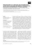
Type-specificity of HPV-reactive antibodiesFigure 1
Type-specificity of HPV-reactive antibodies. A) Infection of human 293TT cells with HPV16 and HPV33 pseudovirions in
the presence of type-specific neutralizing antibodies. Infectious events unaffected by HPV16-specific (mAb H16.56E) or HPV33-
specific (mAbs H33.B6, H33.J3) monoclonal antibodies were monitored 72 hrs post infection. B) Interaction of type-specific
antibodies with HPV16 and HPV33 virus-like particles (VLPs) in a Heparin-BSA ELISA assay. All three antibodies displayed type-
specificity. Although background binding of mAb H33.E12 is significantly increased, specific binding is also restricted to particles
of HPV type 33.
Virology Journal 2006, 3:83 />Page 4 of 11
(page number not for citation purposes)
Binding of monoclonal antibodies to hybrid L1 protein
was first tested by immunofluorescence under non-dena-
turing conditions (Fig. 3). Reactivity of H33.J3 with
hybrid particles was lost by exchanging the BC loop but
was retained after replacement of the other loops (Fig.
3A). This suggests that the BC loop is the binding site for
H33.J3 and that exchange of neighboring surface loops
results in conformationally intact L1 assemblies. HPV16
L1 hybrid particles became reactive with this antibody
when the HPV33 BC loop, but not the DE, FG, and HI
loops, were ectopically expressed on HPV16 (Fig 3B).
Reactivity of H16:56E with HPV16:33BC was retained,
suggesting that this antibody recognizes a different
epitope and, in addition, that this hybrid L1 protein also
forms conformationally intact assemblies.
The epitope recognized by the H33.B6 antibody was
shown to be more complex, as exchange of loops DE or
FGb resulted in the loss of reactivity. Vice versa, introduc-
tion of both HPV33 loops into HPV16 L1 transferred reac-
tivity of H33.B6 to the HPV16:33DE/FGb hybrid (Fig.
3B). Surprisingly, exchange of the DE loop alone was suf-
ficient to render HPV16:33DE reactive with this antibody.
However, the concomitant exchange of the DE and FGa
loops abrogated the binding of H33.B6 with
HPV16:33DE/FGa. Therefore, without being part of the
epitope, the FGa loop has significant influence on the
conformation of the DE loop and thus contributes to the
conformation recognized by H33.B6.
The monoclonal antibody H33.E12 binding site also dis-
plays a high level of complexity. Individual swapping of
loops DE, FGa, and FGb results in the loss of binding to
Determinants of type-specificityFigure 2
Determinants of type-specificity. Alignment of amino acid sequences in surface-exposed loops of capsid proteins L1 of
HPV16 and HPV33. Divergent amino acids are listed; identical amino acids are marked by asterisks. On the right, localization of
these hyper variable loops in the L1 monomer is shown. Modeling by RasMol was based on the monomeric structure of the
HPV16 capsid protein L1.
Virology Journal 2006, 3:83 />Page 5 of 11
(page number not for citation purposes)
Epitope mapping of type-specific antibodiesFigure 3
Epitope mapping of type-specific antibodies. A) Elimination of HPV33-specific epitopes by loop exchanges in capsid pro-
tein L1. Recombinant HPV L1 capsid proteins expressed in HUTK
-
cells were tested by immunofluorescence analysis for the
presence of epitopes for antibodies H16.56E, H33.E12, H33.J3 and H33.B6. Loss of reactivity is marked by (-), gain of antibody
reactivity by (+). B) Functional transfer of HPV33-specific epitopes to HPV 16 by loop swapping, leading to reactivity (+) with
the respective HPV33-specific antibody. Note that the correct presentation of corresponding epitopes is also influenced by
neighboring loops.
Virology Journal 2006, 3:83 />Page 6 of 11
(page number not for citation purposes)
HPV33 hybrid L1 proteins (Fig. 3A), whereas the
exchange of the BC and HI loops had no effect. In contrast
to H33.B6, transfer of individual or two combined HPV33
loops onto HPV16 did not result in the reconstruction of
the epitope. Unfortunately, we were not successful in the
construction of hybrid 16L1 protein carrying all three
HPV33 loops required for binding of H33.E12. Using our
HPV16:33 chimeric particles, we could also show that the
FGa loop is an important part of the H16.56E epitope,
since only HPV33:16FGa particles were recognized by this
antibody. Vice versa, the fact that all HPV16:33 chimeras
were still recognized by this antibody demonstrates that
the H16.56E binding site is not a one-loop epitope but
rather formed by discontiguous sequences of the L1 pro-
tein.
To confirm the validity of our immunofluorescence
approach for measuring conformation-dependent anti-
body binding, we generated and purified hybrid
HPV33:16BC VLPs, using recombinant vaccinia viruses
and HPV16:33BC after transfection of codon-optimized
L1. Reactivity of the monoclonal antibodies with VLPs
was measured in a heparin-BSA ELISA (Fig. 4). Swap of
the BC loop resulted in the loss of reactivity of hybrid
HPV33:16BC with H33.J3 and a gain of reactivity with
H16:33BC. Binding of H33.B6 and H16.56E were not
affected by this exchange and solely dependent on the
backbone (33L1 for HPV33:16BC and 16L1 for
HPV16:33BC) of the chimeric L1 molecules.
Neutralization of hybrid pseudoviruses
To exemplarily demonstrate that the transfer of HPV33-
specific epitopes is functional, hybrid pseudovirions
HPV16:33BC were generated that contain the HPV33 BC
loop in the context of HPV16, following a published pro-
tocol [24]. The mutant was cotransfected with the HPV16
wtL2 expression plasmid and a GFP-expressing marker
plasmid to be packaged. The mutant protein efficiently
assembled with the L2 protein and the marker plasmid
into pseudoviruses that were used in subsequent neutrali-
zation assays. As shown in Fig. 5, HPV16:33BC and wt
HPV33, but not wt HPV16 pseudovirions, were efficiently
neutralized by H33.J3. Hybrid viruses were not neutral-
ized by H33.B6 and H33.E12. These data clearly demon-
strate the functional expression of the heterotypic epitope
on HPV16.
Discussion
A variety of neutralizing epitopes are expressed on the cap-
sid surface of human papillomaviruses. So far, neutraliz-
ing antibody binding sites for HPV6, 11, 16, 31, and 52
have been mapped to the hyper variable surface loops BC,
DE, FG, and HI of the major capsid protein L1
[17,19,20,25-27]. In addition, one neutralizing epitope
has been recently identified in the carboxyl-terminal arm
of HPV16 (aa 430–450) [28]. The complexity of these
epitopes differs considerably among the monoclonal anti-
bodies analyzed so far. We have now demonstrated the
involvement of the BC, DE, and FG surface loops of
HPV33 L1 in the induction of type-specific immune
responses. H33.J3 recognizes a conformation which
solely depends on the presence of the BC loop (Fig. 6A,
D). This seems to be a rare event, since most epitopes of
Neutralization of HPV pseudovirus infection of 293TT cells by type-specific antibodiesFigure 5
Neutralization of HPV pseudovirus infection of
293TT cells by type-specific antibodies. In contrast to
wt HPV16 and HPV33 pseudovirions, HPV16:33BC pseudo-
virions are neutralized by the HPV16-specific H16.56E as well
as the HPV33-specific H33.J3 antibodies. Infection was moni-
tored 72 h post infection.
Heparin-BSA ELISAFigure 4
Heparin-BSA ELISA. Analysis of epitope expression on
wild type (HPV33) and chimeric (HPV33:16BC and
HPV16:33BC) VLPs bound to Heparin-coated ELISA plates
using type-specific antibodies H16.56E, H33.J3 and H33.B6.
Exchange of aa 51–58 (BC-loop of capsid protein L1) results
in the loss or gain of reactivity with antibody H33.J3.
Virology Journal 2006, 3:83 />Page 7 of 11
(page number not for citation purposes)
neutralizing antibodies recognize conformations depend-
ing on more than one loop. By swapping BC loops, the
binding and neutralization capacity of this HPV33-spe-
cific antibody was easily transferable onto HPV16. The
H33.J3 epitope is determined by amino acids 51–58 and
is located at the vertices of capsomeres. Only very few anti-
bodies specific for HPV6 and 11 have been reported to
bind this loop [27], and no HPV high-risk type-specific
antibody other than H33.J3 has been mapped to this
region so far. This may explain the unique properties of
this antibody, which does not interfere with binding of
particles to the primary HPV attachment receptor,
heparan sulfate proteoglycan, and its characteristic feature
to preferentially neutralize cell-bound rather than free
pseudoviruses [29].
We demonstrated that a more complex epitope is recog-
nized by H33.B6 (Fig. 6B, E). Both the DE and the FGb
loop are necessary for binding. Our data also suggest that
the FGa loop contributes to the conformation recognized
by H33.B6 without being part of the binding site. This is
not surprising since all three loops are in intimate proxim-
ity to each other and other monoclonal antibodies have
also been shown to be influenced by more than one of
these loops [20]. The H33.E12 antibody is dependent on
loops DE, FGa, and FGb, since replacement of each of
these loops for HPV16 resulted in the loss of reactivity.
This defines the H33.E12 binding site as an even more
complex epitope (Fig. 6C, F). The previously observed
partial cross-reactivity of H33.J3 with HPV45, 58, and 59
[16] is most likely due to the complex binding site of this
antibody. However, in most cases, cross-reaction might
not be sufficient for cross-protection.
Using the HPV16:33BC chimera in pseudovirus neutrali-
zation assays, we have also shown that the BC hyper vari-
Epitopes of HPV33-specific antibodies on the pentameric L1 capsomereFigure 6
Epitopes of HPV33-specific antibodies on the pentameric L1 capsomere. RasMol pictures showing the epitope pat-
terns for mAb H33.J3 (A), mAb H33.B6 (B) and mAb H33.E12 (C). Variations in the complexity of the epitopes (D-F), rang-
ing from a single loop (D; H33.J3 epitope), two neighboring loops (E, H33.B6 epitope), to at least three loops (F; H33.E12
epitope). Type-specific amino acids are shown in yellow, conserved amino acids in red).
Virology Journal 2006, 3:83 />Page 8 of 11
(page number not for citation purposes)
able loop swap not only transfers the binding ability of
H33.J3 but also the neutralizing capacity to HPV16. This
result suggests that it should be possible to generate HPV
hybrid particles that elicit an immune response directed to
more than one HPV type. Because of the complexity
involving loops DE, FG, and also probably HI [20], which
all can contribute to the conformational binding site of a
given antibody, targeting loops that are clearly separated
seems to be more promising. In addition to the BC loop,
the carboxyl terminal arm is probably a good candidate
for such an approach. Only few antibodies that are
directed against these regions, which were obtained after
experimental immunization of animals, have been
described in the literature so far. This could possibly indi-
cate that these epitopes are not immunodominant. On
the other hand, a recent analysis of the humoral immune
response induced by natural infection with HPV6 and
HPV11 did reveal that all L1 surface loops induced effi-
cient immune responses, and failed to identify any immu-
nodominant epitopes [30], suggesting that each hyper
variable loop may contribute equally to the induction of
virus neutralizing antibodies.
Conclusion
HPV16, 18, 31 and 33 are the four most prevalent HPV
high risk types in cervical cancer. So far, HPV31 and 33 are
not included in current vaccines. Construction of a multi-
valent prophylactic vaccine based on chimeric particles
should be facilitated by selective combination of simple
rather than complex neutralizing epitopes. We have
shown here that various surface exposed hyper variable
loops of the major capsid protein L1 of HPV33 contribute
to the induction of a virus-neutralizing humoral immune
response. The complexity of the identified conforma-
tional epitopes ranges from rather simple structures, con-
sisting of only one loop, e.g. the BC loop, to epitopes to
which several loops contribute. Our data suggest that it
should be possible to generate chimeric polyvalent HPV
particles that could serve as an intertypic vaccine targeting
several HPV types at a time.
Methods
Cell lines and antibodies
The osteosarcoma cell line HuTK
-
143B [31] was grown at
37°C in Dulbecco's modified Eagle medium (DMEM)
supplemented with 10% fetal calf serum and antibiotics.
The human embryonic kidney cell line 293TT [24] was
maintained in DMEM/10% FCS with 1% Glutamax I and
1% non-essential amino acids (Invitrogen). Three confor-
mation-dependent, neutralizing mouse monoclonal anti-
bodies, H33.B6 (IgG2a), H33.E12 (IgG2a) and H33.J3
(IgG2b), respectively, with specificity for HPV33 were
kindly provided by N. D. Christensen, Hershey, PA. The
HPV16-neutralizing mAb H16.56E, was generated by
immunization of mice with HPV16 VLPs, and used as pre-
viously reported [32,33].
Construction of hybrid L1 capsomers by site-directed
mutagenesis
Type-specific amino acids in hypervariable loops of the
HPV33- and HPV16 L1 capsid proteins were identified by
CLUSTAL amino acid sequence alignment [34]. For gener-
ation of HPV33:16 hybrid virus-like-particles, various
loop sequences of the HPV33 L1 capsid protein (BC, DE,
FGa, FGb, HI; Fig. 2) were exchanged by the correspond-
ing amino acids of HPV16 by introducing codon-modi-
fied sequences from p16L1h [35] into pTM33L1 [12].
HPV16:33 hybrids were generated reciprocally, using the
codon-modified pUF3hu16L1 vector and codon-modi-
fied loop sequences of HPV33 L1. Overlap extension PCR
[36] was used to introduce multiple substitutions simulta-
neously. Pairs of PAGE-purified mutagenesis primers with
100 % complementarity (Table 1) were purchased from
Invitrogen and PCR was carried out using puReTaq Ready-
to-go PCR-beads (Amersham Biosciences). In a first step
two separate PCR reactions were prepared to generate
fragments in forward and reverse orientations, both carry-
ing the desired mutations. Thereby, the reverse mutagene-
sis primer was used together with an outer forward
primer, the forward mutagenesis primer in combination
with an outer reverse primer. L1 expression plasmids were
used as template and PCR was performed for 40 cycles
with denaturation at 95°C for 45 seconds, annealing at
42°C for 1 min and elongation at 72°C for 2 min. PCR
fragments generated by these PCRs were purified by agar-
ose gel electrophoresis, followed by Jetsorp gel extraction
prior to their use in subsequent reactions. Because of an
average overlap of 60 bp between appropriate fragments,
these sequences were hybridized by pre-extension PCR
[37], in which the 3'overlap of each strand acts as a primer
for the extension of the complementary strand. This was
done by 2 cycles with denaturation at 95°C for 5 min and
annealing at 72°C for 2 min. Resulting products were
PCR-amplified by addition of the outer primers of step 1
(conditions: denaturation at 95°C, 45 sec; annealing at
50–56°C, 1 min; elongation at 72°C, 2 min; 35 cycles).
Subsequently, the gel-purified mutant L1 amplimers
(sized between 800–1900 bp) were cloned into singular
restriction sites in the transfer vectors pUF3hu16L1 or
pTM33L1 to generate the HPV16/HPV33 or HPV33/
HPV16 hybrid L1-constructs. Ligation mixtures were
transfected into chemically competent cells of E. coli
(DH5α). Colonies containing the desired mutations were
identified by their newly introduced restriction sites or
directly by sequencing. If only one of the two fragments
could be generated in the first PCR round, the purified
fragment was used in a following PCR as a megaprimer.
The fragment was added in excess over the plasmid tem-
plate and combined with a counter-directed common
Virology Journal 2006, 3:83 />Page 9 of 11
(page number not for citation purposes)
primer, using the following conditions for a total of 35
cycles: denaturation at 95°C for 45 sec, annealing at 65°C
for 1 min, elongation at 72°C for 2 min. Generation of
HPV16:33-hybrids with double loop exchanges occurred
successively. One loop was introduced by the approach
described above. To introduce the second loop, a forward
primer was generated using the hybrid L1 as a template.
Subsequently, the fragment served as a megaprimer to
amplify the complete expression plasmid with high-fidel-
ity Pwo DNA polymerase for 18 cycles (denaturation for
30 sec at 95°C, annealing for 1 min at 50°C, elongation
for 14 min at 72°C). The PCR product was then digested
with DpnI to eliminate methylated template DNA and the
remaining mutant plasmids were expressed in E. coli.
Immunofluorescence analysis
HuTK
-
cells were grown on glass coverslips overnight,
infected with the vaccinia helper virus VTF7-3 for 1 h
(MOI of 5) and subsequently transfected using Lipo-
fectamin plus (Invitrogen) and 1 μg transfer plasmid
pTM1 carrying wt or mutated HPV33L1 sequences under
the control of a T7-promotor. Expression of the pUF3 vec-
tor-based wt or hybrid HPV16 L1-constructs occurred by
lipofection without any helper viruses. After an incuba-
tion period of 10 – 24 h at 37°C cells were fixed with 2 %
paraformaldehyde for 20 min at room temperature, per-
meabilized with 0.1 % Nonident P-40 for 15 min and
subsequently blocked in 5 % goat serum dissolved in PBS.
Incubations with primary mAbs and secondary Cy2-con-
jugated Affinipure goat anti-mouse IgG (Jackson Immu-
noresearch Products) were carried out for 1 h at 37°C.
Thereafter, coverslips were washed with PBS several times,
stained with 0.2 μg/ml Bis-benzimide trihydrochloride
(Hoechst 33342; Sigma) and mounted onto slides by
using Fluoprep mounting medium (BioMérieux). Pictures
were taken using a Zeiss Axiovert 200 M microscope and
a Zeiss Axiocam digital camera. The appropriate Axiovi-
sion Software 3.0 was used for merging pictures.
Preparation of pseudovirions and VLPs
HPV33-VLPs and pseudovirions were produced in HuTK
-
cells by infection with recombinant vaccinia viruses
vac33L1, vac33L2 and helper virus VTF7-3, as described
previously [12,38]. For generation of pseudovirions, cells
were transfected 24 h prior to infection with a marker
plasmid encoding a dimeric green fluorescent protein
(GFP), resulting in HPV particles containing the GFP
reporter DNA. Forty-four hours post infection VLPs/PsV
were extracted from nuclei by sonication in hypotonic
buffer supplemented with 0.5% NP-40 and purified by
buoyant caesium chloride density gradients. HPV16 pseu-
dovirions were prepared as described previously [24] by
co-transfection of 293TT cells with pUF3hu16L1 wt or
pUF3hu16/33L1-hybrid plasmids, together with
pUF3hu16L2 wt and the pEGFPGFP marker plasmid. Sub-
sequent to incubation at 37°C for 48 h cells were lysed
and pseudovirions were purified on an OptiPrep gradient.
Thereby, lysis of cells was achieved by adding the non-
ionic detergent Brij58 (Sigma) at a final concentration of
0.5 % in DPBS supplemented with 9.5 mM MgCl
2
. Lysates
were digested over night at 37°C with 2 U of Benzonase
(Sigma) to complete virus maturation [39]. Subsequently
the lysate was mixed with a 0.17 volume of 5 M NaCl,
clarified by centrifugation at 1500 × g for 10 min, loaded
Table 1: Codon optimized sequences of mutagenesis primers
Constructs Sequences for primers (listed 5' to 3')
HPV33:BC For GGCCATCCATATTTTCCCATCAAGAAGCCCAACAACAACAAATTATTGGTACCC
Rev GGGTACCAATAATTTGTTGTTGTTGGGCTTCTTGATGGGAAAATATGGATGGCC
HPV33:DE For TTTGATGACATCGAAAACGCCAGCGCCTACGCCGCCAACGCCGGTGCTGATAATAGG
Rev CCTATTATCAGCACCGGCGTTGGCGGCGTAGGCGCTGGCGTTTTCGATGTCATCAAA
HPV33:FGa For ATGTTTGTAAGACACCTGTTCAACAGGGCCGGCGCCTACGGCGAGAACGTTCCCGATGACCTG
Rev CAGGTCATCGGGAACGTTCTCGCCGTAGGCGCCGGCCCTGTTGAACAGGTGTCTTACAAACAT
HPV33:FGb For ATTAAAGGTTCAGGAAGCACCGCCAACCTGGCCAGCAGCAACTACTTTCCCACTCCTAGTGG
Rev CCACTAGGAGTGGGAAAGTAGTTGCTGCTGGCCAGGTTGGCGGTGCTTCCTGAACCTTTAAT
HPV33:HI For AATATGACTTTATGCGCCGCCATCAGCACCAGCGAGACCACCTACAAGAACAACAATTTTAAAGAATATATAAG
Rev CTTATATATTCTTTAAAATTGTTGTTCTTGTAGGTGGTCTCGCTGGTGCTGATGGCGGCGCATAAAGTCATATT
HPV16:BC For GGCCACCCCTACTTCAGCATCAAGAACCCCACCAACGCCAAGAAGATCCTGGTGCCC
Rev GGGCACCAGGATCTTCTTGGCGTTGGTGGGGTTCTTGATGCTGAAGTAGGGGTGGCC
HPV16:DE For ACCGGCAACAAGTACCCCGGCCAGCCCGGCGTGGACAACAGGGAGTGCATCAGCATGGAC
Rev CCTGTTGTCCACGCCGGGCTGGCCGGGGTACTTGTTGCCGGTCTCGGTGTCGTCCAG
HPV16:FGa For ATGTTCGTGAGGCACTTCTTCAACAGGGCCGGCACCCTGGGCGAGGCCGTGCCCGACGACCTG
Rev CAGGTCGTCGGGCACGGCCTCGCCCAGGGTGCCGGCCCTGTTGAAGAAGTGCCTCACGAACAT
HPV16:FGb For ATCAAGGGCAGCGGCACCACCGCCAGCATCCAGAGCAGCGCCTTCTTCCCCACCCCCAGC
Rev GCTGGGGGTGGGGAAGAAGGCGCTGCTCTGGATGCTGGCGGTGGTGCCGCTGCCCTTGAT
HPV16:HI For AACATGAGCCTGTGCACCCAGGTGGCCAGCGACAGCACCTACAAGAACGAGAACTTCAAGGAGTACCTG
Rev CAGGTACTCCTTGAAGTTCTCGTTCTTGTAGGTGCTGTCGCTGGCCACCTGGGTGCACAGGCTCATGTT
Virology Journal 2006, 3:83 />Page 10 of 11
(page number not for citation purposes)
on top of an OptiPrep step gradient (27%/33%/39%
OptiPrep in DPBS-800 mM NaCl) and centrifuged for 4h
at 234.000 × g. After centrifugation, 250 μl-fractions were
collected by bottom puncture of the tubes and 1 μl of each
fraction was tested in a pseudovirus infection assay.
Infection and neutralization assays
Human embryonic kidney 293TT cells were grown over-
night in 24-well plates and infected with 1 μl of HPV pseu-
dovirions (PsV) in a total volume of 500 μl DMEM. Cells
were grown at 37°C for 72 h and infectious events were
monitored by counting cells with green nuclear fluores-
cence. To perform virus neutralization assays, PsV were
bound to cells for 1 h at 4°C, unbound virions were
removed and various dilutions of HPV-specific neutraliz-
ing antibodies were added to cells in a total volume of 250
μl DMEM. After 1 h at 37°C the culture medium was
replaced and incubation was continued for 72 h.
Heparin-based enzyme-linked immunosorbent assays
(Hep-BSA ELISA)
VLP-ELISAs were used to study the interaction of confor-
mationally intact VLPs with heparin and performed as
previously described [29,40]. Briefly, polysorb microtiter
plates (NUNC, Wiesbaden, Germany) were coated over-
night with 100 ng of heparin-BSA/well in phosphate-buff-
ered saline (PBS), washed and subsequently blocked with
BSA (50 μg/ml) for 30 minutes. Plates were again washed,
100 μl VLPs (1 μg/ml) were added and incubated for 1 h
at 37°C. Unbound particles were eliminated by washing.
HPV type-specific antibodies H16.56E, H33.B6, H33.J3
and H33.E12 were added for 1 h at 37°C at the indicated
concentrations (1:100 – 1:5000). After washing three
times with PBS-Tween 20 (PBS-T), 100 μl horseradish per-
oxidase-coupled secondary antibodies (goat anti-mouse
IgG; 1:10.000 in PBS-T) obtained from Jackson Immuno-
chemicals were added and incubated for additional 30
min at 37°C. Plates were washed and developed with
ready to use trimethyl benzidine (KPL). The reaction was
stopped after 10 min at 37°C with 100 μl 1N HCl.
Absorbance was measured at 450 nm using a Multiscan
EX (Thermo Life Sciences).
Visualization of epitopes by RasMol
The RasMol program is a molecular graphics visualisation
tool for macromolecular structures [41]. Localization of
amino acids in loops structures of capsid protein L1 from
HPV16 or HPV33 was based on the atomic coordinates of
the HPV16 major capsid protein L1 [17] and visualized
using the PDB file 1DZL in the RasMol program.
Competing interests
The author(s) declare they have no competing interests
with this publication.
Acknowledgements
We gratefully acknowledge Neil D. Christensen, (Penn State Hershey Med-
ical Center, PA, USA) for providing HPV33-specific monoclonal antibodies,
Kirsten Freitag (University of Mainz) for technical help and Gilles Spoden,
Maren Knappe and Luise Florin (University of Mainz) for support or critical
reading of the manuscript.
References
1. Walboomers JM, Jacobs MV, Manos MM, Bosch FX, Kummer JA, Shah
KV, Snijders PJ, Peto J, Meijer CJ, Munoz N: Human papillomavirus
is a necessary cause of invasive cervical cancer worldwide. J
Pathol 1999, 189:12-19.
2. Lorincz AT, Reid R, Jenson AB, Greenberg MD, Lancaster W, Kurman
RJ: Human papillomavirus infection of the cervix: Relative
risk associations of 15 common anogenital types. Obstet Gyne-
col 1992, 79:328-337.
3. Munoz N, Bosch FX, de Sanjose S, Herrero R, Castellsague X, Shah
KV, Snijders PJ, Meijer CJ: Epidemiologic classification of human
papillomavirus types associated with cervical cancer. N Engl
J Med 2003, 348:518-527.
4. Koutsky LA, Ault KA, Wheeler CM, Brown DR, Barr E, Alvarez FB,
Chiacchierini LM, Jansen KU: A controlled trial of a human pap-
illomavirus type 16 vaccine. N Engl J Med 2002, 347:1645-1651.
5. Harper DM, Franco EL, Wheeler C, Ferris DG, Jenkins D, Schuind A,
Zahaf T, Innis B, Naud P, de Carvalho NS, Roteli-Martins CM, Teixeira
J, Blatter MM, Korn AP, Quint W, Dubin G: Efficacy of a bivalent
L1 virus-like particle vaccine in prevention of infection with
human papillomavirus types 16 and 18 in young women: A
randomised controlled trial. Lancet 2004, 364:1757-1765.
6. Villa LL, Costa RL, Petta CA, Andrade RP, Ault KA, Giuliano AR,
Wheeler CM, Koutsky LA, Malm C, Lehtinen M, Skjeldestad FE, Ols-
son SE, Steinwall M, Brown DR, Kurman RJ, Ronnett BM, Stoler MH,
Ferenczy A, Harper DM, Tamms GM, Yu J, Lupinacci L, Railkar R, Tad-
deo FJ, Jansen KU, Esser MT, Sings HL, Saah AJ, Barr E: Prophylactic
quadrivalent human papillomavirus (types 6, 11, 16, and 18)
L1 virus-like particle vaccine in young women: A randomised
double-blind placebo-controlled multicentre phase II effi-
cacy trial. Lancet Oncol 2005, 6:271-278.
7. Volpers C, Schirmacher P, Streeck RE, Sapp M: Assembly of the
major and the minor capsid protein of human papillomavirus
type 33 into virus-like particles and tubular structures in
insect cells. Virology 1994, 200:504-512.
8. Rose RC, Bonnez W, Reichman RC, Garcea RL: Expression of
human papillomavirus type 11 L1 protein in insect cells: In
vivo and in vitro assembly of virus-like particles. J Virol 1993,
67:1936-1944.
9. Hagensee ME, Yaegashi N, Galloway DA: Self-assembly of human
papillomavirus type 1 capsids by expression of the L1 protein
alone or by coexpression of the L1 and L2 capsid proteins. J
Virol 1993, 67:315-322.
10. Kirnbauer R, Booy F, Cheng N, Lowy DR, Schiller JT: Papillomavi-
rus L1 major capsid protein self-assembles into virus-like
particles that are highly immunogenic. Proc Natl Acad Sci U S A
1992, 89:12180-12184.
11. Roden RB, Greenstone HL, Kirnbauer R, Booy FP, Jessie J, Lowy DR,
Schiller JT: In vitro generation and type-specific neutralization
of a human papillomavirus type 16 virion pseudotype. J Virol
1996, 70:5875-5883.
12. Unckell F, Streeck RE, Sapp M: Generation and neutralization of
pseudovirions of human papillomavirus type 33. J Virol 1997,
71:2934-2939.
13. Pastrana DV, Buck CB, Y Pang YY, Thompson CD, Castle PE, FitzGer-
ald PC, Kruger Kjaer S, Lowy DR, Schiller JT: Reactivity of human
sera in a sensitive, high-throughput pseudovirus-based papil-
lomavirus neutralization assay for HPV16 and HPV18. Virol-
ogy 2004, 321:205-216.
14. Christensen ND, Reed CA, Cladel NM, Hall K, Leiserowitz GS: Mon-
oclonal antibodies to HPV-6 L1 virus-like particles identify
conformational and linear neutralizing epitopes on HPV-11
in addition to type-specific epitopes on HPV-6. Virology 1996,
224:477-486.
15. Giroglou T, Florin L, Schafer F, Streeck RE, Sapp M: Human papil-
lomavirus infection requires cell surface heparan sulfate. J
Virol 2001, 75:1565-1570.
Publish with BioMed Central and every
scientist can read your work free of charge
"BioMed Central will be the most significant development for
disseminating the results of biomedical research in our lifetime."
Sir Paul Nurse, Cancer Research UK
Your research papers will be:
available free of charge to the entire biomedical community
peer reviewed and published immediately upon acceptance
cited in PubMed and archived on PubMed Central
yours — you keep the copyright
Submit your manuscript here:
/>BioMedcentral
Virology Journal 2006, 3:83 />Page 11 of 11
(page number not for citation purposes)
16. Combita AL, Touzé A, Bousarghin L, Christensen ND, Coursaget P:
Identification of two cross-neutralizing linear epitopes
within the L1 major capsid protein of human papillomavi-
ruses. J Virol 2002, 76:6480-6486.
17. Chen XS, Garcea RL, Goldberg I, Casini G, Harrison SC: Structure
of small virus-like particles assembled from the L1 protein of
human papillomavirus 16. Mol Cell 2000, 5:557-567.
18. Roden RB, Armstrong A, Haderer P, Christensen ND, Hubbert NL,
Lowy DR, Schiller JT, Kirnbauer R: Characterization of a human
papillomavirus type 16 variant-dependent neutralizing
epitope. J Virol 1997, 71:6247-6252.
19. Christensen ND, Cladel NM, Reed CA, Budgeon LR, Embers ME,
Skulsky DM, McClements WL, Ludmerer SW, Jansen KU: Hybrid
papillomavirus L1 molecules assemble into virus-like parti-
cles that reconstitute conformational epitopes and induce
neutralizing antibodies to distinct HPV types. Virology 2001,
291:324-334.
20. Carter JJ, Wipf GC, Benki SF, Christensen ND, Galloway DA: Iden-
tification of a human papillomavirus type 16-specific epitope
on the c-terminal arm of the major capsid protein L1. J Virol
2003, 77:11625-11632.
21. Fleury MJ, Touzé A, Alvarez E, Carpentier G, Clavel C, Vautherot JF,
Coursaget P: Identification of type-specific and cross-reactive
neutralizing conformational epitopes on the major capsid
protein of human papillomavirus type 31. Arch Virol 2006,
151:1511-1523.
22. Rose RC, White WI, Li M, Suzich JA, Lane C, Garcea RL: Human
papillomavirus type 11 recombinant L1 capsomeres induce
virus-neutralizing antibodies. J Virol 1998, 72:6151-6154.
23. Fligge C, Schafer F, Selinka H-C, Sapp C, Sapp M: DNA-induced
structural changes in the papillomavirus capsid. J Virol 2001,
75:7727-7731.
24. Buck CB, Pastrana DV, Lowy DR, Schiller JT: Efficient intracellular
assembly of papillomaviral vectors. J Virol 2004, 78:751-757.
25. Ludmerer SW, Benincasa D, Mark GE, Christensen ND: A neutral-
izing epitope of human papillomavirus type 11 is principally
described by a continuous set of residues which overlap a dis-
tinct linear, surface-exposed epitope. J Virol 1997,
71:3834-3839.
26. White WI, Wilson SD, Palmer-Hill FJ, Woods RM, Ghim SJ, Hewitt
LA, Goldman DM, Burke SJ, Jenson AB, Koenig S, Suzich JA: Charac-
terization of a major neutralizing epitope on human papillo-
mavirus type 16 L1. J Virol 1999, 73:4882-4889.
27. McClements WL, Wang XM, Ling JC, Skulsky DM, Christensen ND,
Jansen KU, Ludmerer SW: A novel human papillomavirus type
6 neutralizing domain comprising two discrete regions of the
major capsid protein L1. Virology 2001, 289:262-268.
28. Carter JJ, Wipf GC, Madeleine MM, Schwartz SM, Koutsky LA, Gallo-
way DA: Identification of human papillomavirus type 16 L1
surface loops required for neutralization by human sera. J
Virol 2006, 80:4664-4672.
29. Selinka H-C, Giroglou T, Nowak T, Christensen ND, Sapp M: Fur-
ther evidence that papillomavirus capsids exist in two dis-
tinct conformations. J Virol 2003, 77:12961-12967.
30. Orozco JJ, Carter JJ, Koutsky LA, Galloway DA: Humoral immune
response recognizes a complex set of epitopes on human
papillomavirus type 6 L1 capsomers. J Virol 2005, 79:9503-9514.
31. Moss B, Elroy-Stein O, Mizukami T, Alexander WA, Fuerst TR: New
mammalian expression vectors. Nature 1990, 348:91-92.
32. Sapp M, Kraus U, Volpers C, Snijders PJ, Walboomers JM, Streeck RE:
Analysis of type-restricted and cross-reactive epitopes on
virus-like particles of human papillomavirus type 33 and in
infected tissues using monoclonal antibodies to the major
capsid protein. J Gen Virol 1994, 75:3375-3383.
33. Bergsdorf C, Beyer C, Umansky V, Werr M, Sapp M: Highly effi-
cient transport of carboxyfluorescein diacetate succinimidyl
ester into Cos7 cells using human papillomavirus-like parti-
cles. FEBS Lett
2003, 536:120-124.
34. Thompson JD, Gibson TJ, Plewniak F, Jeanmougin F, Higgins DG: The
ClustalX windows interface: flexible strategies for multiple
sequence alignmend aided by quality analysis tools. Nucl Acids
Res 1997, 24:4876-4882.
35. Leder C, Kleinschmidt JA, Wiethe C, Muller M: Enhancement of
capsid gene expression: Preparing the human papillomavirus
type 16 major structural gene L1 for DNA vaccination pur-
poses. J Virol 2001, 75:9201-9209.
36. Ho SN, Hunt HD, Horton RM, Pullen JK, Pease LR: Site-directed
mutagenesis by overlap extension using the polymerase
chain reaction. Gene 1989, 77:51-59.
37. An Y, Ji J, Wu W, Lv A, Huang R, Wei Y: A rapid and efficient
method for multiple-site mutagenesis with a modified over-
lap extension PCR. Appl Microbiol Biotechnol 2005, 68:774-778.
38. Sapp M, Selinka H-C: Pseudovirions as specific tools for investi-
gation of virus interactions with cells. Methods Mol Biol 2005,
295:197-212.
39. Buck CB, Thompson CD, Pang YY, Lowy DR, Schiller JT: Matura-
tion of papillomavirus capsids. J Virol 2005, 79:2839-2846.
40. Rommel O, Dillner J, Fligge C, Bergsdorf C, Wang X, Selinka H-C,
Sapp M: Heparan sulfate proteoglycans interact exclusively
with conformationally intact HPV L1 assemblies: Basis for a
virus-like particle ELISA. J Med Virol 2005, 75:114-121.
41. Homepage for RasMol and OpenRasMol. Molecular graphics
visualization software [
]