báo cáo hóa học:" Glutamate carboxypeptidase activity in human skin biopsies as a pharmacodynamic marker for clinical studies" doc
Bạn đang xem bản rút gọn của tài liệu. Xem và tải ngay bản đầy đủ của tài liệu tại đây (335.97 KB, 8 trang )
RESEARC H Open Access
Glutamate carboxypeptidase activity in human
skin biopsies as a pharmacodynamic marker for
clinical studies
Camilo Rojas
1
, Marigo Stathis
1
, Michael Polydefkis
2
, Michelle A Rudek
3
, Ming Zhao
3
, Gigi J Ebenezer
2
,
Barbara S Slusher
1,2*
Abstract
Background: Glutamate excitotoxicity is thought to be involved in the pathogenesis of neurodegenerative disease.
One potential source of glutamate is N-acetyl-aspartyl-glutamate (NAAG) which is hydrolyzed to glutamate and
N-acetyl-aspartate (NAA) in a reaction catalyzed by glutamate carboxypeptidase (GCP). As a result, GCP inhibition is
thought to be beneficial for the treatment of neurodegenerative diseases where excess glutamate is presumed
pathogenic. Both pharmacological and genetic inhibition of GCP has shown therapeutic utility in preclinical models
and this has led to GCP inhibitors being pursued for the treatment of nervous system disorders in human clinical
trials. Specifically, GCP inhibitors are currently being developed for peripheral neuropathy and neuropathic pain.
The purpose of this study was to develop a pharmacodynamic (PD) marker assay to use in clinical development.
The PD marker will determine the effect of GCP inhibitors on GCP enzymatic activity in human skin as measure of
inhibition in peripheral nerve and help predict drug doses required to elicit pharmacologic responses.
Methods: GCP activity was first characterized in both human skin and rat paw pads. GCP activity was then
monitored in both rodent paw pads and sciatic nerve from the same animals following peripheral administration
of various doses of GCP inhibitor. Sign ificant differences among measurements were determined using two-tailed
distribution, equal variance student’s t test.
Results: We describe for the first time, a direct and quantifiable assay to evaluate GCP enzymatic activity in human
skin biopsy samples. In addition, we show that GCP activity in skin is responsive to pharmacological manipulation;
GCP activity in rodent paws was inhibited in a dose response manner following peripheral administration of a
potent and selective GCP inhibitor. Inhibition of GCP activity in rat paw pads was shown to correlate to inhibition
of GCP activity in peripheral nerve.
Conclusion: Monitoring GCP activity in human skin after administration of GCP inhibitors could be readily used as
PD marker in the clinical development of GCP inhibitors. Enzymatic activity provides a simple and direct
measurement of GCP activity from tissue samples easily assessable in human subjects.
Background
Excess glutamate has been shown to be neurotoxic in
many degenerative diseases of the central and peripheral
nervous system [1]. One potential source of glutamate is
N-acetyl-aspartyl-glutamate (NAAG), a dipeptide found
in the brain and peripheral nerves [2]. Glutamate
carboxypeptidase (GCP) catalyzes the hydrolysis of
NAAG to glutamate and N-acetyl-aspartate (NAA) [3].
There are two known GCP enzymes in the nervo us sys-
tem with similar pharmacological profiles: GCPII and
GCPIII. GCPII, the more widely studied homolog, exhi-
bits a high level of expression and it is found on the cell
surface of astrocytes and non-myelinating Schwann cells
[4-6]. GCPIII message on the other hand, is expressed
in mouse cortical and cerebellar neurons in culture [7].
Inhibition of the GCP-catalyzed reaction should be
* Correspondence:
1
Brain Science Institute, Johns Hopkins School of Medicine, 855 North Wolfe
Street, Baltimore, MD 21205, USA
Full list of author information is available at the end of the article
Rojas et al. Journal of Translational Medicine 2011, 9:27
/>© 2011 Rojas et al; licensee BioMed Central Ltd. This is an Open Access article distributed under the terms of the Creative Commons
Attribution License (http://cre ativecommons.org/licenses/by/2.0), which permits unrestricted use , distribution, and reproduction in
any medium, provided the original work is properly cited.
beneficial for the treatment of degenerative diseases
associated with excess glutamate. In fact, both genetic
and pharmacological inhibition of GCP has been found
to be neuroprotective in a variety of cell and animal
models of disease involving excess glutamate [8-17].
Based on these data, GCP inhibitors are currently being
pursued in the clinic as therapeutics for the treatment
of peripheral neuropathy and neuropathic pain [18].
Clinical development of a drug can be aided by phar-
macodynamic (PD) marker assays to predict drug doses
required to elicit pharmacologic responses. Until
recently, monitor ing NAAG levels in biological matrices
(e.g. CSF, plasma, and urine) was considered the PD
marker of choice to monitor GCP inhibition [19]. For
clinical studies, the best biological matrix to evaluate
CNS/PNS penetration is cerebrospinal fluid. However,
sample collection requires considerable skill and it is
uncomf ortable to patients. In addition, NAAG measure-
ments involve the use of HPLC or LC-MS/MS [19] and
are only a surrogate marker of enzyme inhibition. Quan-
tifying GCP enzymatic activity on the other hand, pro-
vides a direct measurement for monitoring enzyme
inhibition and is relative ly straightforward to carry out.
Until recently, GCP activity measurements were thought
to be unfeasible as PD marker assays in the clinic
because GCP was thought to be present only in nervous
tissue, prostate, intestinal tract, and kidney, tissues that
are not easily accessible for collection during clinical
studies [20]. Howeve r, local administration of GCP inhi-
bitors have been shown to be analgesic in peripheral
pain in rats [21] and NAAG is known to be synthesized
and localized in spinal sensory ganglia [22]. Further,
GCP is located in Schwann cells [4,5] which exist in the
epidermis [23]. Consequently, we set out to determine if
GCP was measureable in human skin. In this report, we
describe for the first time, quantifiable GCP activity in
humanskinbiopsysamples.Further,todetermineif
GCP activity in skin is amenable to pharmacological
manipulation, we conducted rodent studies on GCP
activity in rat pa ws after dosing with GCP inhibitor. We
report robust GCP activity in rodent paws which is sen-
sitive to inhibition in a dose response manner following
peripheral administration of a GCP inhibitor. Further,
inhibition of GCP activity in rodent paws was shown to
correlate to GCP inhibition in peripheral nerve.
Methods
Human skin biopsy collection
Punch skin biopsies (3 mm) were obtained from the distal
thigh of healthy volunteers after anesthesia with 0.5 cc 2%
lidocaine subcutaneous injection [24]. The protocol was
approved by the Johns Hopkins Institutional Review Board
in compliance with the Helsinki declaration. Samples were
placed in cold Tris buffer (pH 7.4) and GCP enzymatic
activity was carried out within 1 h of collection.
Rodent drug dosing and paw and sciatic nerve sample
collection
All e xperimental protocols were approved by the Insti-
tutional Animal Care and Use Committee of SoBran,
Inc., Baltimore and adhered to all of the applicable insti-
tutional and g overnmental guidelines f or the humane
treatment of laboratory animals. Rats (male Wistar)
were administered vehicle (HEPES saline, pH 7, 50 mM)
or 2-PMPA (1, 10 and 100 mg/kg, i.p.) using a dosing
volume of 2 mL/kg. There were 10 animals in each
group. Animals were sacrificed 1 h after 2-PMPA or
vehicle administration. 2-PMPA brain concentrations
were previously shown to be highest 50 - 75 min after
i.p. administration [12]. Skin was collected from the
planter hindpaw by 3 mm skin biopsy dissection and
stored at -80°C until ready for analysis. In order to
obtain sciatic nerve, 1-2 cm incisions were made on the
skin on top of the mid thigh so that sciatic nerve,
gluteus superficialis muscle a nd biceps femoris muscle
became exposed. The three were then separated and
5 mm of sciatic nerve was dissected out.
Human skin biopsy and rodent paw and sciatic nerve
sample preparation
Human skin biopsies were sonicated in Tris buffer (pH
7.4, 40 mM, 0.5 mL) for 1 min in ice. The mixture was
centrifuged for 2 min at 16000 × g; the supernantant (con-
taining cytosolic fraction) was removed and the resulting
pellet (containing plasma membrane) was reconstituted in
70 μL assay buffer (Tris pH 7.4, 40 mM containing 1 mM
CoCl
2
) and used as source of GCP in the activity assay.
Rat paw pads and sciatic nerve iso lated from vehicle and
2-PMPA treated animal s were sonicated for 2 min in ice.
The mixture was centrifuged for 2 min at 16000 × g and
the resulting pellet was reconstituted similar to the pellets
obtained from the human skin dissections.
Measurement of GCP activity in human skin biopsies and
rodent paw pads
GCP activity measurements were carried out following
published procedures [3,25]. Briefly, the reaction mix-
ture contained [
3
H]-NAAG (70 nM, 50 Ci/mmol) and
reconstituted pellet (human skin, paw pad, or sciatic
nerve) in Tris-HCl containing 1 mM CoCl
2
in a t otal
volume of 90 μL. The reaction was carried out at 37°C
at different times as indicated, and stopped with ice-
cold sodium phosphate buffer (pH 7.4, 0.1 M, 90 μL).
When human skin was used as GCP source, the reaction
was carried out in the presence and absence of the
selective GCP inhibitor 2-PM PA (1 μM) . When rat
Rojas et al. Journal of Translational Medicine 2011, 9:27
/>Page 2 of 8
tissue w as used from t he ex vivo study, 2-PMPA was
administered i.p. and the animals were sacrifi ced and
their paw pads removed for GCP enzymatic determina-
tions. In both cases, blanks were obtained by incubating
the reaction mixture without pellet. Duplicate aliquots of
90 μL from each terminated reaction was transferred to a
well in a 96-well spin column containing AG1X8 ion-
exchange resin; the plate was centrifuged at 1000 rpm for
5 minutes using a Beckman GS-6R centrifuge equipped
with a PTS-2000 rotor. [
3
H]-NAAG bound to the resin
and [
3
H]-glutamate eluted in the flow through. Columns
were then washed twice with formate (1 M, 90 μL) to
ensure complete elution of [
3
H]-glutamate . The flow
through and the washes were collected in a deep 96-well
block; from each well with a total volume of 270 μL, a
200 μL aliquot was transferred to a glass scintillation vial,
to which 10 ml of Ul tima-Gold (Perkin Elmer) was
added. The radioactivity in each vial corresponding to
[
3
H]-glutamate was determined via a Beckman LS-
6000IC scintillation counter. Radioactivity values in dpm
were converted to fmoles of glutamate using the relation
1 pCi/2.2 dpm and the specific activity of [
3
H]-glutamate
(same as that of [
3
H]-NAAG:1fmole/50pCi).Asa
result, if 16711 dpm [
3
H]-glutamate were measured after
incubating 10 mg tissue for 1 h, the normalized activity
would be: 16711 dpm × (1 pCi/2.2 dpm) × (1 fmole/50
pCi)/10 mg tissue = 15 fmole/h/mg tissue.
Statistical Analysis
Significant differences among measurements were deter-
mined using two-tailed distribution, equal variance stu-
dent’s t test.
Determination of 2-PMPA concentration in rodent paws
by LC-MS/MS
Frozensampleswerethawedinawaterbathatambient
temperature and subjected to a liquid extraction using
MeOH. Samples were placed in brown glass vials con-
taining 500 μL of 100% MeOH. The vial was capped
and mixed vigorously for 10 sec on a vortex-mixer fol-
lowed by 30 min on an automated multitude shaker, fol-
lowed by incubation for 24 h at 4°C. The top organic
layer was transferred to a disposable borosilicate glass
culture tube (13 × 100 mm) and evaporated to dryness
at 40°C under a gentle stream of nitrogen. The residue
was reco nstituted in 100 μL acetonitrile-water (1:1, v/v)
containing the internal standard, temazepam (50 μg/
mL), by vortex mixing (30 sec) and immersion in an
ultrasound bath (5 min). The sample was transferred to
a250μL polypropylene auto sampler vial sealed with a
Teflon crimp cap, and a volume of 50 μL w as injected
onto the HPLC instrument for quantitative analysis
using a temperature-controlled auto sampling device
operating at 10°C.
Chromatographic analysis was p erformed using a
Waters ACQUITY UPL C (Milford, MA, USA). Separa-
tion of t he analytes from potentially interfering material
was achieved at ambient temperature using a Waters
Altantis column (100 × 2.1 mm i.d.) packed with a
3 μm ODS stationary phase, protected by a guard col-
umn packed with 3.5 μm RP18 material (Milford, MA,
USA). The mobile phase used for the chromatographic
separation was composed of acetonitrile-water (60:40, v/
v) containing 0.1% formic acid, and was delivered isocra-
tically at a flow rate of 0.3 mL/min. The column effluent
was monitored using an AB SCIEX TRIPL E QUAD
5500 triple-quadrupole mass-spectrometric detector
(Applied Biosystems, Foster City, CA, USA). The instru-
ment was equipped with an electrospra y interface, oper-
ated in a positive mode and controlled by t he Analyst
ver sion 1.5 software (Applied Biosystems). The spectro-
meter was programmed to allow the [MH
+
]ionof
2-PMPA at m/z 226.8 and that of the internal standard
at m/z 301.1 pass through the first quadrupole (Q1) and
into the collision cell (Q2). The daughter ions for
2-PMPA (m/z 191.1) and the internal standard (m/z
255.1) were monitored through the third quadrupole
(Q3). Calibration curves were generated over the range
of 200 to 10,000 ng/mL. Mouse paw pad samples were
then quantitated in μg/g as: nominal concentration
(ng/mL) × 0.0625 (standardized dilution) × sample
weight (in mg).
Results and Discussion
GCP II activity is present in human skin biopsies
Skin biopsies from human volunteers were homoge-
nized, the homogenate was centrifuged and the pellet
was used as source of GCP in the enzyme activity assay.
Reconstituted pellet was then incubated with [
3
H]
NAAG and production of glutamate was determined in
the presence and absence of 2-PMPA, a highly selective
GCP inhibitor (Metho ds) [26]. When pellets obtained
from human skin biopsy were used, conversion to gluta-
mate was 11 ± 0.2 fmole glutamate generated/h/mg tis-
sue. GCP activity monitoring in human skin was
attempted previously, but reported to exist below the
limit of detection [27]. In this study, according to pre-
vious findings [27], we found that homogenate prepara-
tions of human skin exhibited a very low GCP activity
that was difficult to measure. However, when using pel-
let preparations (methods) as source of GCP, we found
significant measurable a ctivity in human skin biopsies
that was inhibited by 90% when 2-PMPA, a highly speci-
fic GCP inhibitor, was added to the assay mixture.
A time course of glutamate production after different
incubation times ( 0.5, 1, 2, 3, 5, 7.5, 14, 18 and 24 h)
was carried out. Due to the limited number of samples
that can be obtained from one person at a time, samples
Rojas et al. Journal of Translational Medicine 2011, 9:27
/>Page 3 of 8
from different patients were used in this study. Conse-
quently, each time point was an independent determina-
tion; pellets were prepared from separate skin biopsies
from different volunteer donors over two separate days.
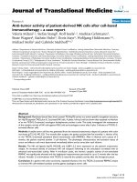
GCP activity was found to be linear for the first 7.5 h of
incubation (F igure 1). [
3
H]-NAAG at 70 nM (~770,000
dpm) provided robust sensitivity to follow GCP activity;
there were approximately 5,000 and 80,000 dpm of
[
3
H]-glutamate after 0.5 and 7.5 h incubation respec-
tively. These values corresponded to 0.6 and 10% con-
version of reactant to product indicating that sufficient
substrate concentration was used and that if additional
GCP activity had been present, additional activity would
have been observed. The linear relationship with respect
to time using samples from different donors suggests
that GCP levels among normal volunteers are relatively
similar.
GCP activity is present in rodent paw pads
A parallel determination of GCP activity was carried out
using male Wistar rat paw pads. Reconstituted pellet
preparations from rat paw pads were used as source of
GCP II and incubated with [
3
H] NAAG. The amount of
GCP activity in rat paw pads was found to be 15 ± 0.2
fmole glutamate generated/h/mg tissue). Interest ingly,
the amount of GCP activity found in rat paw pads (15 ±
0.2 fmole/h/mg tissue) was similar to that obtained from
human skin (11 ± 0.2 fmole/h/mg tissue).
Peripheral administration of 2-PMPA inhibits GCP activity
in rat paw pads in a dose response manner
To be useful as clinical PD marke r, GCP activity in ski n
needs to be amenable to inhibition by peripheral admin-
istration of GCP inhibitors in a dose response manner.
In order to determine if GCP activity in paw pads
in vivo could be inhibited by peripheral administration
of 2-PMPA, rats were treated with 1, 10 and 100 m g/kg
2-PMPA (i.p.) as well as vehicle control. Animals were
sacrificed 1 h after compound administration, paw pads
isolated and GCP activity determined (Methods). GCP
activity in paw pad preparations from animals t reated
with 1 mg/kg 2-PMPA was similar to that of controls.
On the other hand, paw pads from animals treated with
10 and 100 mg/kg exhibited significantly reduced GCP
activity: 60 ± 11 and 47 ± 11% respectively when com-
pared to control animals (Figure 2A). Importantly, these
are the doses of 2-PMPA previously shown to exhibit
therapeutic benefit [13].
Peripheral administration of 2-PMPA inhibits GCP activity
in sciatic nerve in a dose response manner and it
correlates to inhibition observed in rat paw pads
Sciatic nerve is the target tissue for GCP inhibitors in
clinical trials for peripheral neuropathy and neuropathic
pain. Consequently, it is important to demonstrate that
there is a correlation of GCP inhibition in skin and per-
ipheral nerve after administration of different doses of
GCP inhibitor. GCP activity in sciatic nerve preparations
from animals treated with 1, 10 and 100 mg/kg 2-PMPA
was reduced to 92 ± 11, 35 ± 6 and 10 ± 4% respectively
compared to activity in sciatic nerve isolated from con-
trol animals (Figure 2B). Albeit to a different extent,
GCP i nhibition in sciatic nerve is attained at similar 2-
PMPA doses (10 and 100 mg/kg i.p.) as in footpad tis-
sue. Taken together, these results suggested that it will
be possible to follow GCP inhibition in the skin as a
marker of GCP inhibition in peripheral nerve.
2-PMPA is measurable in rat paw pads
GiventhatinhibitionofGCPwasobservedinpawpads,we
wanted to confirm the pres ence of 2-PMPA in paw pads
after peripheral administration of 2-PMPA. Animals were
given 2-PMPA (100 mg/kg, i.p.), sacrificed 1 h after com-
pound administration and paw pads isolated for direct
determination of 2-PMPA levels by L C-MS/MS (Methods).
Since 2-PMPA detection by mass spectrometry has low
sensitivity due to ion suppression, we chose to analyze
samples from animals that had received 100 mg/kg
2-PMPA rather than 10 mg/kg to increase the probability
of detecting 2-PMPA. The characteristic fragmentation
pattern for 2-PMPA was readily detected (Figure 3A) and
the chromatographic peaks of 2-PMPA and internal stan-
dard (Figure 3B) allowed for quantitation of material in the
0
10 20 30
0
50000
100000
150000
Time (h)
GCP Activity (dpm)
0 2 4
6
8
0
50000
100000
Time
(
h
)
GC
P Activity
[
3
H]-glutamate production (dpm)
Figure 1 Dependence of GCP activity in human skin biopsy on
time of incubation - Human skin biopsies were sonicated for 2
min in ice. The resulting mixture was centrifuged at 16000 × g;
precipitate from each preparation was used as GCP source in the
activity assay. Incubations with [
3
H] NAAG (70 nM) at 37°C were
carried out at 0.5, 1, 2, 3, 5, 7.5, 14, 18 and 24 h. Time points
correspond to incubations carried out with biopsies obtained from
different donors. Major plot illustrates the correspondence of
enzyme activity ([
3
H]-glutamate production in dpm) with time while
linearity was observed. Inset illustrates GCP activity measured at
times up to 24 h.
Rojas et al. Journal of Translational Medicine 2011, 9:27
/>Page 4 of 8
(A)
CONTROLS
1 mg/kg
10 mg/kg
100 mg/kg
0
2000
4000
6000
*
**
2-PMPA dose
GCP activity
[
3
H]-glutamate (dpm)/10 mg tissue
(
B)
CONTROLS
1mg/kg
10 mg/kg
100 mg/kg
0
6000
12000
**
**
2-PMPA dose
GCP Activity
[
3
H]-glutamate (dpm)/10 mg tissue
Figure 2 GCP activity in rat paw pads and sciatic nerve is inhibited by peripheral administration of 2-PMPA - Rats were treated with 2-
PMPA (1, 10 and 100 mg/kg i.p.) as well as vehicle control. Animals were sacrificed 1 h after compound administration, paw pads and sciatic
nerve isolated and GCP activity determined (Methods). (A): GCP activity ([
3
H]-glutamate production in dpm/10 mg tissue) in paw pads; * p < 0.05
(B): GCP activity in sciatic nerve. **p < 0.01.
Rojas et al. Journal of Translational Medicine 2011, 9:27
/>Page 5 of 8
(
A
)
(
B)
Figure 3 Measurement of 2-PMPA in rat paw pads using LC-MS/MS - (A) Daughter-scan product ion spectrum of 2-PMPA. Monitoring was
carried out at m/z 226.8 ® 191.1 (B) Select rodent paw pad obtained 1 hour after 2-PMPA (100 mg/kg, i.p.) administration. Retention times for
2-PMPA and internal standard (temazepan) were approximately 1.3 and 2.0 min respectively. When rodent paw pads from untreated animals
were used, only the internal standard peak was observed.
Rojas et al. Journal of Translational Medicine 2011, 9:27
/>Page 6 of 8
sample. Paw pads from animals that were treated with
compound showed 38 ± 5 μg/g tissue (n = 9) (Figure 3B) a
concentration high enough to inhibit GCP activity [25]
while t he compound was undetectable in paw pads isolated
from vehicle-treated animals.
Conclusions
As a biomarker of GCP inhibition in the clinic, skin
biopsy measurements of GCP activity has three areas of
improvement over the prior NAAG bioassay including
simpler sample co llections, less expensive and time con-
suming sample analyses, and the a bility to quantitate
direct vs. indirect measurement of GCP activity. Sample
collection for NAA G bioanalysis involves CSF collection
which requires considerable skill and can be uncomfor-
table to patients; the newly described procedure uses
skin biopsies which is readily acce ssible and can be col-
lected multiple times from a single subject permitting
the ability to evaluate GCP activity before and after
administration of the drug. NAAG analysis uses mass
spectrometry which requires a specialized laboratory
and expensive instrumentation. The new procedure
monitors GCP activity in the skin ex vivo by following
the conversion of [
3
H]-NAAG to [
3
H] glutamate in a
simple enzymatic assay that can be carried out in a stan-
dard biochemistry laborat ory. Finally, the older proce-
dure involved measurements of NAAG levels as
surrogate markers of GCP activity; the new procedure
monitors GCP enzymatic activity directly. In short,
monitoring of GCP activity in human skin after admin-
istration of GCP inhibitors can be readily utilized as a
PD marker in the clinical development of GCP inhibi-
tors. The activity assay provides a simple and direct
measurement of GCP activity from tissue samples easily
assessable in human subjects.
Abbreviations
GCP: glutamate carboxypeptidase; NAAG: N-acetyl-aspartyl-glutamate; NAA:
N-acetyl-aspartate; CSF: cerebrospinal fluid; CNS: central nervous system; PNS:
peripheral nervous system; HPLC: High Pressure Liquid Chromatography; LC-
MS/MS: liquid chromatography-tandem mass spectrometry; PD:
pharmacodynamic; 2-PMPA: 2-(phosphonomethyl) pentanedioic acid.
Acknowledgements and Funding
This work was supported in part by the Analytical Pharmacology Core of the
Sidney Kimmel Comprehensive Cancer Center at Johns Hopkins (NIH grants
UL1 RR025005; MAR and MZ), the Shared Instrument Grant (1S10RR026824-
01; MAR), and the Juvenile Diabetes Research Foundation (MP, RO, and GE).
Author details
1
Brain Science Institute, Johns Hopkins School of Medicine, 855 North Wolfe
Street, Baltimore, MD 21205, USA.
2
Department of Neurology, Johns Hopkins
School of Medicine, 1550 Orleans Street, Baltimore, MD 21231, USA.
3
Department of Oncology, Johns Hopkins School of Medicine, 1650 Orleans
Street, Baltimore, MD 21231, USA.
Authors’ contributions
CR helped with study design and writing of the manuscript. MS carried out
GCP activity measurements in the different biological matrices. MP and GJE
organized the collection of human skin. MAR and MZ carried out 2-PMPA
analysis by LC-MS/MS. BSS conceived the study and study design and
guided the writing and editing of the manuscript. All authors read and
approved the final manuscript.
Competing interests
CR, MS and BSS are former Eisai employees; Eisai is currently working on the
development of a GCP inhibitor.
Received: 27 October 2010 Accepted: 9 March 2011
Published: 9 March 2011
References
1. Doble A: The role of excitotoxicity in neurodegenerative disease:
implications for therapy. Pharmacol Ther 1999, 81:163-221.
2. Neale JH, Olszewski RT, Gehl LM, Wroblewska B, Bzdega T: The
neurotransmitter N-acetylaspartylglutamate in models of pain, ALS,
diabetic neuropathy, CNS injury and schizophrenia. Trends Pharmacol Sci
2005, 26:477-484.
3. Robinson MB, Blakely RD, Couto R, Coyle JT: Hydrolysis of the brain
dipeptide N-acetyl-L-aspartyl-L-glutamate. Identification and
characterization of a novel N-acetylated alpha-linked acidic dipeptidase
activity from rat brain. J Biol Chem 1987, 262:14498-14506.
4. Berger UV, Carter RE, McKee M, Coyle JT: N-acetylated alpha-linked acidic
dipeptidase is expressed by non-myelinating Schwann cells in the
peripheral nervous system. J Neurocytol 1995, 24:99-109.
5. Carozzi VA, Canta A, Oggioni N, Ceresa C, Marmiroli P, Konvalinka J, Zoia C,
Bossi M, Ferrarese C, Tredici G, Cavaletti G: Expression and distribution of
‘high affinity’ glutamate transporters GLT1, GLAST, EAAC1 and of GCPII
in the rat peripheral nervous system. J Anat 2008, 213:539-546.
6. Cassidy M, Neale JH: N-acetylaspartylglutamate catabolism is achieved by
an enzyme on the cell surface of neurons and glia. Neuropeptides 1993,
24:271-278.
7. Bzdega T, Crowe SL, Ramadan ER, Sciarretta KH, Olszewski RT, Ojeifo OA,
Rafalski VA, Wroblewska B, Neale JH: The cloning and characterization of a
second brain enzyme with NAAG peptidase activity. J Neurochem 2004,
89:627-635.
8. Bacich DJ, Wozniak KM, Lu XC, O’Keefe DS, Callizot N, Heston WD, Slusher BS:
Mice lacking glutamate carboxypeptidase II are protected from peripheral
neuropathy and ischemic brain injury. J Neurochem 2005, 95:314-323.
9. Carozzi VA, Chiorazzi A, Canta A, Lapidus RG, Slusher BS, Wozniak KM,
Cavaletti G: Glutamate carboxypeptidase inhibition reduces the severity
of chemotherapy-induced peripheral neurotoxicity in rat. Neurotox Res
2009, 17:380-391.
10. Chen SR, Wozniak KM, Slusher BS, Pan HL: Effect of 2-(phosphono-methyl)-
pentanedioic acid on allodynia and afferent ectopic discharges in a rat
model of neuropathic pain. J Pharmacol Exp Ther 2002, 300:662-667.
11. Ghadge GD, Slusher BS, Bodner A, Canto MD, Wozniak K, Thomas AG,
Rojas C, Tsukamoto T, Majer P, Miller RJ, Monti AL, Roos RP: Glutamate
carboxypeptidase II inhibition protects motor neurons from death in
familial amyotrophic lateral sclerosis models. Proc Natl Acad Sci USA 2003,
100:9554-9559.
12. Nagel J, Belozertseva I, Greco S, Kashkin V, Malyshkin A, Jirgensons A,
Shekunova E, Eilbacher B, Bespalov A, Danysz W: Effects of NAAG
peptidase inhibitor 2-PMPA in model chronic pain - relation to brain
concentration. Neuropharmacology 2006, 51:1163-1171.
13. Slusher BS, Vornov JJ, Thomas AG, Hurn PD, Harukuni I, Bhardwaj A,
Traystman RJ, Robinson MB, Britton P, Lu XC, Tortella FC, Wozniak KM,
Yudkoff M, Potter BM, Jackson PF: Selective inhibition of NAALADase,
which converts NAAG to glutamate, reduces ischemic brain injury.
Nat
Med 1999, 5:1396-1402.
14.
Xi ZX, Li X, Peng XQ, Li J, Chun L, Gardner EL, Thomas AG, Slusher BS,
Ashby CR Jr: Inhibition of NAALADase by 2-PMPA attenuates cocaine-
induced relapse in rats: a NAAG-mGluR2/3-mediated mechanism. J
Neurochem 2010, 112:564-576.
15. Zhang W, Murakawa Y, Wozniak KM, Slusher B, Sima AA: The preventive
and therapeutic effects of GCPII (NAALADase) inhibition on painful and
sensory diabetic neuropathy. J Neurol Sci 2006, 247:217-223.
16. Zhang W, Slusher B, Murakawa Y, Wozniak KM, Tsukamoto T, Jackson PF,
Sima AA: GCPII (NAALADase) inhibition prevents long-term diabetic
neuropathy in type 1 diabetic BB/Wor rats. J Neurol Sci 2002, 194:21-28.
Rojas et al. Journal of Translational Medicine 2011, 9:27
/>Page 7 of 8
17. Zhou J, Neale JH, Pomper MG, Kozikowski AP: NAAG peptidase inhibitors
and their potential for diagnosis and therapy. Nat Rev Drug Discov 2005,
4:1015-1026.
18. van der Post JP, de Visser SJ, de Kam ML, Woelfler M, Hilt DC, Vornov J,
Burak ES, Bortey E, Slusher BS, Limsakun T, Cohen AF, VanGerven JM: The
central nervous system effects, pharmacokinetics and safety of the
NAALADase-inhibitor GPI 5693. Br J Clin Pharmacol 2005, 60:128-136.
19. Thomas AG, Rojas CJ, Hill JR, Shaw M, Slusher BS: Bioanalysis of NAAG as a
Marker of GCP II Inhibition. Anal Biochem 2010, 404:94-96.
20. Slusher BS, Robinson MB, Tsai G, Simmons ML, Richards SS, Coyle JT: Rat
brain N-acetylated alpha-linked acidic dipeptidase activity. Purification
and immunologic characterization. J Biol Chem 1990, 265:21297-21301.
21. Yamamoto T, Saito O, Aoe T, Bartolozzi A, Sarva J, Zhou J, Kozikowski A,
Wroblewska B, Bzdega T, Neale JH: Local administration of N-
acetylaspartylglutamate (NAAG) peptidase inhibitors is analgesic in
peripheral pain in rats. Eur J Neurosci 2007, 25:147-158.
22. Cangro CB, Namboodiri MA, Sklar LA, Corigliano-Murphy A, Neale JH:
Immunohistochemistry and biosynthesis of N-acetylaspartylglutamate in
spinal sensory ganglia. J Neurochem 1987, 49:1579-1588.
23. Ebenezer GJ, McArthur JC, Thomas D, Murinson B, Hauer P, Polydefkis M,
Griffin JW: Denervation of skin in neuropathies: the sequence of axonal
and Schwann cell changes in skin biopsies. Brain 2007, 130:2703-2714.
24. Griffin JW, McArthur JC, Polydefkis M: Assessment of cutaneous
innervation by skin biopsies. Curr Opin Neurol 2001, 14:655-659.
25. Rojas C, Frazier ST, Flanary J, Slusher BS: Kinetics and inhibition of
glutamate carboxypeptidase II using a microplate assay. Anal Biochem
2002, 310:50-54.
26. Jackson PF, Slusher BS: Design of NAALADase inhibitors: a novel
neuroprotective strategy. Curr Med Chem 2001, 8:949-957.
27. Rovenska M, Hlouchova K, Sacha P, Mlcochova P, Horak V, Zamecnik J,
Barinka C, Konvalinka J: Tissue expression and enzymologic
characterization of human prostate specific membrane antigen and its
rat and pig orthologs. Prostate 2008, 68:171-182.
doi:10.1186/1479-5876-9-27
Cite this article as: Rojas et al.: Glutamate carboxypeptidase activity in
human skin biopsies as a pharmacodynamic marker for clinical studies.
Journal of Translational Medicine 2011 9:27.
Submit your next manuscript to BioMed Central
and take full advantage of:
• Convenient online submission
• Thorough peer review
• No space constraints or color figure charges
• Immediate publication on acceptance
• Inclusion in PubMed, CAS, Scopus and Google Scholar
• Research which is freely available for redistribution
Submit your manuscript at
www.biomedcentral.com/submit
Rojas et al. Journal of Translational Medicine 2011, 9:27
/>Page 8 of 8