báo cáo hóa học:" Differentiation associated regulation of microRNA expression in vivo in human CD8+ T cell subsets" pptx
Bạn đang xem bản rút gọn của tài liệu. Xem và tải ngay bản đầy đủ của tài liệu tại đây (666.12 KB, 8 trang )
RESEARCH Open Access
Differentiation associated regulation of microRNA
expression in vivo in human CD8
+
T cell subsets
Bruno Salaun
1†
, Takuya Yamamoto
2†
, Bassam Badran
3,4
, Yasuko Tsunetsugu-Yokota
2
, Antoine Roux
5
, Lukas Baitsch
1
, Redouane Rouas
3
, Hussein Fayyad-Kazan
3
, Petra Baumgaertner
1
, Estelle Devevre
1
, Anirudh Ramesh
6
,
Marion Braun
1
, Daniel Speiser
1
, Brigitte Autran
5
, Philippe Martiat
3
, Victor Appay
5†
and Pedro Romero
1*†
Abstract
Background: The differentiation of CD8
+
T lymphocytes following priming of naïve cells is central in the
establishment of the adaptive immune response. Yet, the molecular events underlying this process are not fully
understood. MicroRNAs have been recently shown to play a key role in the regulation of haematopoiesis in mouse,
but their implication in peripheral lymphocyte differentiation in humans remains largely unknown.
Methods: In order to explore the potential implication of microRNAs in CD8
+
T cell differentiation in humans,
microRNA expression profiles were analysed using microarrays and quantitative PCR in several human CD8
+
T cell
subsets defining the major steps of the T cell differentiation pathway.
Results: We found expression of a limited set of microRNAs, including the miR-17~92 cluster. Moreover, we reveal
the existence of differentiation-associated regulation of specific microRNAs. When compared to naive cells, miR-21
and miR-155 were indeed found upregulated upon differentiation to effector cells, while expression of the miR-
17~92 cluster tended to concomitantly decrease.
Conclusions: This study establishes for the first time in a large panel of individuals the existence of differentiation
associated regulation of microRNA expression in human CD8
+
T lymphocytes in vivo, which is likely to impact on
specific cellular functions.
Background
CD8
+
T cells are major players of the immune response
against viruses and cancers. Even though they represent
a heterogeneous population, the expression of specific
surface molecules characterizes distinct subsets (i.e. cen-
tral memory, early, intermediate or late effector memory
cells), which define the major steps of a process of
memo ry T cell differentiation [1,2]. These multiple sub-
sets present specific transcriptional programs, and there-
fore distinct range of receptors and intracellular
proteins, indicating quite different requirements for sti-
mulation and survival, homing po tential and effector
functions (reviewed in [3]). For instance, expression o f
effector molecules such as perforin or granzymes is
restricted to the late stages of different iation [4,5], while
central memory cells are known for the ir superior pro-
liferative capacity and often considered as the memory
“precursors”. However, the molecular mechanisms con-
trolling peripheral CD8
+
T lymphocyte differentiation,
and therefore the generation of immunological memory,
remain poorly understood in humans.
MicroRNA (miRNA) are 18-22 nucleotide long RNA
molecules that regulate gene expression at the post-tran-
scriptional level through base pairing to partially comple-
mentary sites in the 3’ UTR of the messanger RNA and
integration into RNA induced silencing complexes (RISC)
(reviewed in [6]). Inhibition of translation or degradation
of the m iRNA-bound mRNAs modulate protein output,
inducing profound physiological effects. MicroRNA, which
expression is tightly regulated during lymphopoiesis [7],
rec ently emerged as key regulators of gene expression in
the mammalian immune system (reviewed in [8,9]).
Although the biological functions of most miRNAs are
not yet fully understood, unequivocal evidence for their
* Correspondence:
† Contributed equally
1
Division of Clinical Onco-Immunology, Ludwig Center for Cancer Research
of the University of Lausanne, Switzerland
Full list of author information is available at the end of the article
Salaun et al. Journal of Translational Medicine 2011, 9:44
/>© 2011 Salaun et al; licensee BioMed Central Ltd. Th is is an Open Access article distributed under the terms of the Creative Commons
Attribution License (htt p://creativecommons.org/licenses/by/2.0 ), which permits unrestricted use, distributio n, an d reproduction in
any medium, provided the original work is properly cited.
role in lymphocyte development have been gathered in
mouse models in recent years. For instance, the condi-
tional deficiency in mouse lymphocytes of Dicer, the key
enzyme in miRNA biogenesis, impaired lymphocytic dif-
ferentiation [10,11]. Moreover, it was recently shown
that abolishing Dicer expression in mature mouse CD8
+
T cells strongly impairs their response to pathogens in
vivo [12], demonstrating that miRNAs are not only
important for lymphocytes differentiation, but also for
their functions in the periphery. Interestingly, single
miRNA such as miR-181 have also been shown to be
crucial in mouse haematopoiesis [13]. Thus, mouse
models and/or studies on in vitro differentiated cells
suggested a role for miRNAs in lymphocytes functions
[14-16]. However, the modulation of their expression
along antigen-driven differentiation of peripheral human
CD8
+
T lymphocytes in vivo has not been studied yet.
We thus addressed this issue using microarrays and
quantitative PCR to investigate microRNA expression
profiles in different subsets of ex vivo sorted human
CD8
+
T lymphocytes. Our results show the first e vi-
dence that these cells express a limited set of micro-
RNAs, some of which displayed differential expression
in differentiated CD8
+
T cells when compared to naive
cells. The microRNA expression regulation uncovered
here is likely to strongly influence the gene signature of
these different subsets, and therefore to directly impact
on their functional properties.
Methods
CD8 T cell subsets purification
Peripheral blood CD8
+
T lymphocytes were purified
from healthy volunteers in agreement with local ethics
committees (Cantonal Commission of Ethics in
Research, State of Vaud, Switzerland, authorization #87/
06) as described [4]. Briefly, total CD8
+
T cell prepara-
tions were obtained from leukapheresis by magnetic
bead enrichment (Mylteny i Biotech, Bergish Gladbach,
Germany ) and stained with a combination of antibodies
to CD3, CD8, CD45RA, CCR7, CD27 and CD28. All
antibodies were from BD Ph armingen (San Diego, CA)
except anti-CD45RA purch ased from Beck man Coulter
(Paris, France). The different subsets were then sepa-
rated on a FACS Aria device (BD Bioscience, Allschwil,
Switzerland) to routinely over 99% purity cell suspen-
sions. Cells were either sor ted directly to RNA lysis buf-
fer (RNAlater, Qiagen) and frozen (for single specific
qPCR assays) or into cold medium (for TaqMan Low
Density Array experiments), before washings in ice-cold
PBS and lysis (miRVana kit, Ambion).
MicroRNA expression analysis
The TaqMan
®
Low -density arrays (TLDA, Applied Bio-
systems, Foster City, CA) were used following
manufacturer’ s instructions to simultaneously detect
expression of 364 individual microRNAs. Briefly, total
RNA was extracted with the miRVana kit (Ambion),
quantified with a Nanodrop Spectrophotometer, and
microRNA mature forms were reverse transcribed (30
min at 16°C, 30 min at 42°C, 5 min at 85°C) with Multi-
plex RT human primers and 100 mM dNTPs, 50 U
MutliScribe reverse transcriptase, 20 U RNase inhibitor,
10 × RT buffer (8 reactions with pools of 48 stem-lo op
RT primers, 100 ng RNA per reaction). cDNA were
then amplified on microfluidic cards in an ABI Prism
7900 HT with single microRNA specific primers (40
cycles of 15 s at 95°C and 1 min at 60°C). The same
proto col was applied for single microRNA specific qRT-
PCR (miR-17-3p, miR-17 -5p, miR-19b, miR-20a, miR-
92, miR-21, miR-155, miR-142-3p, miR-142-5p, and
RNU44) from 10 ng total RNA, reverse transcribed with
TaqMan Reverse transcription MicroRNA kit ( Applied
Biosystems) and amplified using Universal Fast Start
Rox Probe Master Mix (Roche) and microRNA Assay
kits in 384 well plates (Applied Biosystems) on a ABI
Prism 7900 HT device (Applied Biosystems). Ct
(RNU44) was subtracted to Ct (microRNA) to calculate
relative expression (ΔCt).
Statistical analysis
Statistical significance was evalu ated with the GraphPad
Prism software on non normalized expression data
using the Friedman test (for non-parametric, paired data
set) with Dunn’ s multiple compariso n correction
applied. Results were considered significant for p values
< 0.05.
Results and Disc ussion
MicroRNA expression profiling in mature CD8
+
T
lymphocyte subsets
Different subsets of human CD8
+
T lymphocytes can be
defined based on the expression of CCR7, CD45RA and
CD28 [4]. Naïve cells are CCR7
+
(and CD45RA
+
CD28
+
) while the effector memory subsets are CCR7
-
. Among
the latter, the CD45RA
+
CD28
-
cells representing the
end stage of differentiation (late effector memory, L-
EM), while CD45RA
-
cells can be further divided in two
functionally distinct subsets, with CD28
+
cells (early
effector memory (E-EM) closer to central memory cells)
being less differentiated than CD28
-
subsets (I-EM),
which share features with fully differentiated effectors
(L-EM).
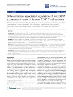
These human CD8
+
T lymphocyte subsets (naive, E-
EM, I-EM and L-EM, ordered from least to mos t differ-
entiated) were purified from peripheral blood leukocytes
from 3 h ealthy donors as indicat ed (Figure 1A), and the
relative expression of 365 microRNAs was analyzed in
these subpopulations using TaqMan Low Density Arrays
Salaun et al. Journal of Translational Medicine 2011, 9:44
/>Page 2 of 8
(TLDA). Unsupervised clustering of r elative expression
levels in all subsets identified 4 main groups of micro-
RNAs (Figure 1B): highly expressed (group A, 22 micro-
RNAs, see Table 1), intermediat e ( group B, 31
microRNAs, see Table 1), low (group C, 46 microRNAs)
and rare/absent (group D, 267 m icroRNAs). In total, 97
microRNAs were expressed well over detection limit, a
figure similar to the 113 microRNAs recently detected
in B cells using the same TLDA [17]. This list is likely
not comprehensive, since the set of microRNAs detected
with TLDA does not cover the whole range of micro-
RNAs cloned in human. However, the low number of
microRNAs considered expressed at high levels (group
A) is in agreement with previous cloning studies in
mouse CD8
+
lymphocytes [15,18] showing that a few
highly expressed microRNAs prevailed in frequency.
Interestingly, these microRNAs include miR16, miR-21,
miR-142-3p and miR-142-5p, wh ich were also found in
the group A in human CD8
+
T lymphocytes, with miR-
142-3p showing the highest relative expression levels
(Table 1). MiR-26a/b and miR-146a/b also clustered to
this group. In addition, 7 microRNAs of the 17~92 and
paralog 106b~25 clusters (namely miR-19a, miR-19b,
miR-20a, miR-25, miR-92, miR-93 and miR-106b) were
identified among the 53 most expressed microRNAs
(groups A and B, see Table 1). MiR-17-5p and miR-17-
3p are expressed at lower levels (belong to group C),
indicating intra-cluster differential expression.
Transgenic overexpression of miR-17~92 cluster in
mouse lymphocytes was shown to induce lymphoproli-
ferative disease [16]. High expression levels are therefore
likely to be tightly controlled. The high expression levels
foundforalargesetofmicroRNAsfromthe17~92
cluster in primary human CD8
+
T lymphocytes had not
been reported yet, and strongly suggests a role for this
set of microRNAs in the biology of CD8
+
T cells.
MiR-21 and miR-155 are upregulated during CD8
+
T
lymphocyte differentiation
To investigate if defined microRNA profiles can be asso-
ciated with CD8
+
T cell differentiation status, the rela-
tive expression levels of the microRNAs from groups A,
B and C were calculated for each CD8
+
lymphocyte sub-
set relative to those found in the naive cells. Global
unsupervised clustering did not identify clear microRNA
clusters, and grouped subpopulations from the same
donor together, due to strong inter-donor hete rogeneity.
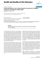
However, specific patterns could be highlighted when
the analysis was focused on a more restricted set of
microRNAs (Figure 2A), chosen for their suggested role
in immunity. The well expressed miR-21, miR-155 and
miR-146a clustered together as consistently upregulated,
while the abundant microRNAs of the miR17~92 clus-
ters (miR-19b, miR-20a and miR-92) showed a clear
trend towards decreased expression in differentiated
cells, as did miR-26a (Figure 2A).
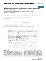
Figure 1 Sorting of human CD8
+
T lymphocytes and microRNA la rge scale expression profiling.a)CD8
+
T cells from healthy volunteers
were FACS-sorted based on their expression of CD45RA, CCR7 and CD28 to isolate Naive (CD45RA
+
CCR7
+
, CD28
+
), E-EM (CD45RA
-
CCR7
-
, CD28
+
), I-EM (CD45RA
-
CCR7
-
, CD28
-
) and L-EM (CD45RA
+
CCR7
-
, CD28
-
). Data shown are gated on CD3
+
CD8
+
cells. b) Unsupervised clustering of the
expression levels (ΔCt, normalized to RNU44) of 223 unique microRNAs (detected in at least one sample, i.e. Ct < 36) in CD8
+
T lymphocyte
subsets from 3 healthy donors (HD1, HD2, HD3) as measured by TLDA (green: low expression/red: high expression).
Salaun et al. Journal of Translational Medicine 2011, 9:44
/>Page 3 of 8
MiR-21 has been shown to be upregulated in mouse
CD8
+
T cells upon in vitro activation [15], but its func-
tion in these cells remain s unknown. MiR-155, encoded
by the BIC transcript, is induced upon antigen-receptor
triggering in lymphocytes and was shown to be instru-
mental for the generation of adaptive immunity in
mouse [19,20]. Our TLDA analysis suggested that both
miR-21 and miR-155 were more expressed in in vivo
differentiated human primary CD8
+
T cells than in their
naive counterparts (Figure 2A). These resul ts were con-
firmed by single specific RT-qPCR on eight additional
donors, which all clearly showed higher expression of
both miR-21 and miR-155 in antigen-experienced cells
(Figure 2B).
Altogether these results demonstrate that an increase
of miR-21 and miR-155 expression occurs upon in vivo
differentiation of human CD8
+
T lymphocytes. Dynamic
regulation of these microRNAs in mouse CD8
+
T cells
has been described upon in vitro activation [15]. By con-
trast, the data presented here on ex vivo sorted lympho-
cytes revealed for the first time modulations that
occurred during in vivo differentiation of human periph-
eral CD8
+
T cell subsets.
The miR-17~92 cluster tends to be downregulated during
CD8
+
T cell differentiation
Since central memory (CM) CD8
+
T cells are present at
extremely low frequency in peripheral blood, a new sort-
ing strategy was then designed to include this subset in
our analysis. Addition o f anti-CD27 to the panel of cell
surface molecule specific antibodies allowed the isola-
tion of naïve (CCR7
+
CD45RA
+
CD27
+
CD28
+
), CM
(CCR7
+
CD45RA
-
CD27
+
CD28
+
), E-EM (CCR7
-
CD45RA
+/-
CD27
+
CD28
+
), I-EM (CCR7
-
CD45RA
+/-
CD27
+
CD28
-
) and L-EM (CCR7
-
CD45RA
+/-
CD27
-
CD28
-
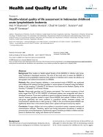
) subsets. Consistent with our previous analysis
(Figure 2B), upregulation of miR-155 in antigen experi-
enced cells versus their naïve counterparts was con-
firmed in the new set of 9 independent donors, with a
clear trend toward highest expression levels in the most
differentiated CD8
+
T cells (L-EM) (Figure 3A). Since
the TLDA analysis showed that both miR-142-3p and
miR-142-5p were among the most expressed m icro-
RNAs in CD8
+
T cells (Table 1), their expression levels
were assessed in the different subsets. No significant
regulation of expression could be found, likely suggest-
ing a constitutive role rather than a differentiation
induced function for these microRNAs (Figure 3B).
Seven members of the 17~92 cluster, which recently
were shown to be critical regulators of lymphocyte
development and proliferation [16], were identified
among the 53 most expressed microRNAs in human
CD8
+
T cells (Table 1), with a trend toward lower
expression in more differentiated cells (Figure 2A).
Expression levels of miR-17-3p, miR-17-5p, miR-19b,
miR-20a and miR-92 were therefore determined by sin-
gle s pecific qPCR in differentiated C D8
+
T cell subsets,
andcomparedtothelevelsfoundinnaïvecells.In
agreement with the TLDA data, and despite a relatively
important inter-donor variability, the expression of
some of these miRNAs was found to be preferentially
associated with specific subsets of CD8
+
Tcelldifferen-
tiation. Interestingly, the expression level of miR-20a
was significantly increased in the central memory subset,
in contrast to more differentiated subsets. Along the
same lines, miR-17-3p expression was significantly
Table 1 Human CD8
+
T cells express a limited set of
microRNA
Group A GROUP B
rank miR rank miR
1 hsa-miR-142-3p 23 hsa-miR-31
2 hsa-miR-26a 24 hsa-miR-125a
3 hsa-miR-16 25 hsa-miR-565
4 hsa-miR-26b 26 hsa-let-7b
5 hsa-miR-146b 27 hsa-let-7a
6 hsa-miR-29a 28 hsa-miR-181b
7 hsa-miR-19b * 29 hsa-miR-30e-3p
8 hsa-miR-92 * 30 hsa-miR-423
9 hsa-miR-342 31 hsa-miR-328
10 hsa-miR-30c 32 hsa-miR-101
11 hsa-miR-20a * 33 hsa-miR-223
12 hsa-let-7g 34 hsa-miR-27a
13 hsa-miR-142-5p 35 hsa-miR-25 *
14 hsa-miR-146a 36 hsa-miR-195
15 hsa-miR-24 37 hsa-miR-425-5p
16 hsa-miR-21 38 hsa-miR-222
17 hsa-miR-29c 39 hsa-miR-197
18 hsa-miR-30b 40 hsa-miR-28
19 hsa-miR-30a-5p 41 hsa-miR-186
20 hsa-miR-191 42 hsa-miR-331
21 hsa-miR-484 43 hsa-miR-192
22 hsa-miR-140 44 hsa-miR-103
45 hsa-miR-594
46 hsa-miR-15b
47 hsa-miR-30e-3p
48 hsa-miR-155
49 hsa-miR-374
50
hsa-miR-93 *
51 hsa-miR-19a
*
52 hsa-miR-106b *
53 hsa-miR-30d
MicroRNA ranking according to relative expression in CD8
+
T cells. Groups A
and B are as identified by unsupervised clustering in Figure 1b. *: microRNA
belonging to the miR-17~92 cluster.
Salaun et al. Journal of Translational Medicine 2011, 9:44
/>Page 4 of 8
decreased in late effector memory cells. There were also
non significant trends towards preferential expression of
miR-19b and miR-92 in the central memory cells. MiR-
17-5p expression showed no association with CD8
+
T
cell differentiation. On the whole, expression of several
members of the miR-17~92 cluster appeared to be
found preferentially during early memory differentiation.
Interestingly, not all members are regulated concomi-
tantly, indicating a differential intra-cluster regulation in
agreement with the TLDA results (Table 1).
Altogether, the data presented here demonstrate for
the first time that human CD8
+
T cell subsets express a
Figure 2 miR-21 and miR-155 are upreg ulated in differentiated CD8
+
lymphocyte s ubsets. a) Unsupervised h eatmap clustering of the
expression levels of a selected set of microRNAs in each subset normalized to levels in naive cells (log2 fold change) (green: downregulated/red:
upregulated). b) Expression levels of miR-21 and miR-155 in sorted human CD8
+
T cell subsets were determined by single specific qPCR
performed in triplicates, and are expressed as fold change relatively to levels in naive cells (n = 8; *: p < 0,05; **: p < 0,01).
Salaun et al. Journal of Translational Medicine 2011, 9:44
/>Page 5 of 8
limited set of microRNAs, which include several mem-
bers of the 17~92 and related clusters. Moreover, this
study shows that the in vivo differentiation from naive
cells is associated with a stage-spec ific regulation of di s-
tinct microRNA expression, with miR-21 and miR-155
being upregulated in an tigen-experienced subsets, the
more differentiated subsets expressing higher levels of
these microRNA (Figures 2B and 3A). MiR-155 has
been suggested to be part of a negative feedback loop
downstream the Toll-like receptor/Interleukin 1 recep-
tor pathway [21]. The results presented here may sug-
gest similar functions in human CD8
+
T cells
downstream of the TCR. Moreover, miR-155 has been
recently shown to target SOCS-1 expression, both in
tumor cells [22] and in mouse CD4
+
regulatory T cells,
in which it modulates the cellular responses to IL-2.
Whether miR-155 plays a similar role in CD8
+
T cells
remains an interesting issue to investigate, as it might
shed new light on the relationship between cytokine sig-
naling and lymphocytes differentiation.
In contrast to the clear upregulation observed for
miR-155, the expression of the miR-17~92 cluster (espe-
cially miR-17-3p and miR-20a) tended to decrease along
differentiation. Our results are in line with a recent
report by Hackl and colleagues [23] that shows downre-
gulation of this cluster in CD2 8 negative CD8+ T cells.
Figure 3 The miR-17~92 cluster is preferentially downregulated in differentiate d CD8
+
Tcellsubsets. Expression levels of miR-155 (a),
miR-142.3p and miR-142-5p (b) and of members of the miR-17~92 cluster (c) were determined by single specific qPCR in the indicated sorted
human CD8
+
T cell subsets (a), and are expressed as fold change relatively to levels in naive cells (n = 9; *: p < 0,05; **: p < 0,01).
Salaun et al. Journal of Translational Medicine 2011, 9:44
/>Page 6 of 8
Considering the role of the miR-19~92 cluster in lym-
phocyte development and proliferation, it is tempting to
speculate that its expression may be r elated to the
greater proliferative potential and memory precursor-
like capacity, characteristic of central memory cells com-
pared to more differentiated s ubsets (in particular late
effector memory cells). Interestingly, expression of this
cluster is induced by the transcription factor c-Myc [24],
which has been shown in the mouse to be a component
of the IL-15-dependent pathway controlling homeostatic
proliferation of memory CD8
+
T cells [25]. In addition,
the 17~92 cluster has been experimentally shown to
inhibit Bim expression [16], which plays a key role in
activation induced cell death in lymphocytes [26].
Whether 17~92 induced regulation of Bim plays a func-
tion in CD8
+
T cells survival potential (greater in less
differentiated cells) is an interesting issue to be further
investigated.
Conclusions
Mouse models have shown that the limited set of micro-
RNAs expressed in mature CD8
+
T cells critically sup-
port their functions in response to pathogens [12, 15].
Similarly, the differential microRNA expression profile
observed here for the first time in human CD8
+
T cell
subsets is likely to have functional relevance in lympho-
cyte biology, in view of proteomic experiments demon-
strating that a single miRNA can directly repress the
production of hundreds of proteins in mammalian cells,
through both downregulation of mRNA levels and
translation inhibition [27]. Further investigation is war-
ranted to dissect the precise role of individual differen-
tially expressed microRNA in the regulation of CD8
+
T
cell functions. We are currently pursuing analyses on
human CD8+ T cell clones and in mouse models to bet-
ter understand the functional relevance of the results
presented here. Understanding this novel aspect of lym-
phocyte biology will help to better define CD8
+
T cell
differentiation, and might shed light on the relationship
between differentiation and functional properties. In
that respect, it might therefore be helpful to elaborate
better vaccination strategies for induction of CD8
+
T
cells with appropriate differentiation and functions.
Acknowledgements
The authors would like to thank Ms. Céline Beauverd for excellent technical
assistance, Dr Donata Rimoldi for critical reading of the manuscript, and Drs.
Tatsuo Shioda, Tetsuro Matano, Kazuo Kobayashi and Jun-ichiro Inoue for
their support to international collaborations. This work was supported by
grants from the Fondation MEDIC and the European Framework Program
FP6 “Cancer immunotherapy” to B.S. and P. R. These sponsors had no role in
the design of the study and the analysis of the results.
Author details
1
Division of Clinical Onco-Immunology, Ludwig Center for Cancer Research
of the University of Lausanne, Switzerland.
2
Department of Immunology,
National Institute of Infectious Diseases, Shinjuku-ku, Tokyo, Japan.
3
Laboratory of Experimental Hematology, Bordet Institute, University of
Brussels (ULB) Brussels, Belgium.
4
Department of Biochemistry, Laboratory of
Immunology, Lebanese University, Faculty of Sciences, Hadath Beirut,
Lebanon.
5
Infections and Immunity, INSERM U945, Avenir Group, Hôpital
Pitié-Salpêtrière, Paris, France.
6
Department of Biological Sciences, Cornell
University, Ithaca, NY.
Authors’ contributions
BS, TY, PM, YTY BA, VA, PR designed the study. BS, TY, BB, RR, HFK, PB, ED,
AR, MB generated the data. BS, TY, BB, LB, YTY, DS, VA, PR analyzed the data.
BS, VA, PR wrote the paper. All authors have read and approved the final
manuscript.
Competing interests
The authors declare that they have no competing interests.
Received: 21 January 2011 Accepted: 20 April 2011
Published: 20 April 2011
References
1. Appay V, Rowland-Jones SL: Lessons from the study of T-cell
differentiation in persistent human virus infection. Semin Immunol 2004,
16:205-212.
2. van Lier RA, ten Berge IJ, Gamadia LE: Human CD8(+) T-cell differentiation
in response to viruses. Nat Rev Immunol 2003, 3:931-939.
3. Appay V, van Lier RA, Sallusto F, Roederer M: Phenotype and function of
human T lymphocyte subsets: consensus and issues. Cytometry A 2008,
73:975-983.
4. Romero P, Zippelius A, Kurth I, Pittet MJ, Touvrey C, Iancu EM, Corthesy P,
Devevre E, Speiser DE, Rufer N: Four functionally distinct populations of
human effector-memory CD8+ T lymphocytes. J Immunol 2007,
178:4112-4119.
5. Rufer N, Zippelius A, Batard P, Pittet MJ, Kurth I, Corthesy P, Cerottini JC,
Leyvraz S, Roosnek E, Nabholz M, Romero P: Ex vivo characterization of
human CD8+ T subsets with distinct replicative history and partial
effector functions. Blood 2003, 102:1779-1787.
6. Bartel DP: MicroRNAs: genomics, biogenesis, mechanism, and function.
Cell 2004, 116:281-297.
7. Kuchen S, Resch W, Yamane A, Kuo N, Li Z, Chakraborty T, Wei L,
Laurence A, Yasuda T, Peng S, et al: Regulation of microRNA expression
and abundance during lymphopoiesis. Immunity 2010, 32:828-839.
8. Baltimore D, Boldin MP, O’Connell RM, Rao DS, Taganov KD: MicroRNAs:
new regulators of immune cell development and function. Nat Immunol
2008, 9:839-845.
9. Xiao C, Rajewsky K: MicroRNA control in the immune system: basic
principles. Cell 2009, 136:26-36.
10. Cobb BS, Hertweck A, Smith J, O’Connor E, Graf D, Cook T, Smale ST,
Sakaguchi S, Livesey FJ, Fisher AG, Merkenschlager M: A role for Dicer in
immune regulation. J Exp Med 2006, 203:2519-2527.
11. Muljo SA, Ansel KM, Kanellopoulou C, Livingston DM, Rao A, Rajewsky K:
Aberrant T cell differentiation in the absence of Dicer. J Exp Med 2005,
202:261-269.
12. Zhang N, Bevan MJ: Dicer controls CD8+ T-cell activation, migration, and
survival. Proc Natl Acad Sci USA 2010, 107(50):21629-21634.
13. Chen CZ, Li L, Lodish HF, Bartel DP: MicroRNAs modulate hematopoietic
lineage differentiation. Science 2004, 303:83-86.
14. Pang KC, Dinger ME, Mercer TR, Malquori L, Grimmond SM, Chen W,
Mattick JS:
Genome-wide identification of long noncoding RNAs in CD8+
T
cells. J Immunol 2009, 182:7738-7748.
15. Wu H, Neilson JR, Kumar P, Manocha M, Shankar P, Sharp PA, Manjunath N:
miRNA profiling of naive, effector and memory CD8 T cells. PLoS ONE
2007, 2:e1020.
16. Xiao C, Srinivasan L, Calado DP, Patterson HC, Zhang B, Wang J,
Henderson JM, Kutok JL, Rajewsky K: Lymphoproliferative disease and
autoimmunity in mice with increased miR-17-92 expression in
lymphocytes. Nat Immunol 2008, 9:405-414.
17. Zhang J, Jima DD, Jacobs C, Fischer R, Gottwein E, Huang G, Lugar PL,
Lagoo AS, Rizzieri DA, Friedman DR, et al: Patterns of microRNA
expression characterize stages of human B cell differentiation. Blood
2009, 113(19):4586-94.
Salaun et al. Journal of Translational Medicine 2011, 9:44
/>Page 7 of 8
18. Neilson JR, Zheng GX, Burge CB, Sharp PA: Dynamic regulation of miRNA
expression in ordered stages of cellular development. Genes Dev 2007,
21:578-589.
19. Haasch D, Chen YW, Reilly RM, Chiou XG, Koterski S, Smith ML, Kroeger P,
McWeeny K, Halbert DN, Mollison KW, et al: T cell activation induces a
noncoding RNA transcript sensitive to inhibition by immunosuppressant
drugs and encoded by the proto-oncogene, BIC. Cell Immunol 2002,
217:78-86.
20. Rodriguez A, Vigorito E, Clare S, Warren MV, Couttet P, Soond DR, van
Dongen S, Grocock RJ, Das PP, Miska EA, et al: Requirement of bic/
microRNA-155 for normal immune function. Science 2007, 316:608-611.
21. Ceppi M, Pereira PM, Dunand-Sauthier I, Barras E, Reith W, Santos MA,
Pierre P: MicroRNA-155 modulates the interleukin-1 signaling pathway in
activated human monocyte-derived dendritic cells. Proc Natl Acad Sci
USA 2009, 106:2735-2740.
22. Jiang S, Zhang HW, Lu MH, He XH, Li Y, Gu H, Liu MF, Wang ED:
MicroRNA-155 functions as an OncomiR in breast cancer by targeting
the suppressor of cytokine signaling 1 gene. Cancer 2010, 70:3119-3127.
23. Hackl M, Brunner S, Fortschegger K, Schreiner C, Micutkova L, Muck C,
Laschober GT, Lepperdinger G, Sampson N, Berger P, et al: miR-17, miR-
19b, miR-20a, and miR-106a are down-regulated in human aging. Aging
Cell 2010, 9(2):291-296.
24. O’Donnell KA, Wentzel EA, Zeller KI, Dang CV, Mendell JT: c-Myc-regulated
microRNAs modulate E2F1 expression. Nature 2005, 435:839-843.
25. Bianchi T, Gasser S, Trumpp A, MacDonald HR: c-Myc acts downstream of
IL-15 in the regulation of memory CD8 T-cell homeostasis. Blood 2006,
107:3992-3999.
26. Sandalova E, Wei CH, Masucci MG, Levitsky V: Regulation of expression of
Bcl-2 protein family member Bim by T cell receptor triggering. Proc Natl
Acad Sci USA 2004, 101:3011-3016.
27. Baek D, Villen J, Shin C, Camargo FD, Gygi SP, Bartel DP: The impact of
microRNAs on protein output. Nature 2008, 455:64-71.
doi:10.1186/1479-5876-9-44
Cite this article as: Salaun et al.: Differentiation associated regulation of
microRNA expression in vivo in human CD8
+
T cell subsets. Journal of
Translational Medicine 2011 9:44.
Submit your next manuscript to BioMed Central
and take full advantage of:
• Convenient online submission
• Thorough peer review
• No space constraints or color figure charges
• Immediate publication on acceptance
• Inclusion in PubMed, CAS, Scopus and Google Scholar
• Research which is freely available for redistribution
Submit your manuscript at
www.biomedcentral.com/submit
Salaun et al. Journal of Translational Medicine 2011, 9:44
/>Page 8 of 8