báo cáo hóa học:" Prostate transglutaminase (TGase-4) antagonizes the anti-tumour action of MDA-7/IL-24 in prostate cancer" potx
Bạn đang xem bản rút gọn của tài liệu. Xem và tải ngay bản đầy đủ của tài liệu tại đây (11.23 MB, 9 trang )
RESEARCH Open Access
Prostate transglutaminase (TGase-4) antagonizes
the anti-tumour action of MDA-7/IL-24 in prostate
cancer
Richard J Ablin
1*
, Howard G Kynaston
2
, Malcolm D Mason
2
and Wen G Jiang
2
Abstract
Background: Transglutamiase-4 (TGase-4), also known as prostate transglutaminase, belongs to the TGase family
and is uniquely expressed in the prostate gland. The functions of this interesting protein are not clearly defined. In
the present study, we have investigated an unexpected link between TGase-4 and the melanoma differentiation-
associated gene-7/interleukin-24 (MDA-7/IL-24), a cytokine known to regulate the growth and apoptosis of certain
cancer and immune cells.
Methods: Frozen sections of normal and malignant human prostate tissues and human prostate cancer (PCa) cell
lines PC-3 and CA-HPV-10, cell lines expressing low and high levels of TGase-4, and recombinant MDA-7/IL-24
(rhMDA-7/IL-24) were used. Expression construct for human TGase-4 was generated using a mammalian expression
vector with full length human TGase-4 isolated from normal human prostate tissues. PC-3 cells were transfected
with expression construct or contr ol plasmid. Stably transfected cells for control transfection and TGase-4 over
expression were created. Similarly, expression of TGase-4 in CA-HPV-10 cells were knocked down by way of
ribozyme transgenes. Single and double immunofluorescence microscopy was used for localization and co-
localization of TGase-4 and MDA-7/IL-24 in PCa tissues and cells with antibodies to TGase-4; MDA-7/IL-24; IL-
20alpha; IL-20beta and IL-22R. Cell-matrix adhesion, attachment and migration were by electric cell substrate
impedance sensing and growth by in vitro cell growth assay. A panel of small molecule inhibitors, including Akt,
was used to determine signal pathways involving TGase-4 and MDA-7/IL-24.
Results: We initially noted that MDA-7 resulted in inhibition of cell adhesion, growth and migration of human PCa
PC-3 cells which did not express TGase-4. However, after the cells over-expressed TGase-4 by way of transfection,
the TGase-4 expressing cells lost their adhesion, growth and migratory inhibitory response to MDA-7. On the other
hand, CA-HPV-10 cells, a cell type naturally expressing high levels of TGase-4, had a contrasting response to MDA-7
when compared with PC-3 cells. Inhibitor to Akt reversed the inhibitory effect of MDA-7, only in PC-3 control cells,
but not the TGase-4 expressing PC-3 cells. In human prostate tissues, TGase-4 was found to have a good degree of
co-localization with one of the MDA-7 recep tor complexes, IL-20Ra.
Conclusion: The presence of TGase-4 has a biological impact on a prostate cancer cell’s response to MDA-7.
TGase-4, via mechanism(s ) yet to be identified, blocked the action of MDA-7 in prostate cancer cells. This has an
important implication when considering the use of MDA-7 as a potential anticancer cytokine in prostate cancer
therapies.
* Correspondence:
1
Department of Pathology, University of Arizona College of Medicine,
Arizona Cancer Center and BIO5 Institute, Tucson, Arizona, AZ 85724-5043
USA
Full list of author information is available at the end of the article
Ablin et al. Journal of Translational Medicine 2011, 9:49
/>© 2011 Ablin et al; licensee BioMed Cent ral Ltd. This is an Open Access article distributed under the terms of the Creative Commons
Attribution License ( ), which permits unrestricted use, distribution, and reproduction in
any medium, provided the original work is properly cited.
Background
Transglutaminases (EC 2.3.2.13) catalyze the posttransla-
tional modification of proteins by the formation of epsi-
lon-(gamma-glutamyl) lysine isopeptide bonds [ 1]. A
number of human transglutaminases (TGases), as
reviewed [2] have been identified and shown to have rela-
tively restrict distribution patterns. The intracellular
forms are: tissue TGase (TGase-2), keratinocyte TGase,
and hair follicle TGase; extracellular TGases include fac-
tor XIIIa (plasma TGase) and prostate TGase (TGase-4,
or TGaseP). In the case of TGase-4, the focus of this
study, the gene is located to 3p22-p21.33 [3] and by ana-
lysis of somatic cell hybrids, mapped to chromosome 3
[3-5]. TGase-4 has a strong pattern of distribution in the
prostate [6-8].
The function of the TGase-4 is not clear. The rat
homologue homologue of TGase-4 (dorsal prostate
TGase or Dorsal protein 1 [DP 1]) has been suggested to
be responsible for the cross-linking during the copulatory
plug [9] formation and may be involved in sperm cell
mobility and immunogenicity to some degree [10,11]. In
initial studies by others [6,7], TGase-4 expression was
restricted to luminal epithelial cells. The expression pat-
tern as observed for TGase-4 has not been found thus far
for any other p rostate-specific marker [6]. However, the
function of this enzyme in prostate cancer is unclear.
Recently, it has been shown that TGase-4 is linked to the
invasivenes s of prostate cancer ce lls [12] and participates
in the regulation of the interactions between prostate
cancer cells and endothelial cells, the later involving the
Rock signalling pathway [13]. In addition, variants of
TGase-4 have been recently reported in benign and
malignant human prostate tissues [14].
As part of our continuing studies to investigate proteins
interacting with TGase-4 using immunoprecipitation of
proteins from the prostate gland, we i dentified a small
panel of proteins that interacted with TGase- 4, including
RON (the HGF-like protein receptor) [15]. MDA-7 was
one of the other proteins precipitated with TGase-4.
MDA-7 (melanoma differentiation associated gene-7),
also known as IL-24, was initially identified from cancer
cells and found to be up-regulated in melanoma cells [16].
Forced expression of MDA-7 in cancer cells was found to
be growth inhibitory [17]. The human MDA-7 gene,
mapped to 1q32.2-q41, encodes a protein with a predicted
size of 23.8 kD. The secreted mature MDA-7 is a 35-40
kDa phosphorylated glycoprotein. Cell types known to
express MDA-7 are diverse, including B cells, NK cells,
dendritic cells, monocytes, melanocytes and melanoma
cells. It is now known that MDA-7 is a differentiation-,
growth-, and apoptosis-associated gene with potential uti-
lity for the gene-based thera py of diverse human cancers.
The location of the MDA-7 gene is closely linked to the
IL-10, IL-19, and IL-20 genes within a 195-kb region -the
IL-10 fami ly cytokine cluster. MDA-7/IL-24 functions in
cells via its receptor, MDA-7R/IL-24R. The MDA-7 recep-
tor complexes include at least the IL-20alpha and IL-
20beta complex and the IL-22R and IL-20Rbeta complex.
Limited information is available on the effect of MDA-7
on prostate cancer ce lls. Studies of adenoviral vector-
induced expression o f MDA-7 in human prostate cancer
cells demonstrated varying degree of inhibition of growth
and induction of apoptosis. It is interesting to note that
Bcl-2 and Bcl-xL may differentially prot ect human pros-
tate cancer cells from MDA-7 induced apoptosis [18].
In the present study, we have evaluated the biological
impact of TGase-4 and MDA-7 and herein report a link
between MDA-7 and TGase-4 in prostate cancer cells
and tissu es. In the course thereof, we have further found
that the effect of MDA-7 on prostate cancer cells is
dependent on the presence of TGase-4 in the cell.
Materials and methods
Materials and cell lines
Human prostate cancer cells, PC-3 and CA-HPV-10 were
from ATCC (American Type Cell Collection, Manassas,
VA, USA). Fresh frozen human pro state tissues were col-
lected from University Hospital of Wales under the
approval of the local ethical committee, obtained imme-
diately after surgery and stored at -80°C until use.
Recombinant human MDA-7/IL-24 was purchased from
R&D Systems Euro pe (Abingdon, Oxon, UK). Antibodies
to human MDA-7/IL-24, anti-IL-20Ralpha, anti-IL-
20Rbeta, and anti-IL-22R we re from Santa-Cruz Bio-
technologies, Inc. (Santa Cruz, CA, USA) Two antibodies
to human TGase-4 were respectively purchased from Cov-
alab (Axxora Platform, Nottingham, UK) and ABCAM
(Cambridge,UK).ROCKinhibitorwasfromSanta-Cruz
Biotechnologies, Inc. (Santa Cruz, CA, USA), Akt inhibi-
tor, SIS3 inhibitor, PLC-gamma inhibitor, JNK inhibitor,
JAK inhibitor, MET inhibitor, Wortmannin, and Wiskos-
tatin were from Calbiochem (Nottingham, UK). Matrigel
(reconstituted baseme nt membrane) was purchased from
Collaborative Research Products (Bedford, MA, USA).
Transwell plates equipped with a porous insert (pore size
8 μm) were from Becton Dickinson Labware (Oxfo rd,
UK). DNA gel extraction and plasmid extraction kits were
from Sigma (St. Louis, MO, USA).
Construction of hammerhead ribozyme transgenes
targeting the human TGase-4 and mammalian expression
vector for human TGase-4
Hammerhead ribozymes that specifically target a GTC site
of human TGase-4 (GenBank accession NM_003241),
based on the secondary structure of TGase-4, were gener-
ated as previously described [12,19]. Touch-down PCR
Ablin et al. Journal of Translational Medicine 2011, 9:49
/>Page 2 of 9
was used to generate the ribozymes with the respective
primers (Table 1). This was subsequently cloned into a
pEF6/V5-His vector (Invitrogen, Paisley, Scotland, UK;
selection markers: ampicillin and blasticidin, for prokaryo-
tic and mammalian cells, respectively), and amplified in E.
coli, purified, verified and used for electroporation of pros-
tate cancer cells. Following selection of transfected cells
with blasticidin (used at 5 μg/ml) and verification, the fol-
lowing stably transfected cells were established: TGase-4
knock-down cells (designated here as CA-HPV-10
ΔTGase4
in this manuscript), plasmid only control cells (CA-HPV-
10
pEFa
), and the wild type, CA-HPV-10
WT
. The CA-HPV-
10
ΔTGase4
and the CA-HPV-10
pEFa
cells thus created were
always kept in a maintenance medium which contained
0.5 μg/ml blasticidin. A mammalian TGase-4 expression
construct was prepared as previously reported [15]. PC-3
cells which express little TGase-4 were transfected with
either the control vector or TGase-4 expression vector.
Stably transfected cells were designated as PC-3
pEF/His
and
PC-3
TGase4exp
, for control transfection and TGase-4
expression, respectively. Pooled populations of genetically
manipulated cells from multiple clones wer e used in the
subsequent studies.
RNA preparation and RT-PCR
RNA from cells was extracted using an RNA extraction kit
(AbGene Ltd, Surrey, UK) and the concentrati on quanti-
fied using a spectrophotometer (Wolf Laboratories, York,
UK). cDNA was synthesised using a first strand synthesis
with an oligo
dt
primer (ABgene, Surrey, UK). PCR was
performed using sets of primers (Table 1) with the follow-
ing conditions: 5 min at 95°C, and then 20 sec at 94°C-25
sec at 56°C, 50 sec at 72°C for 36 cycles, and finally 72°C
for 7 min. ß-actin was amplified and used as a house keep-
ing control. PCR products were then separated on a 0.8%
agarose gel, visualized under UV light, photographed
using a Unisave™ camer a (Wolf Laboratories, York, UK)
and documented with Photoshop software.
Quantitative analysis of TGase-4
The level of the TGase-4 transcripts in the above-prepared
cDNA was also determined using a real-time quantitative
PCR, based on the Amplifluor™ technology modified as
previously reported [19,20]. Briefly, pairs of PCR primers
were designed using the Beacon Designer™ software (ver-
sion 2, Palo Alto, CA, USA), but added to one of the
primers was an additional sequence, known as the Z
sequence (5’actgaacctgaccgtaca’3) which is complementary
to the universal Z probe (Intergen Inc., Oxford, UK). A
Taqman detection kit for ß-actin was purchased from Per-
kin-Elmer. The reaction was carried out using the follow-
ing: Hot-start Q-master mix (ABgene, Surrey, UK), 10
pmol of specific forward primer, 1 pmol reverse primer
which has the Z sequence (underlined [Table 1]), 10 pmol
of FAM-tagged probe, and cDNA generated from approxi-
mate 50 ng RNA. The reaction was carried out using Icy-
clerIQ™ (Bio-Rad, Hammel H emstead, UK) which was
equipped with an optic unit that allows real time detection
of 96 reactions. The following condition was used: 94°C
for12min,50cyclesof94°Cfor15sec,55°Cfor40sec
and 72°C for 20 sec. The levels of the transcripts were gen-
erated from an internal standard that was simultaneously
amplified with the samples.
In vitro cell growth assay
Cells were plated into 96-well plate d at 2,000 cells/well
followed by a period of incubation. Cells were fixed in
10% formaldehyde on the day of plating and daily for the
subsequent 5 days. 0.5% crystal violet (w/v) was used to
stain cells. Following washing, the stained crystal violet
was dissolved with 10% (v/v) acetic acid and the absor-
bance was determined at a wavelength of 540 nm using
an ELx800 spectrop hotometer. Absorb ance represents
the cell number.
Electric Cell-substrate Impedance Sensing (ECIS) based cell
adhesion assay
Two models of ECIS instrument were used: ECIS 9600
for screening and ECIS1600R for modeling. In both sys-
tems, 8W10 arrays were used (Applied Biophysics Inc,
Troy, NY, USA) [21,22]. Following treatment of the array
surface with a Cysteine solution, the arrays were incu-
bated with complete medium for 1 hr. The same number
of prostate cancer cells, PC-3
pEF/His
,PC-3
TGase4exp
,or
PC-3
wt
when appropriate CA-HPV-10
ΔTGase4
,CA-HPV-
10
pEF/His
or CA-HPV-10
wt
(300,000 per well) were added
Table 1 Primer and oligo sequences for PCR, ribozyme and amplification of full coding sequence of prostate
transglutaminase (TGase-4)
Sense (5’ -’3) AntiSense (5’ - ‘3)
TGase-4 expression Atgatggatgcatcaaaaga Ctacttggtgatgagaacaatcttctga
TGase-4 (position 62) Atggatgcatcaaaagagc Aggtgaaacacctgtcctc
(Aactgaacctgaccgtacaaggtgaaacacctgtcctc [for Q-PCR])
TGase-4 (position 1957) Ataaaatgcaccccaataaa Ctacttggtgatgagaacaatc
(Actgaacctgaccgtacacctacttggtgatgagaacaatc [for Q-PCR])
GAPDH Agcttgtcatcaatggaaat Cttcaccaccttcttgatgt
GAPDH for Q-PCR Ctgagtacgtcgtggagtc Actgaacctgaccgtacacagagatgatgacccttttg
Ablin et al. Journal of Translational Medicine 2011, 9:49
/>Page 3 of 9
to each well. Elec tric changeswerecontinuouslymoni-
tored for up to 24 hr. In the 9600 system, the monitoring
was at fixed 3 0 Hz. In the 1600R system, two conditions
were recorded: 400 Hz, 4,000 Hz, 40,000 Hz for screening
the nature of resistance changes and 4,000 Hz fix fre-
quency for cell modeling. For cell adhesion and motility
modeling, we employed the Rb modeling methods pro-
vided by the software of ECIS-160 0R, based on a method
previously reported [23]. After recording adhesion and
migration at 4,000 Hz, cell behaviour was mo deled using
the Rb method by using a cell free well as a reference
unit. Cell migration and adhesion are shown here as the
resistance.
Immunofluorescence co-staining of TGase-4 and MDA-7 or
MDA-7 receptors in cells and tissues
Frozen sections of human prostate tissues (normal and
tumour) were sectioned at a thickne ss of 6 μmusinga
cryostat. The se ctions were mounted on super frost plus
microscope slides, air dried and then fixed in a mixture of
50% acetone and 50% methanol. The sections were then
placed in “Opt imax” wash buffer for 5 -10 min to rehy-
drate. Sections were incubated for 20 min in a 10% horse
serum blocking solution and probed with the primary
antibodies (1:50 for anti-TGase-4, 1:100 for anti-MDA-7,
anti-IL-20Ralpha, and 1:150 for anti-IL-20Rbeta and anti-
IL-22R). Following extensive washings, sections were incu-
bated for 30 min in the secondary FITC- and TRITC con-
jugated in the presence of HOESCHT-33258 at 10 μg/ml
(Sigma, St. Louis, MO, USA). Following extensive wash-
ings, the slides were mounted using Fluorosave™ mount-
ing media (Calbiochem, Nottingham, UK) and allowed to
harden overnight in the refrigerator, before being exam-
ined. Slides were examined using an Olympus fluorescence
microscope and photographed using a Hamamatsu digital
camera. The images were documented using the Cellysis
software (Olympus, Bristol, England, UK).
Statistical analysis was carried out using Minitab. For
normality test: Anderson-Darling test and for statistical
difference Student’s “t” test.
Results
Over-expression of TGase-4 in prostate cancer cells
diminishes the action of MDA-7/IL-24 in prostate cancer
cells -Adhesion assays
We first created a set of cell sublines to over-express
human TGase-4(PC-3
TGase4exp
), from the prostate cancer
cell line, PC-3, whose wild typ e had little expression of
TGase-4. Using Quantitative RT-PCR analysis, PC-3
TGa-
se4exp
cells were found to express significantly higher
levels of TGase-4 transcript (16.9 ± 2.2 copies), compared
with PC-3
pEF6
and PC-3
wt
(1.8 ± 0.12 for PC-3
wt
and 2.1
± 0.53 copies for PC-3
pEF6
,p<0.001vsPC-3
TGase4exp)
.
The stably transfected cells were subject to testing for
their adhesiveness. Figure 1 shows traces of Electric Cell-
Substrate Impedance Sensing (ECIS) from an adhesion
assay (A and B-left 9600 and C- right 1600R modeling).
Two c ell types were direc tly compared: PC-3 o ver-
expressing TGase4 (PC-3
TGase4exp
) and control trans-
fected cells (PC-3
pEF6
). In control cells (A-t op left),
rhMDA-7/rhIL-24 resulted in a substantial inhibition of
adhesion at 50 ng/ml. PC-3
TGase4exp
, which had rapidly
increased its adhesion, failed to respond to rhMDA-7 (B-
left bottom). Using the 1600R and Rb based cell model-
ing (C-right), the same was clearly demonstrated.
Over-expression of TGase-4 in prostate cancer cells
diminishes the action of MDA-7/IL-24 in prostate cancer
cells -Motility assays
Here, an ECIS based wounding assay was used. Confluent
monolayer cells were wounded at 6V for 30 sec which
resulted in complete death of the cells over the electrode.
Themigrationofhealthycellsfromtheedgeofthe
wounding to the wounding space was tracked. Similar to
the changes seen with adhesion, over- expression of
TGase-4 in PC-3 cells (PC-3
TGase4exp
) rendered cells, lost
their response to rhMDA-7 as shown in Figure 2. PC-3
cells showed a reduced motility in the presence of
rhMDA-7 (50 ng/ml), however, the response was lost in
PC-3
TGase4exp
.
A cell line naturally expressed TGase-4 responded to
rhMDA7/IL-24 differently from PC-3
Of all the prostate cancer cell lines in our collection, CA-
HPV-10 is one that naturally expressed high levels of
TGase-4 (TGase-4 transcript level in wild type being 15. 8
± 2.3 copies) [12]. We therefore tested if this cell
responded differently from PC-3 cells, to the treatment
of MDA-7. Unexpectedly, the CA-HPV-10 displayed, as
shown in Figure 3, a very different response as evident in
the two traces from 9600 (adhesion) and 1600R model
(motility - wounding model). It is clear that CA-HPV-10
cells, which have high levels of TGase-4 responded to
rhMDA-7 in a virtually reverse manner to PC-3, with an
increased adhesion (top) and partly motility (wounding
migration assay, bottom) (Figure 3).
Effects of TGase-4 and MDA-7 on the growth of prostate
cancer cells
MDA-7 is known to have an inhibitory effect on the
growth of certain cells, including some cancer cells. This
was indeed seen with PC-3
wt
and PC-3
pEF6
cells, as shown
in Figure 4 (left). It is interesting to observe that the PC-
3
TGase4exp
cells have lost their response to rhMDA-7.
Effects of TGase-4 expression and signalling pathways
In order to determine the potential pathways by which
TGase-4 may interrupt the action of MDA-7, we used a
panel of small molecule inhibitors that are e ither
Ablin et al. Journal of Translational Medicine 2011, 9:49
/>Page 4 of 9
downsteam of the MDA-7 receptor pathways or known
to be involved in the regulation of cell motility and
growth. No significant effects were seen with the JNK
inhibitor, JAK3 inhibitor, piceatannol, Wortmannin,
MET inhibitor and SIS3. However, it is interesting to
note that the Akt inhibitor reversed the inhibitory
effects of rhMDA-7 on control PC-3 cells, but had no
effect on PC-3
TGase4exp
cells (Figure 4 right).
Cellular co-distribution of TGase-4 and MDA-7/IL-24 in
prostate cancer cells
We have stained MDA-7 in prostate cancer cells. Shown
in Figure 5A, PC-3 wild type cells stained for M DA-7,
mostly in the cytosolic region and perinucleus areas.
Over-expression of TGase-4 in the cells appeared to
reduce the cytosolic staining of MDA-7 (Figure 5A).
Tissue co-localization of TGase-4 and MDA-7/IL-24 in
prostate cancer tissues
By application of double-immunofluorescent staining,
we also attempted to determine if TGase-4 and MDA-7,
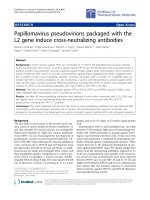
Figure 2 Inhibition of cell migration by rhMDA-7 was reverted
by TGase-4 expression. PC-3
pEF6
control cells had a slower pace of
migration in the presence of rhMDA-7. However, PC-3
TGase4exp
cells
migrated rapidly and had no response to rhMDA-7.
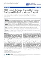
Figure 1 TGase-4 expression and the cells response to rhMDA-7. Left panel: Adhesion assay using ECIS 9600 system (A - PC-3
pEF6
control
cells; B - PC-3
TGase4exp
cells). Right panel (C): Cell adhesion assay using Rb modeling (ECIS 1600R, 4000 Hz). PC-3
TGase4exp
cells showed a
significant increase in cell migration when compared with the PC-3
pEF6
control cells (**, p < 0.01 vs the PC-3
pEF6
control cells). MDA-7 inhibited
cell adhesion in PC-3
pEF6 cells
(top left), a response lost in PC-3
TGase4exp
cells. * p < 0.01 vs no MDA-7 control PC-3
pEF6
cells.
Ablin et al. Journal of Translational Medicine 2011, 9:49
/>Page 5 of 9
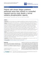
Figure 3 Response to rhMDA-7 in cell adhesion (top) and migration (bottom) by CA-HPV-10 wild type cells, a cell with high levels of
expression of TGase-4.
Figure 4 Effects of rhMDA-7 on the in vitro growth of PC-3 cells (left) and the effects of the Akt inhibitor on the motility of PC-3 cells
(right). In Array-A are PC-3
pEF6
cells and in Array- B are PC-3
TGase4exp
cells. Cells were treated with or without rhMDA-7 (shown at 10 ng/ml), in
the presence or absence of the Akt inhibitor (shown at 5 μM).
Ablin et al. Journal of Translational Medicine 2011, 9:49
/>Page 6 of 9
and indeed, the MDA-7 receptor, may co-localize in
normal and malignant human prostate tissues. Shown in
Figure 5 (B, C and D), strong staining of TGase-4 was
seen in the matrix and epithelial cells. Prostate tissues
also showed staining of MDA-7 (Figure 5B and 5D) and
IL-20Ra (Figure 5C). These observations demonstrated a
good degree of co-localization between TGase-4, IL-
20Ra and MDA-7.
Discussion
The present study has shown that TGase-4 in human
prostate cancer cells has a direct impact on the adhe-
sive, motility and growth properties of the cell’ s
response to rhMDA-7. Specifi cally, when not expressing
TGase-4, cells responded well to rhDMA-7 by exhibiting
a reduction of adhesion, motility and growth. However,
cells expressing TGase-4 (either natur ally - CA-HPV-10
or by forced expression -PC-3
TGase4exp
), had either no
response to rhMDA-7 or had a marginal response oppo-
site to those cells without TGase-4.
MDA-7/IL-24, although initially found to be up-regu-
lated in melanoma cells [16,17], has been shown to have
a growth inhibitory role in certain cancer cells [17]
which include ovarian [24], colorectal [25] and glioma
cancer cells [26]. The present study has shown that the
MDA-7/IL-24 cytokine also inhibits the adhesion, moti-
lity and growth of p rostate cancer cells. These observa-
tions place MDA-7/IL-24 within the context of a limited
number of cytokines that inhibit the adhesiveness,
growth and migration of cancer cells.
The most int riguing finding of the present study was
that the function of MDA-7 in prostate cancer cells
appears to be dependent upon the presence of TGase-4.
Using two cell models, i.e., the TGase-4 expressing CA-
HPV-10 and TGase-4 non-expressing PC-3 cells, we
have shown that when TGase-4 is not present, MDA-7
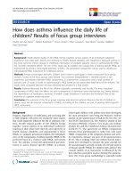
Figure 5 Staining of MDA-7 in PC-3 cells (A) and co-localization of TGase-4 and MDA-7/MDA-7 receptor in human prostate tissues (B,
C and D). A: Staining of MDA-7 in wild type (top left), control transfected cells (bottom left) and in TGase-4 expression vector transfected PC-3
cells (right two micrographs). PC-3 stained positive for MDA-7. However, after over-expressing TGase-4, staining intensity of MDA-7 is reduced. B
and D: co-localization of TGase-4 and MDA-7 in normal (B) and tumour (D) prostate tissues. TGase-4 staining appears in both stroma and in the
cells. C: co-localization of TGase-4 and MDA-7 receptor, IL20R, in prostate tissue. TGase-4 and IL20R have a good degree of co-localization. HOE:
Nuclear staining using Hoescht 33258. Comp.: Double immunofluorescent staining reaction obtained with each of the respective antibodies in B,
C and D. Magnification was ×100.
Ablin et al. Journal of Translational Medicine 2011, 9:49
/>Page 7 of 9
inhibits the migration of the cells (i.e., PC-3 wild type
and control cells). When TGase-4 is expressed (in CA-
HPV-10 and PC-3
TGase4exp
), cells no longer respond to
MDA-7.
The mechanism(s) by which TGase-4 affects MDA-7
is not clear. MDA-7/IL-24 acts via its receptor -MDA-
7R/IL-24R. Receptor complexes include at least the IL-
20alpha and IL-20beta complex and the IL-22R and IL-
20Rbeta complex. Intracellular signalling pathways
downstream of these receptors are not clear. MAPK
pathways and the Fas-FasL pathway [26] have been
implicated.
The present study has shown that blocking the Akt
pathway using an Akt inhib itor abolishes MDA-7
induced inhibition of migration, thus indicating that Akt
may be a potential pathway downstream of MDA-7. It is
interesting to note that PC-3 cells over-expressing
TGase-4 did not respond to MDA-7 nor the Akt inhibi-
tor. Furthermore, inhibitors to pathways including the
PLC-g , JAK, PKC pathway, and WASP pathways, have
no obvious impact on the action of MDA-7. Together,
this may suggest that TGase-4 interferes with the action
of MDA-7 at a stage before receptor activation. From
the immunofluorescent staining of TGase-4 and MDA-7
receptor, it is clear that there is a good degree of co-
localization between the TGase-4 and IL-20R a. A possi-
bility thus exists that TGase-4 may interact with IL-
20Rs masking the site for MDA-7 to interact. More
work is required t o clarify the interaction of this
possibility.
MDA-7 has been tested for its clinical application as
an anti-cancer treatment option. Using an adenoviral-
based delivery method, MDA-7 has been shown to have
an anti-tumour effect in ovarian, lung, and hepatoma
cancer models. MDA-7 has also been shown to increase
the efficiency bevacizumab and Herceptin. Information
on the effect of MDA-7 on prostate cancer cells is
rather limited. However, it has been demonstrated that
expression of MDA-7 in prostate cancer cells inhibits
growth and induction of apoptosis [18]. Albeit, at an
early stage, observations from the present study are
interesting and have important clinical implications, e.g.,
therapeutic consideration of the use of MDA-7 would
be dependent on the degree of expression of TGase-4.
MDA-7 may be more sensitive in t umours that express
low levels of TGase-4 and vice versa. This is an interest-
ing point to consider in future pre-clinical and clinical
studies.
Conclusion
This study reports for the first time that the presence of
TGase-4, a prostate specific TGase-4, has an overriding
effect on a cells response to MDA-7, a potential anti-
cancer cytokine. TGase-4 , via mechanism(s) yet to be
identified, blocked the action of MDA-7 in prostate
cancer cells. This has an important implication when
considering the use of MDA-7 in prostate cancer
therapies.
Acknowledgements
The authors wish to thank Cancer Research Wales, Robert Benjamin Ablin
Foundation for Cancer Research, and Albert Hung Foundation for supporting
their work.
Author details
1
Department of Pathology, University of Arizona College of Medicine,
Arizona Cancer Center and BIO5 Institute, Tucson, Arizona, AZ 85724-5043
USA.
2
Metastasis and Angiogenesis Research Group, Card iff University School
of Medicine, Cardiff, UK.
Authors’ contributions
RJA and WGJ contributed to the study design, experimental work, and
manuscript preparation. MDM and HGK contributed to sample collection
and manuscript preparation. All of the authors read and approved the final
manuscript.
Competing interests
The authors declare that they have no competing interest s.
Received: 19 November 2010 Accepted: 28 April 2011
Published: 28 April 2011
References
1. Folk JE: Transglutaminases. Annu Rev Biochem 1980, 49:517-531.
2. Chen JSK, Mehta K: Tissue transglutaminase: an enzyme with a split
personality. Int J Biochem Cell Biol 1999, 31:817-836.
3. Gentile V, Grant FJ, Porta R, Baldini A: Localization of the human prostate
transglutaminase (type IV) gene (transglutaminase-4) to chromosome
3p21.33-p22 by fluorescence in situ hybridization. Genomics 1995,
27:219-220.
4. Grant FJ, Taylor DA, Sheppard PO, Mathewes SL, Lint W, Vanaja E,
Bishop PD, O’Hara PJ: Molecular cloning and characterization of a novel
transglutaminase cDNA from a human prostate cDNA library. Biochem
Biophys Res Commun 1994, 203:1117-1123.
5. Dubbink HJ, de Waal L, van Haperen R, Verkaik NS, Trapman J, Romijn JC:
The human prostate-specific transglutaminase gene (TGM4): genomic
organization, tissue-specific expression, and promoter characterization.
Genomics 1998, 51:434-44.
6. Dubbink HJ, Hoedemaeker RF, van der Kwast TH, Schroder FH, Romijn JC:
Human prostate-specific transglutaminase: a new prostatic marker with
a unique distribution pattern. Lab Invest 1999, 79:141-150.
7. An G, Meka CS, Bright SP, Veltri RW: Human prostate-specific
transglutaminase gene: promoter cloning, tissue-specific expression, and
down-regulation in metastatic prostate cancer. Urology 1999,
54:1105-1111.
8. Cho SY, Jeon JH, Kim CW, Shin DM, Jang GY, Jeong EM, Lee SE, Song KY,
Kim IG: Monoclonal antibodies to human transglutaminase 4. Hybridoma
(Larchmt) 2010, 29:263-267.
9. Ho KC, Quarmby VE, French FS, Wilson EM: Molecular cloning of rat
prostate transglutaminase (type IV) gene (transglutaminase-4) to
chromosome 3p21.33p22 by fluorescence in situ hybridization. Genomics
1995, 27:219-220.
10. Williams-Ashman HG: Transglutaminases and the clotting of mammalian
seminal fluids. Mol Cell Biochem 1984, 58:51-61.
11. Ablin RJ, Whyard TC: Identification and biological relevance of
spermatozoal transglutaminase. Experientia 1991, 47:277-279.
12. Davies G, Ablin RJ, Mason MD, Jiang WG: Expression of the prostate
transglutainase (TGase-4) in prostate cancer cells and its impact on the
invasiveness of prostate cancer. J Exp Therapeut Oncol 2007, 6 :257-264.
13. Jiang WG, Ablin RJ, Kynaston HG, Mason MD: The Prostate
Transglutaminase (TGase-4, TGaseP) regulates the interaction of prostate
cancer and vascular endothelial cells, a potential role for the ROCK
pathway. Microvas Res 2009, 77:150-157.
Ablin et al. Journal of Translational Medicine 2011, 9:49
/>Page 8 of 9
14. Cho SY, Choi K, Jeon JH, Kim CW, Shin DM, Lee JB, Lee SE, Kim CS, Park JS,
Jeong EM, Jang GY, Song KY, Kim IG: Differential alternative splicing of
human transglutaminase 4 in benign prostate hyperplasia and prostate
cancer. Exp Mol Med 2010, 42:310-318.
15. Jiang WG, Ablin RJ, Ye L, Kynaston , Mason MD: The prostate
transglutaminase, TGase-4, coordinate with the HGFL/MSP-RON system
in stimulating the migration of prostate cancer cells. Int J Oncology 2010,
37:413-418.
16. Jiang H, Lin JJ, Su ZZ, Goldstein NI, Fisher PB: Subtraction hybridization
identifies a novel melanoma differentiation associated gene, mda-7,
modulated during human melanoma differentiation, growth and
progression. Oncogene 1995, 11:2477-2486.
17. Jiang H, Su ZZ, Lin JJ, Goldstein NI, Young CSH, Fisher PB: The melanoma
differentiation associated gene mda-7 suppresses cancer cell growth.
Proc Nat Acad Sci USA 1996, 93:9160-9165.
18. Lebedeva IV, Sarkar D, Su ZZ, Su Z-Z, Kitada S, Dent P, Stein CA, Reed JC,
Fisher PB: Bcl-2 and Bcl-X
L
differentially protect human prostate cancer
cells from induction of apoptosis by melanoma differentiation
associated gene-7, mda-7/IL-24. Oncogene 2003, 22:8758-8773.
19. Jiang WG, Davies G, Martin TA, Parcc C, Watkins G, Mason MD, Mokbel K,
Mansel RE: Molecular targeting of matrilysin and its impact on tumour
growth in vivo, the potential implications in breast cancer therapy. Clin
Cancer Res 2005, 11:6012-6019.
20. Jiang WG, Watkins G, Douglas-Jones A, Mokbel K, Mansel RE, Fodstad O:
Expression of Com-1/p8 in human breast cancer, and its relevance to
clinical outcome and ER status. Int J Cancer 2005, 117:730-737.
21. Giaever I, Keese CR: Micromotion of mammalian cells measured
electrically. Proc Natl Acad Sci USA 1991, 88:7896-7900.
22. Jiang WG, Martin TA, Russell-Lewis J, Ye L, Douglas-Jones A, Mansel RE:
Eplin-alpha expression in human breast cancer, the impact on cellular
migration and clinical outcome. Mol Cancer 2008, 7:71.
23. Keese CR, Wegener J, Walker SR, Giaever I: Electrical wound-healing assay
for cells in vitro. Proc Natl Acad Sci USA 2004, 101:1554-1559.
24. Gopalan B, Litvak A, Sharma S, Mhashilkar AM, Chada S, Ramesh R:
Activation of the Fas-FasL signaling pathway by MDA-7/IL-24 kills
human ovarian cancer cells. Cancer Res 2005, 65:3017-3024.
25. Zhao L, Dong A, Gu J, Liu Z, Zhang Y, Zhang W, Wang Y, He L, Qian C,
Qian Q, Liu X: The antitumor activity of TRAIL and IL-24 with replicating
oncolytic adenovirus in colorectal cancer. Cancer Gene Ther 2006,
13:1011-1022.
26. Yacoub A, Mitchell C, Lebedeva IV, Sarkar D, Su ZZ, McKinstry R,
Gopalkrishnan RV, Grant S, Fisher PB, Dent P: MDA-7 (IL-24) Inhibits
growth and enhances radiosensitivity of glioma cells in vitro via JNK
signaling. Cancer Biol Ther 2003, 2:347-353.
doi:10.1186/1479-5876-9-49
Cite this article as: Ablin et al.: Prostate transglutaminase (TGase-4)
antagonizes the anti-tumour action of MDA-7/IL-24 in prostate cancer.
Journal of Translational Medicine 2011 9:49.
Submit your next manuscript to BioMed Central
and take full advantage of:
• Convenient online submission
• Thorough peer review
• No space constraints or color figure charges
• Immediate publication on acceptance
• Inclusion in PubMed, CAS, Scopus and Google Scholar
• Research which is freely available for redistribution
Submit your manuscript at
www.biomedcentral.com/submit
Ablin et al. Journal of Translational Medicine 2011, 9:49
/>Page 9 of 9