báo cáo hóa học:" Increased human defensine levels hint at an inflammatory etiology of bisphosphonateassociated osteonecrosis of the jaw: An immunohistological study" docx
Bạn đang xem bản rút gọn của tài liệu. Xem và tải ngay bản đầy đủ của tài liệu tại đây (2.45 MB, 8 trang )
RESEARC H Open Access
Increased human defensine levels hint at an
inflammatory etiology of bisphosphonate-
associated osteonecrosis of the jaw:
An immunohistological study
Philipp Stockmann
1*
, Falk Wehrhan
1
, Stephan Schwarz-Furlan
2
, Florian Stelzle
1
, Susanne Trabert
1
,
Friedrich W Neukam
1
and Emeka Nkenke
1
Abstract
Background: Human b-defensins (hBD) are antimicrobial peptides that are an integral part of bone innate
immunity. Recently, it could be shown that expression of hBD-1, -2 and -3 were upregulated in cases of
osteomyelitis of the jaws. In order to gain insight into the possible impairment of hBD metabolism in
bisphosphonate-associated osteonecrosis of the jaws (BONJ), the present exploratory study was designed so as to
determine the qualitative and quantitative expression of afore mentioned hBDs in BONJ and infected
osteoradionecrosis (ORN), both of which represent inflammatory bone diseases.
Methods: Bone samples were collected from patients with BONJ (n = 20) and ORN (n = 20). Non-infected healthy
bone samples (n = 20) were included as controls. Immu nohistological staining in an autostainer was carried out by
the (Strept-ABC)-method against hBD-1,-2,-3. Specific positive vs. negative cell reaction of osteocytes (labeling
index) near the border of bony resection was determined and counted for quantitative analysis. Number of vital
osteocytes vs. empty osteocytes lacunae was compared between groups.
Results: hBD-1,-2 and -3 could be detected in BONJ as well as ORN and healthy bone samples. Immunoreactivity
against hBD-2 and -3 was significantly higher in BONJ than in ORN and healthy jaw bone samples. Number of
empty osteocyte lacunae was significantly higher in ORN compared with BONJ (P = 0.001).
Conclusion: Under the condition of BONJ an increased expression of hBD-1,-2,-3 is detectable, similarly to the
recently described upregulation of defensins in chronically infected jaw bones. It remains still unclear how these
findings may relate to the pathoetiology of these diseases and whether this is contributing to the development of
BONJ and ORN or simply an after effect of the disease.
Keywords: antimicrobial peptide, bisphosphonate-associated osteonecrosis, osteoradionecrosis, human beta defen-
sins, innate immunity
Background
Bisphosphonates are an important component of treat-
ment in metastatic bon e disease and the management of
osteoporosis. An increasing number of reports have
associated the use of bisphosphonates with the occur-
rence of osteonecrosis of the jaw.
The clinical sympto ms of bispho sphonate-associated
osteonecrosis of the jaw (BONJ) are rather similar to the
lesions seen in patients with infect ed osteoradione crosis
(ORN) [1]. The lesio ns are surrounded by inflammatory
soft tissue reactions and show symptoms and radiologi-
cal signs of bone sequestration and/or osteomyelitis [2].
Microorganisms like Actinomyces spp. seem to play an
etiological role in the development of both ORN and
BONJ [3-5].
* Correspondence:
1
Department of Oral and Maxillofacial Surgery, University of Erlangen-
Nuremberg, Erlangen, Germany
Full list of author information is available at the end of the article
Stockmann et al. Journal of Translational Medicine 2011, 9:135
/>© 2011 Stoc kmann e t al; licen see BioMed Central Ltd. This is an Open Access article distributed unde r the terms of the Creative
Commons Attribution License ( which perm its unrestricted use, distribution , and
reproduction in any medium, provided the original work is properly cit ed.
Defensins are antimicrobial pe ptides that are an inte-
gral part of innate and antigen-specific acquired i mmu-
nity [6,7]. These small cationic and cysteine-rich
peptides (3.5 to 6.5 kDA) have the potency to disrupt
membranes and interfere with intracellular functions of
various gram-positive and gram-negative bacteria as well
as fungal and encapsulated viral pathogens [8]. Even the
potential role of defensins in pathogenesis of oral cancer
is under discussion [9,10]. To date, various numbers of
defensins subdivided into a-andb-andθ-defensins
have been discovered in humans [11,12]. They are char-
acterized and distinguished owing to their sequence
homology and disulfide pairing. Human b-defensins
(hBD) -1, -2 and -3 have a broad spectrum antimicrobial
activity and structural similarities [13]. They are mainly
produced b y epithelial cell s, but quite recently Warnke
and co-workers adduced evidence that they are
expressed by osteocyt es in jaw bone as well. These find-
ings might explain the relatively rare occurrence of
osteomyelitis after exposure of jaw bone e.g. after surgi-
cal dent oalveol ar procedures with expo sure of jaw bone
to the oral cavity and hint to the important role of these
peptides in the pathophysiological mechanism of inflam-
matory jaw bone diseases [14].
As elevated human hBD -1, -2, -3 levels have been
detected in osteomyelitis of the jaw, this leads us to the
hypothesis that an impaired hBD expression in the bone
might contribute to the development of BONJ or other
inflammatory jaw bone diseases like the infected osteor-
adionecrosis (ORN). Therefore, it was the aim of the
present exploratory study to prove and quantify b-defen-
sin expression in BONJ and to compare it with ORN
and healthy jaw bone as a control.
Methods
After approval by the ethical committee of the Univer-
sity of Erlangen-Nuremberg bone biopsies of patients
suffering from BONJ (n = 20) and ORN (n = 20) as well
as control samples (n = 20) of healthy jaw bone were
used for evaluation. All bone samples were harvested in
the molar region of the mandible. The samples of BONJ
and ORN comprised non-necrotic bone adjacent to
necrotic zones. Controls were surplus of resected unin-
fected bone during orthognathic surgery. All patients
hadbeeninformedaboutthisstudyandgavetheir
informed consent for participation.
The average age at surgery was 70 ± 11 years in the
BONJ group, 59 ± 8 years in the ORN group and 46 ±
13 years in the control group.
BONJ was defined as an area of exposed bone in the
maxillofacial region that did not heal within eight weeks
of identification by a health care provider, in a patient
who was receiving or had been exposed to a bispho-
sphonate and had not had radiation therapy to the
craniofacial region [15,16]. 6 patients in the BONJ group
suf fered from metastatic prostate canc er, 8 patients had
breast cancer and 6 patients had plasmocytoma. 13
patients received IV zoledronate and 6 pat ients pami-
dronate and 1 patient received ibandronate after zole-
dronic acid on a monthly basis. The mean duration of
bisphosphonate therapy was 3 4.3 ± 23.5 months before
surgery was carried out. All patients in this group
underwent o steotomy of the necrotic bone followed by
primary wound closure [17].
According to Marx, ORN is defined as exposed irra-
diated bone tissue that fails to heal over a period of
three months without a residual or recurrent tumor
[18]. Clinica l signs of inflammation and bacterial super-
infection lead to a diagnosis of infected ORN.
Bone samples of BONJ and ORN were only included
in the study when the region of exposed bone showed
signs of infection as evidenced by pain and erythema
with or without purulent drainage (stage 2 of BONJ
[15]) and the patients were not under permanent medi-
cation with steroids (Figure 1).
Immunohistochemistry
Bone biopsies were fixed in neutral 4% Formalin solu-
tion. Afterw ards, the samples were decalcified in a 25%
ethylenediaminetetraacetic acid (EDTA) solution (pH
7.4). The decalcification lasted 10 days and the EDTA
solution was changed several times during the pro cess.
The dehydration procedure was performed in an
ascending alcohol sequence at room temperature in a
dehydration unit (Shandon Citadel 1000, Shandon
GmbH, Germany). Paraffin-embedded bone samples
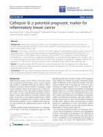
Figure 1 The clinical picture of bisphospho nate-associated
osteonecrosis of the jaw is rather similar to the lesions seen in
patients with infected osteoradionecrosis. Clinical pictures of a
stage 2 bisphosphonate-associated osteonecrosis of the jaw (A) and
infected osteoradionecrosis (B). Bone samples of these clinical
manifestations were included in the study.
Stockmann et al. Journal of Translational Medicine 2011, 9:135
/>Page 2 of 8
were sectioned in cuts of 4 μm thickness with a stan-
dard microtome (Leica RM 2165
®
, L eica Microsystems,
Nussloch GmbH, Germany). Subsequently, the surface
of the samples was blocked to prevent unspecific stain-
ing using a serum-free protein block (DAKO Diagnos-
tics GmbH, Germany).
Immunohistological staining was obtained for detec-
tion of expression of hBD-1, -2, and -3 with the Strepa-
vidin-Biotin-Peroxidase-complex (Strept-ABC)-method
performed for all bone samples with an autostainer
(Autostainer plus
®
, DakoCytomation, Dako Deutschland
GmBH, Germany). To deparaffinize the slices we
washed them in Xylol and then cooked them for 15 min
in EDTA-buffer (Dako Retrieval Puffer, pH 9,0) to
uncover the relevant antigens. W e applied 3% H
2
O
2
to
block endogeneous peroxidase. The slides were washed
in Tris-Buffered Saline (TBS) and incubated with rabbit
antisera to hBD-1 (Biologo, DEF01-A, Kiel, Germany,
dilution 1:500), hBD-2 (DEF02, dilution 1:250) and
hBD- 3 (DEF03-S, dilution 1:500) as well as pre-immune
serum as negative control. Further processing of the
Strept-ABC method was carried out according to the
manufacturer’s manual (Dako, Hamburg, Germa ny).
Finally, the samples were stained with Hematoxylin-
Eosin (Dako S 3301) for light microscopic evaluation.
Negative controls without primary antibody were passed
by in each cycle to verify antibody specificity.
Qualitative and quantitative analysis
Qualitative and quantitative analyses were performed for
the absence of osteocytes in the osteocyte lacunae next
to ea ch Kwire track as a measure of bone necrosis. For
quantification of hBD-1 through three expressions the
immunostained slices were analyzed and digitized with a
light microscope (Axioscope
®
Zeiss, Jena, Germany).
Regions of interest (ROI) were bone areas in spongy
bone which showed equal bone trabecular and bone
marrow cells. Three v isual fields per section for e ach
sample were digitized with a CCD camera. In a 400-fold
magnification the analyzing software enabled cells inside
an ROI to be digitally marked, and measurement para-
meters were determined by means of Bioquant Osteo
®
software V7.10.10 (Nashville, USA). As a sign of bone
necrosis the numbers of empty o steocytes lacunae were
related to the number of total count of osteocytes inside
the ROI. The labeling index was defined as the ratio of
stained osteocytes vs. total number of osteocytes/osteo-
cytes lacunae inside the ROI. The intensity of immunos-
taining was not considered for the labeling index.
Statistics
For statistic al analysis, group means and standard devia-
tions were calculated for each parameter with SPSS soft-
ware (version 16; SPSS Inc., Chicago, USA). Data were
compared with the Mann-Whitney-U-Test. A P-value <
0.05 was considered statistically significant.
Results
In ORN it was evident that there were 75.0% empty
osteocyte lacunae whereas BONJ had only 24.8% empty
lacunaeinsidetheROI.Thehealthybonesamples
showed 2.4% empty osteocyte lacunae. The number of
vital osteocytes w as significantly higher in BONJ than
ORN (P = 0.001).
Specific immunoreactivity was able to identify the pre-
sence of hBD-1, -2 and -3 within jaw bone biop sies in
all tested groups. Expression is especially prominent in
osteoblasts and the osteocytes included in woven bone
while the resting osteocytes in lamellar bone are
negative.
Human b-defensin-1 (Figure 2)
BONJ samples showed high immunoreactivity in stromal
cells including rims of osteoblasts along the endosteal
cell lines. Numerous osteocytes in the mineralized bone
trabeculae showed specific positive cell reactions.
Besides typical cell changes (radiocytes) in ORN the
immunoreactivi ty of osteocytes against hBD-1 was
barely visible and was detectable mostly in stromal cells.
Non-viable bone was demonstrated by lack of nucle oli
in bone lacunae. Large parts of the bone marrow
showed fibronecrotic lesions and fibrosis without any
immunoreactivity against hBD-1.
Healthy bone showed only smooth positive cell reac-
tion mostly in the vicinity of blood vessels and bone mar-
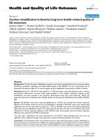
row. The average value of the labeling index revealed that
positive cell reaction to hBD-1 was slightly higher in
BONJ (22.3 ± 20 .3%) than in ORN (7. 2 ± 10.4%) and
healthy jaw bone samples (12.8 ± 14.8%). These differ-
ences were not statistically significant (Figure 3).
Human b-defensin-2 (Figure 4)
Intensity of immunost aining in hBD-2 was weaker com-
pared with hBD-1 in all groups.
Inside bone trabecula o f BONJ sampl es sufficient
immunoreactivity could be often found in the cytoplasm
of osteocytes and stromal cells, which indicates expres-
sion of hBD-2 in these cells. Smooth positive staining
could be detected along osteoblasts lined up at the
endosteal cell lines. Moreover, the highest extensive
immunoreactivity was visible in areas of bone marrow,
which showed infiltration with polymorphonuclear leu-
kocytes, indicating a reaction to acute inflammation.
In ORN samples osteocytes had mainly negative cell
reactions to hBD-2, and enhanced positivity was
observed only in osteoblasts near the endosteal cell line.
Similarly, healthy bone showed weak cytoplasmic posi-
tivity of osteocytes and stromal cells.
Stockmann et al. Journal of Translational Medicine 2011, 9:135
/>Page 3 of 8
Quantitative analysis reveale d an average labeling
index of 29.2 ± 16.4% in BONJ samples. Significantly
lower values for the labeling index could be ob served in
healthy jaw bone (12.3 ± 12. 5%, P = 0.017) and smallest
values in ORN (5.0 ± 7.0, P = 0.002) (Figure 3).
Human b-defensin-3 (Figure 5)
There was distinctive immunoreactivity against hBD-3
in all g roups compared with hBD-1 and -2. The highest
intensity o f immunostaining was detectable beside the
border of bone marrow and mineralized bone. Bands of
osteoblasts at the endosteal cell line showed intensive
dyeing.
As regards the labeling index, the average value for
ORN (8.1 ± 10.3%) and healthy bone biopsies (8.1 ±
7.4%) were appr oximately equal compared to the signifi-
cantly higher measured values for BONJ (30.7 ± 16.4, p
< 0.05) (Figure 3).
Discussion
To date the etiopathogenesis of BONJ is not sufficiently
clarified. Different hypotheses concerning the pathophy-
siology of BONJ are to be found in the literature: The
inhibition of osteoclast and osteoblast activity followed
by an impaired bone turnover with compromised bone
healing [19,20], an inhibition of endothelial cells with
impaired intraosseous angiogenesis, mucosal damage
secondary to toxic exposure of the bone [21-23] and the
A
B
C
Figure 2 Immunohistochemical staining a gainst hBD-1 (400-
fold magnification). A: BONJ. Increased immunoreactivity can be
detected within bone marrow area (BM), mostly in bands along the
endosteal cell lines (arrows). Inside mineralized bone trabecula (BT)
immunostaining demonstrates nuclear expression of hBD-1. B: ORN.
Immunoreactivity is barely visible in cytoplasm of stromal cells.
Specific cells for diagnosis of ORN in terms of radicytes (arrows)
were traceable. C: Healthy bone samples. Detailed enlargement of
mineralized area (BT) of controls shows a Haversian channel (arrow)
in which positive nuclear staining is seen only along the endosteal
cell line. Immunoreactivity of osteocytes is negative in this sample.
Figure 3 Immunoreactivity against hBD-2 and -3 was
significantly higher in bisphosphonate-associated
osteonecrosis of the jaw than in infected osteoradionecrosis
and healthy jaw bone samples. Value distribution of hBD-1 trough
3 expression in bone shown as boxplots divided into groups. The
labeling index was defined as the ratio of stained osteocytes vs.
total number of osteocytes and osteocyte lacunae inside the ROI.
Outliers are marked as circles.
Stockmann et al. Journal of Translational Medicine 2011, 9:135
/>Page 4 of 8
A
B
C
Figure 4 Immunohistochemical staining a gainst hBD-2 (400-
fold magnification). A: BONJ. Smooth positive staining could be
detected along the endosteal cell lines. Inside bone trabecula (BT)
scattered positive cell reactions of osteocytes are visible, which
indicate expression of hBD-2. B: ORN. Negative empty osteocytes
lacunae (arrow) and enhanced positivity in the endosteal cell line
(star) are evident. C: Healthy bone samples. Partial positivity of
osteocytes (arrow) and weak cytoplasmic positivity of stromal cells
(stars) within the bone marrow area (BM) are visible.
A
B
C
Figure 5 Immunohistochemical staining a gainst hBD-3 (400-
fold magnification). A:. BONJ. Immunoreactivity could be detected
along the endosteal cell lines (stars). Inside bone trabecula (BT)
numerous positive cell reactions of osteocytes are visible (arrows),
which indicate expression of hBD-3. BM, bone marrow area. B: ORN.
Strong positivity in osteoblasts could be detected along the
endosteal cell line (arrows). Inside mineralized bone trabecula (BT)
only weak positivity in osteocytes is evident. C: Healthy bone
controls. Positive staining could be detected along the endosteal
cell lines (arrows) with moderate positivity of osteoblasts, which
indicates the presence of hBD-3. Inside bone trabecula (BT) a no
positive osteocytes are visible.
Stockmann et al. Journal of Translational Medicine 2011, 9:135
/>Page 5 of 8
infectious-immune hypothesis with impaired immune
defense at the mucosal barrier [12,21,22]. All hypotheses
could not yet explain the ra re occurrence of BONJ and
its restriction mainly to the jaws.
Based on the infectious-immune hypothesis and on
the knowledge that hBDs are expressed in osteogenetic
cell lineages, the main focusofthisexploratorystudy
was to determine the e xpression level of antimicrobial
peptides in BONJ, so as to proof the hypothesis, that
there is a possible impairment, which could affect sus-
ceptibility to BONJ.
As part of innate immunity, antimicrobial peptides like
defensins seem to play an important role in protection
of oral cavity integrity against invasion by microbes [24].
b-defensin exhibits a bactericidal effect on pathogens
thatisbasedonaninhibitionofcellproliferation[25]
and extracellula r matrix production [8] and the modula-
tion of cellular immune responses [26]. The localization
of hBD-1-3 in oral mucosa has been confirmed at pro-
tein and mRNA levels [24].
Recently it was shown that hBD-1, -2 and -3 are
expressed in chronically infected as well as healthy jaw
bone [14]. Subsequently, Kraus and coworkers could
demonstrate that hBD-1, -2, -3 were expressed in osteo-
blast-like MG63 cells in vitro. Moreover, they could pro-
vide evidence that hBD-2 stimulates their proliferation
and hBD-2 and -3 positively affected their differentiation
processes [27].
To date, the detailed pathways regulating the expres-
sion of human b-defensins are not completely under-
stood. It seems that hBD-1 may be modulated by
inflammation, while hBD-2 and hBD-3 are expressed by
cells upon stimulation with proinflammatory cytokines
and by microorganisms [24]. hBD-1 can be induced and
upregulated by lipopolysaccharides (LPSs), heat-inacti-
vated Pseudomonas eruginosa and interferon gamma
(IFN-g). hBD-2 expression is induced in response to
gram- and gram+ bacteria as well as Candida albicans
[28]. In contrast with hBD-2, upregulation of hBD-3
expression in keratinocytes was observed in the presence
of inflammatory proteins like transforming growth factor
alpha (TGF- a) a nd insulin-like growth factor 1 (IGF-1)
[29].
There are data to indicate that nitrogen-containing
bisphosphonates affect the function of cells of both
innate and acquired immunity. In particular, these
agents have a profound effect on differentiation and
maturation of human myeloid dendritic cells (DC) [30].
Interestingly, both hBD-1 and -2 seem to possess immu-
noregulatory activity as well, by chemoattraction of
immature dendritic cells and memory T cells through
interaction with beta chemokine receptor [31].
In addition nitrogen-containing bisphosphonates have
been shown to augment the allostimulatory activity of
DC on naive CD4
++
and CD45
+
T cells in terms of their
proliferation and interferon-g production [32]. There is
evidence that hBD-3 expression is inducible by inter-
feron-g [33], which might be the reason why hBD-3
showed the highest immunoreactive values in BONJ
samples in our study.
Also, it has been shown that the activation of Vg9Vδ2
T cells by aminobisphosphonate drugs results in a mas-
sive release of cytokines and chemokines that may
induce expression of defensines. Moreover, that soluble
factors released by aminobisphosphonate -stimulated
Vg9Vδ2 T cells activate granulocytes by inducing their
chemotaxis, phagocytosis, and alpha-defensins release
[34].
A lack of induction of osteoblast-derived hBD-2 in the
presence of immunosuppressive drugs, which are fre-
quently used in chronic inflammatory joint diseases, is
ass umed to be responsible for the increased susceptibil-
ity of these patients to bone and joint infection [35].
So far, no studies a re available that have determined
the expression of defensins in BONJ. Therefore, the pre-
sent study was conducted to determining the expression
of human b- defensins in BONJ quantitatively. Because
of a number of similar clinical and pathological features,
samples o f infected osteoradionecrosis were also exam-
ined in the present study. Although both conditions are
related to bacterial infection (e.g. Actinomyces) and they
share similar clinic al sympto ms, there are differences in
their histologic al appearance. BONJ shows elements of
osteomyelitis and it is not directly comparable to osteor-
adionecrosis of the mandible [12]. In particul ar, areas of
active acute inflammation with the presence of inflam-
matory ce lls were seen in peripheral areas, where orga-
nized bacterial biofilms were present [36,37].
Tothebestofourknowledgethisisthefirstreport
on the expression analysis of hBD in BONJ bone sam-
ples . Our hypothesis that hBD expression is hindered in
BON J bone samples could not b e confirmed in the pre-
sent study. However, the results reveal that immunor-
eactivity for antimicrobial peptides hBD-1, hBD-2 and
hBD- 3 in jaw bone biopsies of BONJ can be found on a
regular basis. The results indicate that jaw bone samples
harvested from BONJ are still able to express defensins
on a higher level than healthy uninfected jaw bone. This
result points out, that there is still an unimpaired meta-
bolic reaction in BONJ bone samples due to an infection
sti mulus. In contrast, the expression of human b-defen-
sins in ORN was significantly reduced. Therefore, it
seems that bone affected by BONJ does not exclusively
show characteristics of necrotic bone l ike ORN samples,
but behaves in a similar fashion to that described pre-
viously for bone suffering from bacterial infection [14].
Some authors have already pointed to the role of infec-
tion in BONJ. Hansen and colleagues showed that 93.5%
Stockmann et al. Journal of Translational Medicine 2011, 9:135
/>Page 6 of 8
of patients suffering from BONJ also had a superinfec-
tion of Actinomyces israelii [1,4]. Sedghizadeh and col-
leagues examined bony sequesters of BONJ by electron
microscopy and identified various species of the genus
Fusobacterium, bacillus, a ctinomyces, staphylococcus,
streptococcus, Selenomonas, and t hree different m or-
photypes of treponemes or spirochetes, which were
organized in complex biofims [36]. Staining of hBD-3
seemed to be distinctly more intense in all samples
compared to hBD-1 and -2. This may indicate that there
is an intrinsic basal level of hBD-3 expression that is
independent on exposure to bacterial stimuli. Similar
results that were seen in healthy periodontal tissues and
tissue samples of healthy bone suggest a potentially
important protective role of defensins in the host
immune response to infection by oral pathogens [38,39].
At the moment, however, it is not clear if infection is
a major etiological factor for BONJ or just a sequela of
this disease. It seems that BONJ is a multifactorial pro-
cess resulting from an alteration in bone homeostasis,
inhibition of angiogenesis and, in particular, bacterial
risk factors [20,21,40].
While these are inte res ting findings it is not clear how
these results may relate to the pathoetiology of BONJ and
ORN and whether this is contributing to the development
of the diseases or simply an after effect of the disease.
Additionally, the methodology of the presented study
leads to no conclusion whether the expression of hBD-1
throug h 3 is associated with the degree of inflammation,
the presence or the amount of bacteria or the severity of
BONJ and ORN. However, the increased expression of
human b-defensins in bone samples of BONJ can be inter-
preted as a sign of unimpaired metabolic activity and can
therefore be seen as a reaction of vital bone to microbial
invasion. In this context, the study could demonstrate a
significant difference between BONJ and ORN concerning
their potency in immunological response. The question
that remains still unanswered is whether the defensins
retain their full functionality in the bisphosphonate-laden
bone. In addition, the present study provides no data
regarding the regulation or induction process of hBD in
BONJ and ORN.
Future research needs to clarify whether the in creased
expression of b-defensins in BONJ suggests that bone
infection is the crucial point in BONJ while osteone cro-
sis only accompanies the disease. It has been proposed
previously that BONJ should rather be termed bispho-
sphonate-associated osteomyelitis of the jaws [12].
Hence, further studies should focus on the discovery of
the detailed functio n of the three hBDs in innate and
adaptive immune system especially in the jaw bone and
the possible impact of bisphosphonates on their immu-
nological pathway. The results of the study, which hint
at an inflammatory etiology , can further help to
optimise preventive measures and existing treatment
regimes, e.g. avoidance of extended exposition of bi sph-
sophonate-laden jaw bone to the oral cavity, the impor-
tance of supportive application of antibiotics and
strengthen of the immune system by influencing the
local immune defence.
Conclusions
Under the condition of BONJ an increased expression of
hBD-1,-2,-3 are detectable, similarly to the recently
described upregulation of defensins in chronically
infected jaw bones. It remains still unclear how these
findings may relate to the pathoetiology of BONJ and
whether this is contributing t o the development o f
BONJ or simply an after effect of the disease.
Future research should focus on evolving the specific
role of hBDs in the innate and adapt ive immune system
of the bone and whether there is a possible impairment
of their antimicrobial activity under the influence of
bisphosphonates. Thereby, knowledge could be derived
regarding the understanding of the etiopathogenesis and
subsequently the prevention and treatment of BONJ.
Acknowledgements
The authors thank Heidemarie Heider and Susanne Schönherr for technical
assistance with the immunohistochemistry autostainer and processing the
bone samples.
Author details
1
Department of Oral and Maxillofacial Surgery, University of Erlangen-
Nuremberg, Erlangen, Germany.
2
Department of Pathology, University of
Erlangen-Nuremberg, Erlangen, Germany.
Authors’ contributions
PS was responsible for the conduction of study, built the hypothesis,
established and conducted the methods and analytic procedures and wrote
the manuscript. SS and FW interpreted the histopathological samples and
performed the immunohistochemistry analysis. ST participated in the design
of the study and performed immunohistochemistry. FS worked on the
statistical analysis. FWN have given final approval of the version to be
published. EN interpreted the data and revised the manuscript. All authors
read and approved the final manuscript.
Competing interests
The authors declare that they have no competing interests.
Received: 9 March 2011 Accepted: 15 August 2011
Published: 15 August 2011
References
1. Hansen T, Kunkel M, Weber A, James Kirkpatrick C: Osteonecrosis of the
jaws in patients treated with bisphosphonates - histomorphologic
analysis in comparison with infected osteoradionecrosis. J Oral Pathol
Med 2006, 35:155-160.
2. Stockmann P, Hinkmann FM, Lell MM, Fenner M, Vairaktaris E, Neukam FW,
Nkenke E: Panoramic radiograph, computed tomography or magnetic
resonance imaging. Which imaging technique should be preferred in
bisphosphonate-associated osteonecrosis of the jaw? A prospective
clinical study. Clin Oral Investig 2009.
3. Nair SP, Meghji S, Wilson M, Reddi K, White P, Henderson B: Bacterially
induced bone destruction: mechanisms and misconceptions. Infect
Immun 1996, 64:2371-2380.
Stockmann et al. Journal of Translational Medicine 2011, 9:135
/>Page 7 of 8
4. Hansen T, Kunkel M, Springer E, Walter C, Weber A, Siegel E, Kirkpatrick CJ:
Actinomycosis of the jaws-histopathological study of 45 patients shows
significant involvement in bisphosphonate-associated osteonecrosis and
infected osteoradionecrosis. Virchows Arch 2007, 451:1009-1017.
5. Hall V: Actinomyces–gathering evidence of human colonization and
infection. Anaerobe 2008, 14:1-7.
6. Ong PY, Ohtake T, Brandt C, Strickland I, Boguniewicz M, Ganz T, Gallo RL,
Leung DY: Endogenous antimicrobial peptides and skin infections in
atopic dermatitis. N Engl J Med 2002, 347:1151-1160.
7. Kagan BL, Ganz T, Lehrer RI: Defensins: a family of antimicrobial and
cytotoxic peptides. Toxicology 1994, 87:131-149.
8. Gallo RL, Huttner KM: Antimicrobial peptides: an emerging concept in
cutaneous biology. J Invest Dermatol 1998, 111:739-743.
9. Wenghoefer M, Pantelis A, Dommisch H, Gotz W, Reich R, Berge S,
Martini M, Allam JP, Jepsen S, Merkelbach-Bruse S, et al: Nuclear hBD-1
accumulation in malignant salivary gland tumours. BMC Cancer 2008,
8:290.
10. Wenghoefer M, Pantelis A, Dommisch H, Reich R, Martini M, Allam JP,
Novak N, Berge S, Jepsen S, Winter J: Decreased gene expression of
human beta-defensin-1 in the development of squamous cell carcinoma
of the oral cavity. Int J Oral Maxillofac Surg 2008, 37:660-663.
11. Pazgier M, Hoover DM, Yang D, Lu W, Lubkowski J: Human beta-defensins.
Cell Mol Life Sci 2006, 63:1294-1313.
12. Wimalawansa SJ: Insight into bisphosphonate-associated osteomyelitis of
the jaw: pathophysiology, mechanisms and clinical management. Expert
Opin Drug Saf 2008, 7:491-512.
13. Schibli DJ, Hunter HN, Aseyev V, Starner TD, Wiencek JM, McCray PB,
Tack BF, Vogel HJ: The solution structures of the human beta-defensins
lead to a better understanding of the potent bactericidal activity of
HBD3 against Staphylococcus aureus. J Biol Chem 2002, 277:8279-8289.
14. Warnke PH, Springer IN, Russo PA, Wiltfang J, Essig H, Kosmahl M, Sherry E,
Acil Y: Innate immunity in human bone. Bone 2006, 38:400-408.
15. AAOMS: American Association of Oral and Maxillofacial Surgeons
position paper on bisphosphonate-related osteonecrosis of the jaws. J
Oral Maxillofac Surg 2007, 65:369-376.
16. Khosla S, Burr D, Cauley J, Dempster DW, Ebeling PR, Felsenberg D,
Gagel RF, Gilsanz V, Guise T, Koka S, et al: Bisphosphonate-associated
osteonecrosis of the jaw: report of a task force of the American Society
for Bone and Mineral Research. J Bone Miner Res 2007,
22:1479-1491.
17. Stockmann P, Vairaktaris E, Wehrhan F, Seiss M, Schwarz S, Spriewald B,
Neukam FW, Nkenke E: Osteotomy and primary wound closure in
bisphosphonate-associated osteonecrosis of the jaw: a prospective
clinical study with 12 months follow-up. Support Care Cancer 2010,
18:449-460.
18. Marx RE: Osteoradionecrosis: a new concept of its pathophysiology. J
Oral Maxillofac Surg 1983, 41:283-288.
19. Marx RE, Sawatari Y, Fortin M, Broumand V: Bisphosphonate-induced
exposed bone (osteonecrosis/osteopetrosis) of the jaws: risk factors,
recognition, prevention, and treatment. J Oral Maxillofac Surg 2005,
63:1567-1575.
20. Wehrhan F, Hyckel P, Guentsch A, Nkenke E, Stockmann P, Schlegel KA,
Neukam FW, Amann K: Bisphosphonate-associated osteonecrosis of the
jaw is linked to suppressed TGFbeta1-signaling and increased Galectin-3
expression: A histological study on biopsies. J Transl Med 2011, 9:102.
21. Reid IR: Osteonecrosis of the jaw - Who gets it, and why? Bone 2008.
22. Reid IR, Bolland MJ, Grey AB: Is bisphosphonate-associated osteonecrosis
of the jaw caused by soft tissue toxicity? Bone 2007, 41:318-320.
23. Otto S, Pautke C, Opelz C, Westphal I, Drosse I, Schwager J, Bauss F,
Ehrenfeld M, Schieker M: Osteonecrosis of the jaw: effect of
bisphosphonate type, local concentration, and acidic milieu on the
pathomechanism. J Oral Maxillofac Surg 2010, 68:2837-2845.
24. Abiko Y, Saitoh M, Nishimura M, Yamazaki M, Sawamura D, Kaku T: Role of
beta-defensins in oral epithelial health and disease. Med Mol Morphol
2007, 40:179-184.
25. Murphy CJ, Foster BA, Mannis MJ, Selsted ME, Reid TW: Defensins are
mitogenic for epithelial cells and fibroblasts. J Cell Physiol 1993,
155:408-413.
26. Fleischmann J, Selsted ME, Lehrer RI: Opsonic activity of MCP-1 and MCP-
2, cationic peptides from rabbit alveolar macrophages. Diagn Microbiol
Infect Dis 1985, 3:233-242.
27. Kraus D, Deschner J, Jager A, Wenghoefer M, Bayer S, Jepsen S, Allam J,
Novak N, Meyer R, Winter J: Human beta-defensins differently affect
proliferation, differentiation, and mineralization of osteoblast-like MG63
cells. J Cell Physiol 2011.
28. Harder J, Bartels J, Christophers E, Schroder JM: A peptide antibiotic from
human skin. Nature 1997, 387:861.
29. Sorensen OE, Cowland JB, Theilgaard-Monch K, Liu L, Ganz T, Borregaard N:
Wound healing and expression of antimicrobial peptides/polypeptides
in human keratinocytes, a consequence of common growth factors. J
Immunol 2003, 170:5583-5589.
30. Wolf AM, Rumpold H, Tilg H, Gastl G, Gunsilius E, Wolf D: The effect of
zoledronic acid on the function and differentiation of myeloid cells.
Haematologica 2006, 91:1165-1171.
31. Yang D, Chertov O, Bykovskaia SN, Chen Q, Buffo MJ, Shogan J,
Anderson M, Schroder JM, Wang JM, Howard OM, Oppenheim JJ: Beta-
defensins: linking innate and adaptive immunity through dendritic and
T cell CCR6. Science 1999, 286:525-528.
32. Chen YJ, Chao KS, Yang YC, Hsu ML, Lin CP, Chen YY: Zoledronic acid, an
aminobisphosphonate, modulates differentiation and maturation of
human dendritic cells. Immunopharmacol Immunotoxicol 2009.
33. Nomura I, Goleva E, Howell MD, Hamid QA, Ong PY, Hall CF, Darst MA,
Gao B, Boguniewicz M, Travers JB, Leung DY: Cytokine milieu of atopic
dermatitis, as compared to psoriasis, skin prevents induction of innate
immune response genes. J Immunol 2003, 171:3262-3269.
34. Agrati C, Cimini E, Sacchi A, Bordoni V, Gioia C, Casetti R, Turchi F,
Tripodi M, Martini F: Activated V gamma 9V delta 2 T cells trigger
granulocyte functions via MCP-2 release. J Immunol 2009, 182:522-529.
35. Varoga D, Tohidnezhad M, Paulsen F, Wruck CJ, Brandenburg L, Mentlein R,
Lippross S, Hassenpflug J, Besch L, Muller M, et al: The role of human
beta-defensin-2 in bone. J Anat 2008, 213:749-757.
36. Sedghizadeh PP, Kumar SK, Gorur A, Schaudinn C, Shuler CF, Costerton JW:
Identification of microbial biofilms in osteonecrosis of the jaws
secondary to bisphosphonate therapy. J Oral Maxillofac Surg 2008,
66:767-775.
37. Favia G, Pilolli GP, Maiorano E: Histologic and histomorphometric features
of bisphosphonate-related osteonecrosis of the jaws: an analysis of 31
cases with confocal laser scanning microscopy. Bone 2009, 45:406-413.
38. Bissell J, Joly S, Johnson GK, Organ CC, Dawson D, McCray PB,
Guthmiller JM: Expression of beta-defensins in gingival health and in
periodontal disease. J Oral Pathol Med 2004, 33:278-285.
39. Varoga D, Wruck CJ, Tohidnezhad M, Brandenburg L, Paulsen F, Mentlein R,
Seekamp A, Besch L, Pufe T: Osteoblasts participate in the innate
immunity of the bone by producing human beta defensin-3. Histochem
Cell Biol 2009, 131:207-218.
40. Migliorati CA: Bisphosphonate-associated oral osteonecrosis. Oral Surg
Oral Med Oral Pathol Oral Radiol Endod 2005, 99:135.
doi:10.1186/1479-5876-9-135
Cite this article as: Stockmann et al.: Increased human defensine levels
hint at an inflammatory etiology of bisphosphonate-associated
osteonecrosis of the jaw: An immunohistological study. Journal of
Translational Medicine 2011 9:135.
Submit your next manuscript to BioMed Central
and take full advantage of:
• Convenient online submission
• Thorough peer review
• No space constraints or color figure charges
• Immediate publication on acceptance
• Inclusion in PubMed, CAS, Scopus and Google Scholar
• Research which is freely available for redistribution
Submit your manuscript at
www.biomedcentral.com/submit
Stockmann et al. Journal of Translational Medicine 2011, 9:135
/>Page 8 of 8