báo cáo hóa học:" In vivo dose-response of insects to Hz-2V infection" pdf
Bạn đang xem bản rút gọn của tài liệu. Xem và tải ngay bản đầy đủ của tài liệu tại đây (374.75 KB, 7 trang )
BioMed Central
Page 1 of 7
(page number not for citation purposes)
Virology Journal
Open Access
Research
In vivo dose-response of insects to Hz-2V infection
John P Burand*
†1,2
and Christopher P Rallis
†1
Address:
1
Department of Entomology, University of Massachusetts at Amherst, Amherst, Massachusetts, USA and
2
Department of Microbiology,
University of Massachusetts at Amherst, Amherst, Massachusetts, USA
Email: John P Burand* - ; Christopher P Rallis -
* Corresponding author †Equal contributors
Abstract
Background: Hz-2V infection of female Helicoverpa zea moths is manifested as insects that are
either sterile "agonadal" individuals with malformed reproductive tissues or fertile asymptomatic
carriers which are capable of transmitting virus on to their progeny. Virus infected progeny arising
from eggs laid by asymptomatic carrier females may themselves be either sterile agonadals or
asymptomatic carriers.
Results: By injecting virus into female moths, a correlation was established between virus doses
administered to the females and the levels of resulting asymptomatic and sterile progeny.
Conclusions: The results of these experiments indicate that high virus doses produced a higher
level of agonadal progeny and lower doses produced higher levels of asymptomatic carriers.
Background
The insect virus, Hz-2V originally named gonad-specific
virus (GSV) [1] was first identified in moths from a colony
of corn earworms, Helicoverpa zea originating at the
USDA-ARS in Stoneville, MS [1,2]. Insects infected with
this virus were found to have malformed and missing
reproductive tissues and were sterile, a condition that has
been referred to as "agonadal". The examination of
infected moths revealed that this virus replicated in a vari-
ety of male and female reproductive tissues including the
common and lateral oviducts. Hence the tropism and rep-
lication of the virus is not specific to gonadal tissues. This
rod shaped, enveloped, DNA virus has been more appro-
priately named Hz-2V since it resembles Hz-1V in size,
pathology in vitro and in genome structure and size [3-5].
While examining progeny from eggs laid by infected
female moths, Hamm et al. [2] identified individuals that
appeared healthy and were capable of transmitting Hz-2V
to their progeny. Using PCR analysis Lupiani et al. [6],
were able to detect viral DNA sequences in feral corn ear-
worms from wild populations that appeared healthy.
These apparently healthy, infected moths are asympto-
matic carriers of Hz-2V. The ability of this virus to persist
in these asymptomatic carriers is a key feature of the biol-
ogy of this virus. Since productive replication of Hz-2V
results in the gross malformation of reproductive tissues
and sterility of infected adult moths, persistence in asymp-
tomatic carrier moths allows the virus to be maintained in
insect populations such as the Stoneville colony.
Hamm et al. [2] presented evidence from experimental
matings involving asymptomatic female moths and unin-
fected males that showed the proportion of agonadal
progeny arising from eggs laid on successive oviposition
days increased rapidly with each oviposition day, suggest-
ing a change in viral activity in the asymptomatic female.
They proposed that the outcome of virus infection in
Published: 21 December 2004
Virology Journal 2004, 1:15 doi:10.1186/1743-422X-1-15
Received: 09 December 2004
Accepted: 21 December 2004
This article is available from: />© 2004 Burand and Rallis; licensee BioMed Central Ltd.
This is an Open Access article distributed under the terms of the Creative Commons Attribution License ( />),
which permits unrestricted use, distribution, and reproduction in any medium, provided the original work is properly cited.
Virology Journal 2004, 1:15 />Page 2 of 7
(page number not for citation purposes)
progeny was related to virus dose, such that eggs laid on
early oviposition days received a low virus dose resulting
in more asymptomatic virus carrier moths, whereas those
arising from later oviposition days received a high virus
dose and developed into agonadal moths. These findings
indicate that Hz-2V is able to exist in a persistent or latent
state in some corn earworms and become induced into
productive replication at a specific time in the develop-
ment of the insect. During their experiments, Hamm et al.
[2] were unable to accurately determine and control the
virus dose female moths received and they were unable to
directly detect females that were asymptomatic carriers of
the virus.
Raina et al. [7] showed that it was possible to inject Hz-2V
into healthy female corn earworm moths, and upon mat-
ing with healthy male moths, produce asymptomatic car-
rier and agonadal progeny. They found that about half of
all of the progeny produced by females that were infected
with a moderate virus dose exhibited the agonadal condi-
tion and that about 90% of the remaining apparently
healthy progeny actually carried viral DNA sequences
detectable by PCR. This data suggests that adult females
can be injected with virus to experimentally produce
females that mimic the asymptomatic carrier females
described by Hamm et al. [2].
In this study we have used the approach of injecting virus
into healthy female moths to examine the relationship
between virus dose and the level of infected, agonadal and
asymptomatic carrier progeny insects hatching from eggs
laid on successive ovipostion days. The results presented
here demonstrate that virus dose affects both the level of
infected progeny and the kind of infection found in
insects hatching from eggs laid by virus infected females,
indicating a direct correlation between virus dose received
by females and the level of infected progeny they produce.
Also demonstrated here is the fact that for each virus dose,
as the level of agonadal insects hatching from eggs laid on
successive oviposition days increase, the level of asympto-
matic carrier progeny decreases.
Results
A total of 1856 progeny moths resulting from
approximately116 eggs laid on each of the first four ovi-
position days by females infected with 2 × 10
5
, 2 × 10
6
, 2
× 10
7
, or 2 × 10
8
TCID
50
units of Hz-2V were dissected and
the reproductive tissues examined for signs of virus
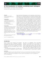
pathology. The PCR products of DNA samples from repro-
ductive tissues of all apparently healthy progeny moths
were examined for the presence of Hz-2V DNA via slot
blot hybridization (figure 1), and the size of the PCR
products of representative samples was determined by
agarose gel electrophoresis. The results of agarose gel elec-
trophoresis of PCR products from representative samples
Slot blot hybridization results of DNA extracted from repro-ductive tissues of corn earworm mothsFigure 1
Slot blot hybridization results of DNA extracted from repro-
ductive tissues of corn earworm moths. DNA was extracted,
amplified via PCR, transferred onto a nylon membrane, and
hybridized with a DIG-labeled viral DNA probe. Dark blots
are indicative of DNA from asymptomatic carrier moths
(As). Blots of DNA samples from insects from the healthy
colony (H) and from insects that were determined to be
apparently healthy (Ah) were blank or very light.
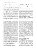
Agarose gel of PCR products from DNA extracted from the reproductive tissues of corn earworm mothsFigure 2
Agarose gel of PCR products from DNA extracted from the
reproductive tissues of corn earworm moths. The first lane
is from a sample containing purified Hz-2V DNA (V). Lanes 2
denotes a sample from normal, healthy moth (H) from our
insect colony. Lane 3 contains a DNA sample from an appar-
ently healthy (AH) progeny moth arising from and infected
female. Lanes 4–8 contain DNA samples extracted from
asymptomatic progeny corn earworm moths (AS).
Virology Journal 2004, 1:15 />Page 3 of 7
(page number not for citation purposes)
of agonadal, asymptomatic carriers and apparently
healthy moths are shown in figure 2.
Moths that had reproductive tissues that appeared to be
normal but tested positive for Hz-2V DNA by PCR analy-
ses were considered asymptomatic carriers of the virus.
For each virus dose tested the number of agonadal moths,
asymptomatic carriers, infected individuals (the sum of
agonadal and asymptomatic carriers), and uninfected
progeny moths hatching from eggs laid on each oviposi-
tion day was recorded.
The analysis of these results showed that the percentage of
total infected progeny (asymptomatic carriers and ago-
nadal moths) at all doses tested increased with each suc-
cessive oviposition day, and the level of infected progeny
increased as virus dose increased from 2 × 10
5
to 2 × 10
8
TCID
50
units (figure 3). For individuals hatching from
eggs on oviposition day one, the highest percentage of
infected progeny (approximately 80%) was produced by
females infected with the two highest virus dose (2 × 10
7
and 2 × 10
8
), whereas the lowest percentage (about 60%)
was produced by females infected with the lowest doses of
virus (2 × 10
5
and 2 × 10
6
TCID
50
).
Virus infected progeny moths arising from eggs laid on
each oviposition day by females infected with different
virus doses were divided into agonadal and asymptomatic
carriers and these results are presented in figure 4. No ago-
nadal insects arose from eggs laid on oviposition day one
by females infected at the lowest virus doses, whereas
approximately 15% of the progeny females from eggs laid
at this time by females infected at the two highest doses
were agonadal. At all of the viruses doses tested, between
70 and 90% of the individuals hatching from oviposition
day one eggs were asymptomatic carriers (figure 4).
For all F1 insects hatching from eggs laid on day two, (fig-
ure 4) the percentage of agonadal moths increased with
increasing virus dose and the percentage of asymptomatic
carriers at each dose declined (figure 4). The highest
number of agonadal moths (approximately 70%) hatch-
ing from eggs laid on day two came from females that
received the highest virus dose. At the two lowest doses
the level of agonadals hatching from day two eggs was
between 5 and 20%. At all doses almost 100% of the eggs
laid on days three and four gave rise to agonadal moths.
In order to better illustrate the relationship between the
two types of infections and to emphasize the effects of
virus dose upon the proportions of asymptomatic and
agonadal infections, percentages of asymptomatic carriers
and agonadal progeny for only the highest and lowest
dose are presented in figure 5. The trend in the two types
of infected progeny insects follows the same general
pattern for both virus doses relative to oviposition day.
That is, at both doses the percentage of agonadal progeny
increases with ovipostion day as the percentage of infected
Mean percentages of infected (agonadal and asymptomatic carriers) progeny arising from eggs laid by female moths infected with 2 × 10
5
, 2 × 10
6
, 2 × 10
7
, or 2 × 10
8
TCID
50
units of Hz-2VFigure 3
Mean percentages of infected (agonadal and asymptomatic
carriers) progeny arising from eggs laid by female moths
infected with 2 × 10
5
, 2 × 10
6
, 2 × 10
7
, or 2 × 10
8
TCID
50
units of Hz-2V.
Mean percentages of all (male and female) agonadal (AG) and asymptomatic carrier (AS) F1 moths arising from eggs laid by females infected with 2 × 10
5
, 2 × 10
6
, 2 × 10
7
, or 2 × 10
8
TCID
50
units of Hz-2VFigure 4
Mean percentages of all (male and female) agonadal (AG) and
asymptomatic carrier (AS) F1 moths arising from eggs laid by
females infected with 2 × 10
5
, 2 × 10
6
, 2 × 10
7
, or 2 × 10
8
TCID
50
units of Hz-2V.
Virology Journal 2004, 1:15 />Page 4 of 7
(page number not for citation purposes)
insects that are asymptomatic carriers of Hz-2V decreases.
At the highest dose, the proportion of agonadal insects
starts out higher (~ 10%) on the first oviposition day than
that of agonadal progeny of females infected at the lowest
dose (0%), and rises more quickly to ~ 70% of the prog-
eny from eggs laid on oviposition day two. This is com-
pared to only about 5% of the progeny arising from day
two eggs laid by females infected at the lowest dose. Inter-
estingly the reverse is the case for asymptomatic progeny
hatching from oviposition day one eggs. Whereas approx-
imately 90% of the infected oviposition day one individ-
uals from females infected at the lowest virus does are
asymptomatic only about 10% of the individuals from
females infected at the highest dose are asymptomatic.
Discussion
Injecting Hz-2V into female moths results in experimen-
tally infected insects that resemble asymptomatic females
and females that have become infected with the virus dur-
ing copulation, not unlike the females infected during
mass-matings by infected males in transmission experi-
ments conducted by Hamm et al. [2]. These infected
moths appear healthy, are fertile, and can transmit the
virus to progeny that result from mating. Some of the
progeny moths arising from these infected females do not
carry any detectable Hz-2V DNA sequences, others are
sterile with malformed reproductive tissues, and still
others are fertile, asymptomatic carriers of the virus. This
variety of infections suggests that the virus is not initially
present in all of the eggs laid by infected females, but is
transmitted transovarially to some of the eggs at some-
time time prior to oviposition. This idea is important in
that it suggests that the dose or titer of virus transmitted
from the parent female moth to the developing oocytes is
not constant, and that the virus dose that each oocyte
receives determines the outcome of the infection when
these progeny insects mature into adult moths. The pre-
cise molecular mechanism that determines which infected
individuals become agonadal and which will maintain
the virus in the population as asymptomatic carriers has
yet to be determined.
The results presented in figure 4, demonstrate that the per-
centage of agonadal progeny resulting from eggs laid on
oviposition day one by female moths infected with Hz-2V
increased as the dose of Hz-2V used to infect female
moths increased. Progeny arising from eggs laid on ovipo-
sition day two also exhibited this correlation between
virus dose and percent agonadal progeny. This indicates
that the titer of the virus present in the experimentally
infected female moths determines the amount of virus
that is transmitted to eggs, and is directly correlated to the
percentage of agonadal progeny arising from eggs laid by
the infected females. As the dose of Hz-2V used to infect a
female moth is increased, a corresponding increase is
observed in total agonadal progeny arising from all eggs
laid by the infected female.
The percentage of agonadal progeny also increases with
each successive oviposition day, approaching 100% ago-
nadal progeny by day three at all virus doses tested, and all
progeny moths arising from eggs laid on oviposition day
four in all groups were agonadal. Based on the correlation
between virus titer and percent agonadal progeny
observed in these experiments, the increase in agonadal
progeny per oviposition day is likely due to an increase in
the titer of virus transmitted to the eggs, suggesting that
the titer of virus increases in the parent female moths with
each successive oviposition day.
Studies of Hz-2V replication in vitro revealed a rapid
increase in virus titer by 24 hours post infection in Tn-368
and Ld-652Y cells [4,5]. Hz-2V replication in vivo in the
epithelial cells of agonadal female oviduct tissue has been
described previously by Rallis and Burand [8]. The level of
detectable virus in these tissues increased dramatically
between 8 days post pupation (dpp), measured from the
day the last larval exuviae was shed, and 10 dpp. It is likely
that the large increase in virus over a 24 hour cycle
observed in vitro also occurs in vivo, resulting in a signifi-
cant daily increase in the titer of Hz-2V in these experi-
mentally infected female moths. Although the precise site
of virus replication in these experimentally infected
females is not known, the increase in virus titer in these
Mean percentages of all (male and female) agonadal (AG) and asymptomatic carrier (AS) F1 moths arising from eggs laid by females infected at the highest and lowest doses of Hz-2VFigure 5
Mean percentages of all (male and female) agonadal (AG) and
asymptomatic carrier (AS) F1 moths arising from eggs laid by
females infected at the highest and lowest doses of Hz-2V.
Virology Journal 2004, 1:15 />Page 5 of 7
(page number not for citation purposes)
individuals almost certainly results in an increase in virus
being transmitted to the progeny with each successive
oviposition day and ultimately in the patterns of infection
reported here.
If, as we have proposed, low virus doses result in asymp-
tomatic carrier moths, and high virus doses produce ago-
nadal progeny, then asymptomatic carrier progeny would
likely arise from eggs produced on the earliest oviposition
days and decrease with each day, as the virus titer in the
egg-laying female moth increases. In fact, the percentage
of asymptomatic carrier progeny in these experiments
does decrease with each successive oviposition day to 0%
by day four. The percent asymptomatic carriers is highest
in progeny that receive the lowest virus dose, specifically
progeny from oviposition day one and progeny arising
from the parent female moths that were experimentally
infected with the lowest dose of virus. This is directly
opposite of what is observed for agonadal progeny, which
is at its highest level at the highest virus dose, specifically
on the later oviposition days (days three or greater) and in
progeny arising from parent female moths that were
infected with the highest virus dose. Interestingly, the low-
est percentage of asymptomatic carrier progeny arose
from eggs laid by the group of female moths that received
the highest virus dose of Hz-2V (figure. 3). These data sug-
gest that the virus dose transmitted by infected female
moths to their developing eggs determines whether the
progeny develop the agonadal condition or become
asymptomatic carriers of Hz-2V.
The results presented here clearly show that there is a
direct correlation between virus dose and the relative per-
centage of agonadal and asymptomatic progeny. That is,
increasing the virus dose causes an increase in the percent-
age of agonadal progeny, but a decrease in the percentage
of asymptomatic progeny. At the present time, it is
unknown how the development of an infected individual
into an agonadal adult or an asymptomatic carrier is
regulated. It is likely that a minimum titer of Hz-2V is
needed at a key point in larval development to produce a
viral factor(s) within the larval tissues at a threshold level
required to reprogram the development and differentia-
tion of the reproductive tissues into the agonadal struc-
tures. If this threshold is equaled or exceeded at this point
in development, the progeny will exhibit the agonadal
condition. However, if this threshold level is not attained,
then the reproductive tissues are not reprogrammed and
the infected insect becomes an apparently healthy, fertile,
asymptomatic carrier of Hz-2V.
Conclusions
The evolution of Hz-2V infection in H. zea has resulted in
the ability of the virus to produce two different types of
infections in the insects that enable the virus to replicate
to high titers in the reprogrammed reproductive tissues in
sterile agonadal moths, while maintaining itself in a
population in asymptomatic carrier moths. This replica-
tion strategy appears to be essential for the continued
existence of Hz-2V, since the development of the sterile,
agonadal condition in all infected moths would lead to
the extinction of the insect host, and the possibly the virus
as well. The production of asymptomatic carrier moths
ensures that some fertile, infected moths exist that can
mate and produce infected progeny, enabling an Hz-2V-
infected population to sustain itself, as in the case of the
Stoneville colony.
Methods
Source of insects and virus
Corn earworm larvae used to start a laboratory colony of
healthy H. zea were obtained from the USDA-ARS in
Stoneville, MS. Insects were reared on artificial diet and
maintained as outlined previously [9].
Hz-2V for infecting female moths was prepared as
described previously and purified via sucrose gradient
centrifugation [4].
Injection of adults
Newly emerged adult female moths were prepared and
injected with Hz-2V as outlined by Rallis and Burand [8].
The female moths were divided into four dose groups, and
9 or 10 insects were infected with Hz-2V at one of the fol-
lowing concentrations of 2 × 10
5
, 2 × 10
6
, 2 × 10
7
, and 2
× 10
8
TCID
50
units.
TCID50 assays
Tn368 cells were cultured as per Burand & Lu [4] and 100
ul of cell culture medium containing 8 × 10
4
Tn368 cells
were seeded into each well of a 96-well plate. Between 6
and 13 serial dilutions were made from each virus sample
assayed and 10 or 20 wells were plated with 10 ul for each
dilution. Plates were incubated at 27°C for 3 to 4 days and
examined for the appearance of cytopathic effect (CPE).
The numbers of wells with CPE were counted and the
TCID
50
calculated [9].
DNA extraction and purification of viral DNA
DNA was extracted from the reproductive tissues of adult
moths by first homogenizing dissected tissues in 200 ul of
TE buffer (10 mM Tris, pH 7.4, 1 mM EDTA, pH 8.0)
followed by a 2-minute incubation in a boiling water
bath. The homogenate was then chilled on ice, after which
Ribonuclease A (10 ug/ul) was added to each sample,
which was then incubated at room temperature for 15
min. The samples were then clarified by centrifugation at
15,600 × g for 2 min.
Virology Journal 2004, 1:15 />Page 6 of 7
(page number not for citation purposes)
Viral DNA used as template for PCR reactions was
extracted from purified virus using 1% SDS in TE contain-
ing 1 mg/ml Protease K as outlined by Burand and Lu [4].
PCR amplification of viral DNA sequences
Two sets of primers were used to amplify Hz-2V genomic
DNA to prepare a probe for use in slot blot analysis of
insect reproductive tissues. The first set (P4-1, 5'-GCAC-
GATTCGTAATGTTC-3'; and P4-2, 5'-GCACACCTAT-
CAATCACC-3') was designed to amplify a 434 bp
sequence of the Hz-2V genome [6]. PCR reactions using
P4-1 and P4-2 primers were brought to a final volume of
20 ul using the Bioneer AccuPower
®
PCR reagent premix
kit with 1 unit of Taq DNA polymerase. Each reaction was
carried out in10 mM Tris-HCl (pH 9.0), 1.5 mM MgCl
2
and 40 mM KCl, containing 250 uM of each of the four
dNTP's, with 100 pM of P4-1 forward and P4-2 reverse
primers, and 10 ng of purified viral DNA as template.
These primer set and reaction conditions were also used to
amplify viral DNA sequences in approximately 100 ng of
DNA from reproductive tissues of moths thought to be
asymptomatic carriers of Hz-2V.
The second set of primers (P4-3, 5'-GCTGTGCTGTA-
CAAGTGC-3'; and P4-4, 5'-CCCTTGACGATCCCTTTTG-
3') was designed to amplify a 350 bp region directly inte-
rior to that of the P4-1 and P4-2 amplified sequence.
These primers were used to generate a DIG-labeled probe
for Hz-2V to be used in slot blot hybridization assays. PCR
reactions for production of the DIG-labeled probe were
carried out in a final volume of 50 ul using the Boehringer
Manheim DIG High Prime DNA Labeling and Detection
Kit, with 1X concentrations of Taq Polymerase buffer (100
mM Tris-HCl pH 8.0, 500 mM KCL pH 8.3, and 25 mM
MgCL
2
), 100 pM of both P4-3 and P4-4 primers, a hexa-
nucleotide mixture containing DIG-labeled dUTP (2 mM
dATP, dCTP, dGTP, 1.3 mM dTTP, and 0.7 mM alkali
labile DIG-11 dUTP pH 7.0), and 100 pM of Hz-2V
genomic DNA. The DIG-labeled PCR product was purified
on a 0.8% agarose gel using the Qiagen gel electrophoresis
purification kit.
Both PCR reactions for amplification of the viral DNA in
tissue samples and for the production of the viral DNA
probe consisted of 30 cycles of a DNA denaturation step
at 95°C for 1 min., a primer annealing step for 1 min. at
55°C, and a 1 min. primer extension step at 72°C.
Detection of a viral DNA sequence by slot blotting
To prepare the DNA for slot blot analysis, 15 ul of the P4-
1 and P4-2 PCR amplified DNA from insect samples was
denatured by incubating with NaOH (0.4 M)/ EDTA (10
mM, pH 8.2) at 100°C for 10 min., then applied to a
Hybon-N+ membrane prewashed with 500 ul 5X SSC
buffer (0.6 M NaCl, 60 mM Na citrate pH 7.0) in a Mani-
fold II slot blotter (Schleicher & Schuell). After applying
the DNA, the membrane was baked at 88°C for 2 hrs
under vacuum and prehybridized for 6 hrs. at 42°C in
50% formamide prehybridization buffer (5X SSC, 0.1%
(w/v) N-laurylsarcosine, 1% (w/v) Na
2
-Dodecylsulfate,
2% Blocking reagent (Boehringer-Manheim), and 50%
Formamide). Slot blots were hybridized with 150 ng DIG-
labeled Hz-2V probe at 37°C for 12–14 hrs. Following
washing, chemiluminescent detection was carried out as
recommended by the DIG High Prime Labeling and
Detection Kit Manual for DNA Hybridization (Boehringer
Mannheim).
Analysis of PCR products by agarose gel electrophoresis
In order to confirm that the PCR products that hybridized
to the viral DNA probe contained an amplified DNA frag-
ment of the appropriate size (434 bp), representative sam-
ples were analyzed by electrophoresis on 0.8% agarose
gels with 0.5X TBE buffer at 100 volts for approximately 1
hr, then stained with EtBr to visualize DNA bands under
ultraviolet light.
Competing interests
The author(s) declare that they have no competing
interests.
Authors' contributions
CPR participated in the design of the study, carried out the
work with the insects, coordinated the project and assisted
in the molecular analysis and drafting of the manuscript.
JPB conceived the study, designed and supervised the
experimental work and drafted the manuscript. All
authors read and approved the final manuscript.
Acknowledgements
This project was funded in part by the Lotta Crabtree Graduate Fellowship
in Agriculture, by USDA NRICGP grant #2001-35302-10885, and by
Project # MAS00802 of the Massachusetts Agricultural Experiment Station.
References
1. Raina AK, Adams JR: Gonad-specific virus of corn earworm.
Nature 1995, 374:770.
2. Hamm JJ, Carpenter JE, Styer EL: Oviposition day effect on inci-
dence of agonadal progeny of Helicoverpa zea (Lepidotera:
Noctuidae) infected with a virus. Ann Entomol Soc Am 1996,
89:266-275.
3. Burand JP: Nudiviruses. In The Insect Viruses Edited by: Miller LK, Ball
LA Ball. New York: Plenum Publishing Corp; 1988:69-90.
4. Burand JP, Lu H: Replication of a gonad-specific virus in TN-368
cells in culture. J Invertebr Pathol 1997, 70:88-95.
5. Lu H, Burand JP: Replication of the gonad-specific virus Hz-2V
in Ld652Y cells mimics replication in vivo. J Invertebr Pathol 2001,
77:44-50.
6. Lupiani B, Raina AK, Huber C: Deveopment and use of a PCR
assay for detection of he reproductive virus in wild popula-
tions of Helicoverpa zea (Lepidopera: Noctuidae). J Invertebr
Pathol 1998, 73:107-112.
7. Raina AK, Adams JR, Lupiani B, Lynn DE, Kim W, Burand JP, Dough-
erty EM: Further characterization of the gonad-specific virus
of corn earworm, Helicoverpa zea. J Invertebr Pathol 2000,
76:6-12.
Publish with BioMed Central and every
scientist can read your work free of charge
"BioMed Central will be the most significant development for
disseminating the results of biomedical research in our lifetime."
Sir Paul Nurse, Cancer Research UK
Your research papers will be:
available free of charge to the entire biomedical community
peer reviewed and published immediately upon acceptance
cited in PubMed and archived on PubMed Central
yours — you keep the copyright
Submit your manuscript here:
/>BioMedcentral
Virology Journal 2004, 1:15 />Page 7 of 7
(page number not for citation purposes)
8. Rallis CP, Burand JP: Pathology and ultrastructure of the insect
virus, Hz-2V, infecting agonadal female corn earworms, Heli-
coverpa zea. J Invertebr Pathol 2002, 81:33-44.
9. Rallis CP, Burand JP: Pathology and ultrastructure of the insect
virus, Hz-2V, infecting agonadal male corn earworms, Heli-
coverpa zea. J Invertebr Pathol 2002, 80:81-89.
10. King LA, Possee RD: The Baculovirus Expression Vector System: A Labo-
ratory Guide London: Chapman and Hall; 1992.