báo cáo hóa học:" Molecular advances in the cell biology of SARS-CoV and current disease prevention strategies" pot
Bạn đang xem bản rút gọn của tài liệu. Xem và tải ngay bản đầy đủ của tài liệu tại đây (520.41 KB, 8 trang )
BioMed Central
Page 1 of 8
(page number not for citation purposes)
Virology Journal
Open Access
Review
Molecular advances in the cell biology of SARS-CoV and current
disease prevention strategies
Caren J Stark and CD Atreya*
Address: Division of Viral Products, Center for Biologics Evaluation and Research, US Food and Drug Administration, Bethesda, MD 20892 USA
Email: Caren J Stark - ; CD Atreya* -
* Corresponding author
AntiviralsCell biologyMolecular virologySARS-CoVVaccines
Abstract
In the aftermath of the SARS epidemic, there has been significant progress in understanding the
molecular and cell biology of SARS-CoV. Some of the milestones are the availability of viral genome
sequence, identification of the viral receptor, development of an infectious cDNA clone, and the
identification of viral antigens that elicit neutralizing antibodies. However, there is still a large gap
in our understanding of how SARS-CoV interacts with the host cell and the rapidly changing viral
genome adds another variable to this equation. Now the SARS-CoV story has entered a new phase,
a search for preventive strategies and a cure for the disease. This review highlights the progress
made in identifying molecular aspects of SARS-CoV biology that is relevant in developing disease
prevention strategies. Authors conclude that development of successful SARS-CoV vaccines and
antivirals depends on the progress we make in these areas in the immediate future.
Introduction
Following reports of the last case of the severe acute respi-
ratory syndrome (SARS) epidemic in July 2003, there has
been remarkable progress in several areas of research on
the molecular identification of the pathogen and its
pathogenesis, replication, genetics, and host immuno-
genicity, as well as elegant epidemiological studies. The
sequence of epidemiological events that unfolded early in
the outbreak gave researchers a glimpse into the first new
pathogen of the era of globalization. As the year 2002
drew to a close, multiple reports of an "infectious atypical
pneumonia" caught public health officials across the
globe by surprise and suggested that a new human patho-
gen had emerged in the Guangdong Province in China
[1]. By the end of February 2003, this outbreak of SARS
had infected almost 800 patients and caused 31 deaths in
the Province [2]. One month later, the disease had spread
throughout Asia and into Europe and North America. This
epidemic eventually affected more than 8000 people and
resulted in approximately 800 deaths worldwide, with
mortality rates reaching over 40% in certain populations
[3,4].
Electron microscope analysis quickly identified the puta-
tive SARS agent as having features associated with corona-
viruses. The SARS agent was later unambiguously
identified as a new coronavirus member and named
SARS-coronavirus (SARS-CoV) [5-7]. Coronaviruses are
enveloped, plus-stranded RNA viruses with the largest
RNA genomes known (on the order of 30 kb). Coronavi-
ruses have long been important in the world of veterinary
viral diseases. However, previously known human
Published: 15 April 2005
Virology Journal 2005, 2:35 doi:10.1186/1743-422X-2-35
Received: 13 April 2005
Accepted: 15 April 2005
This article is available from: />© 2005 Stark and Atreya; licensee BioMed Central Ltd.
This is an Open Access article distributed under the terms of the Creative Commons Attribution License ( />),
which permits unrestricted use, distribution, and reproduction in any medium, provided the original work is properly cited.
Virology Journal 2005, 2:35 />Page 2 of 8
(page number not for citation purposes)
coronaviruses such as HCoV-229E and HCoV-OC43 cause
only minor health problems such as the common cold
and gastrointestinal diseases. In contrast, the SARS-CoV
pathogen causes fever, pulmonary edema, and diffuse
alveolar damage in severely affected individuals (collec-
tively termed severe acute respiratory syndrome) [8].
SARS-CoV is also a unique coronavirus in that, to date, it
is the only member known to cause severe morbidity and
mortality in humans [8]. Demonstration that SARS-CoV
can cause serious public health problems has focused
attention on the need to understand the viral replicative
strategy and devise prophylactic measures.
The clinical symptoms of SARS are those of a lower respi-
ratory tract infection and are accompanied by damage to
the lungs [6,9,10]. Gastrointestinal involvement is also
common, with more than 20% of patients presenting with
watery diarrhea [11]. Fecal samples from SARS patients
taken up to 25 days after onset of disease contain viral
RNA, which suggests viral shedding through the bowels
[5]. Liver dysfunction has also been reported based on
observed necrosis in hepatocytes [9,12]. Post-mortem tis-
sue examination of SARS patients has found the virus
presence in lung, bowel, lymph node, liver, heart, kidney,
and skeletal muscle samples [13]. The primary mode of
SARS-CoV transmission is airborne via droplets [14,15].
However, there are also reports of the presence of replicat-
ing virus in blood cells (peripheral blood mononuclear
cells) and in the small and large intestine [11,16]. Alterna-
tive modes of transmission, such as blood-borne or fecal-
oral are therefore possible.
The virus has been isolated from wild animals (Hima-
layan palm civets and raccoon dogs) found in the animal
markets of Guangdong, China [17]. The actual natural res-
ervoir for SARS-CoV is still unknown. Once transmitted to
humans, SARS-CoV appears to evolve to facilitate to
human-human transmission. Sequence analysis of differ-
ent SARS-CoV isolates from early in the epidemic show
deletion events occurring in open reading frame 8 (Orf 8)
[18]. Identical deletions in Orf 8 have also been seen in
animal coronaviruses supporting the idea that SARS-CoV
was introduced to humans via an animal intermediate. In
addition to deletion events occurring early and late in the
epidemic, a slowing of missense mutations is seen over
time, with the most extensive changes occurring in the S
protein during the early stages of the outbreak [18]. This
suggests the virus has undergone some level of adaptation
but has ultimately stabilized at a time in the epidemic
where SARS-CoV has become more virulent. Deciphering
the evolutionary passage of this virus will undoubtedly
provide valuable information on preventing future
outbreaks.
In the wake of the SARS epidemic, a number of excellent
review articles on the clinical and molecular aspects of
SARS epidemiology have been published. These reviews
have focused primarily on rapid advances made in the
identification and characterization of SARS-CoV genomes
as well as describing the etiology of the virus and clinical
features of the disease [19-21]. Now the SARS-CoV story
has entered a new phase, a search for preventative strate-
gies and a cure. In this review, we highlight the progress
made in revealing the molecular aspects of SARS-CoV
biology and how such information may lead to strategies
for disease prevention.
Brief overview of the SARS-CoV genome
Coronaviruses are subdivided into three groups based on
genetic and serological markers [22]. Groups I, and II
infect mammals while group III is specific for avian spe-
cies. Group I members are the porcine transmissible gas-
troenteritis virus (TGEV) and epidemic diarrhea virus
(PEDV), feline and canine coronavirus (FCoV and CCoV),
and human coronavirus 229E (HCoV-229E). Group II
includes porcine hemagglutinating encephalomyelitis
virus (HEV), murine hepatitis virus (MHV), bovine,
equine, and rat coronavirus (BCoV, ECoV, and RtCoV),
and human coronavirus OC43 (HCoV-OC43). Group III
includes the turkey coronavirus (TCoV), pheasant corona-
virus and avian infectious bronchitis virus (IBV).
Although most closely related to Group II coronaviruses,
SARS-CoV, with some of its unique genetic features, repre-
sents a distinct phylogenetic group [22-24].
To date, approximately 61 SARS-CoV genomic sequences
have been analyzed representing different phases of the
epidemic (early, middle, and late) and two isolates
obtained from palm civets [18]. The SARS-CoV genomic
RNA is approximately 30 kb and is organized into 13 to
15 open reading frames (ORFs) [25-27]. The SARS CoV
structural gene arrangement follows the same pattern as
most coronavirus genomes: 5'- Replicase (ORF 1a)-Pro-
tease (ORF 1b)-Spike (S)-Envelope (E)-membrane (M)-
Nucleocapsid (N)-3' [27]. However, in contrast to other
coronaviruses, two ORFs of unknown function are located
between the S and E ORFs and 3–5 ORFs are located
between M and N. In addition, despite the evolutionary
overlap between SARS-CoV and Group II coronavirus
genome sequences, the SARS genome lacks a gene for
hemagglutinin-esterase (HE) protein, which is common
to a majority of Group II coronaviruses [25]. For an excel-
lent pictorial representation of SARS-CoV genome with
functions (or lack of) assigned to each ORF, please refer to
the recent review by Tan et al [21]. A significant milestone
in SARS-CoV molecular biology was the construction of a
SARS-CoV full-length cDNA-containing plasmid from
which infectious viral RNA can be produced [28]. This
development facilitates the study of SARS-CoV gene
Virology Journal 2005, 2:35 />Page 3 of 8
(page number not for citation purposes)
functions and should promote the elucidation of function
for ORFs whose function is still unknown [29]. Although
it has been the perception that these ORFs are not essen-
tial for viral replication, they may play a role in the mani-
festation or severity of disease.
Progress in SARS-CoV genome-based
evolutionary biology
RNA viruses utilize a variety of mechanisms to exchange
their genetic repertoire. The viral RNA dependent RNA
polymerases (RdRP) have a built in error rate that allows
diversification of the genomic sequence as replication
proceeds. Estimates put the error rate of an RdRp at 10
-3
to
10
-5
per nucleotide [30]. Coronaviruses also undergo high
rates of RNA recombination, providing an additional
mechanism by which the viruses can rapidly amplify
genomic diversity. The SARS-CoV polymerase gene has a
recombination breakpoint, suggesting multiple genetic
origins for this molecule. [31]. These evolutionary mech-
anisms may have facilitated the adaptation of the animal-
borne SARS-CoV ancestor to the human host, suggesting
that such events in the future could lead to a virus with
increased pathogenicity for humans or one capable of
infecting multiple species. Recent evidence indicates that
the human-adapted SARS virus has crossed into another
species. Sequence and epidemiological analyses revealed
that a SARS-CoV isolated from a pig was derived from a
human strain. Complete nucleotide sequencing of the pig
virus isolate (designated TJF) and an S gene-based phylo-
genetic tree analysis revealed a closer relationship with
human SARS-CoV isolates than with animal coronavi-
ruses [32].
Progress in cell biology of SARS-CoV: Signaling
pathways
Successful viral replication depends upon the ability of
the virus to subvert cellular processes to their advantage
and counteract cellular defense mechanisms. Such virus-
cell interactions represent potential targets for the devel-
opment of virus-specific antiviral drugs, therapeutics, and
prophylactic vaccines. Different viruses, based on their
target cell types and entry pathways, differ in their cellular
exploitation mechanisms. The mechanism of SARS virus
pathogenesis in vivo may reflect both the effect of viral rep-
lication in target cells and host immune responses. The
molecular basis for SARS-CoV replication, the signaling
pathways affected, and the inflammatory responses pro-
voked by viral infection are not yet clearly understood.
Progress in these areas should lead to more effective pre-
ventive strategies to counter SARS-CoV infections.
It has been shown that the SARS-CoV N protein selectively
activates the Activator Protein-1 (AP-1) signal transduc-
tion pathway, which regulates a wide variety of cellular
processes including cell proliferation, differentiation, and
apoptosis [33]. Such viral induced modifications of the
AP-1 pathway may play a significant role in the viral rep-
licative strategy. Recently, another group demonstrated
that the S protein alone induces AP-1 activation and that
the region from 324–688 amino acids within the S pro-
tein is essential for AP-1 activation-dependent IL-8 induc-
tion [34]. Another SARS-CoV protein, the U122 ORF of
unknown function (also known as X4), was shown to be
produced in virus infected Vero E6 cells and expression of
this protein alone was shown to induce apoptosis in cell
culture [35,36]. This raises the question of how apoptosis
of SARS-CoV infected cells is balanced in order for the
virus to survive and propagate (Figure 1). This has been
addressed to some extent in recent studies which indicate
that SARS-CoV infection of Vero E6 cells induces both
pro-apoptotic [activation of p38 mitogen-activated pro-
tein kinase (MAPK)] and anti-apoptotic [activation of the
protein kinase B (PKB, also known as Akt)] signaling path-
ways, although Akt induction appears to be insufficient to
prevent the virus-induced apoptosis [37,38]. Exactly how
SARS-CoV manipulates these cellular signaling pathways
to facilitate viral replication remains to be determined.
As mentioned above, IL-8 induction was shown to be
dependent upon AP-1 activation by SARS-CoV S protein
and in this process NF-κB was not involved [34]. This may
partially explain the clinical observation of dramatic
cytokine storm (high serum levels of IL-6 and IL-8) and
inflammation responses observed in SARS patients in the
acute stage associated with lung lesions; it has been also
suggested that the elevations of IL-6 and IL-8 due to SARS-
CoV infection of the respiratory tract can induce the
hyper-innate inflammatory response [39]. It is established
that cellular MAPKs regulate AP-1 activation-dependent
IL-8 induction in viral infections [40-42]. In SARS-CoV
infection, the IL-8 induction signaling pathway is perhaps
related to angiotensin-converting enzyme 2 (ACE2), as
anti-ACE2 antibodies inhibit IL-8 induction/release [34].
ACE2 is the cellular receptor for the SARS-CoV and the
receptor-binding sites on the virion are located in the 12–
672 amino acid region of the S protein [43].
Current advances towards SARS-CoV
prevention strategies
During the SARS outbreak that occurred in 2002–2003,
the spread of the disease was primarily controlled by strict
quarantine protocols and patient-isolation measures as
well as by broad-spectrum antibiotics and antiviral regi-
mens with or without administration of corticosteroids
[44,45]. Since then, the wealth of information that has
emerged on SARS-CoV molecular and cellular biology, as
updated in the preceding sections of this review, now
offers potential avenues for developing more efficient
anti-viral as well as vaccine strategies.
Virology Journal 2005, 2:35 />Page 4 of 8
(page number not for citation purposes)
a. Antiviral agents
Coronavirus genome structure and major gene-product
functions have been known for years, but since they cause
mild disease, selection of the virus-specific antiviral drugs
was not a priority in the past. The SARS-CoV epidemic
changed this selective view. Tan et al, 2004, tabulated a
screen of available antiviral agents against SARS virus in
detail in their recent review [46]. The obvious molecular
targets for SARS-CoV antiviral agents are the viral
polymerase/replicase, protease, receptor, the viral mRNA
cap-1 methyl transferase and NTPase/helicase [47-54]. In
addition, a 32-nucleotide long, highly conserved RNA
structure in the 3' untranslated region of coronaviruses
and astroviruses was identified [55]. This structure resem-
bles the 530 loop of 16s rRNA involved in translation ini-
tiation suggesting a possible role for this element in
sequestering host translation machinery. The tertiary
interactions of this structure create a tunnel lined with
negative charge where Mg
2+
can bind. This unique struc-
ture presents an attractive target for tunnel binding antivi-
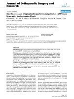
The balance of cell survival and cell death in response to SARS-CoV infectionFigure 1
The balance of cell survival and cell death in response to SARS-CoV infection. SARS-CoV is shown approaching a cell with
ACE2 receptors (blue "Y"s) on the surface. The virus enters the cell, uncoats, and the viral RNA is replicated and translated.
The SARS-CoV U122 protein induces apoptosis in cells. SARS-CoV S and N proteins each can activate the cellular AP-1 pro-
tein, which regulates apoptosis, as well as other cellular processes. AP-1 also activates IL-8, a cellular cytokine. SARS-CoV
infection induces both MAPK (pro-apoptotic) and Akt (anti-apoptotic) pathways. How this balance between cell survival and
apoptosis is maintained is yet unknown. Cellular proteins are labeled in blue, viral proteins in black.
Virology Journal 2005, 2:35 />Page 5 of 8
(page number not for citation purposes)
ral drugs [55]. Finally, since the functional details of most
coronavirus replicase gene products are not known,
random screening of potential antiviral compound librar-
ies will be a key area of drug discovery for SARS virus in
the near future [47].
b. Vaccine development
Vaccines are the best and least expensive prophylactic
measures against pathogens that cause epidemics in
humans. The fact that high titers of virus neutralizing anti-
body to SARS-CoV are found in sera of patients recovering
from infection and that those infected with the virus show
improvement after passive antibody administration sug-
gests a SARS-CoV vaccine is possible and points toward
antibody based treatments for the disease [47,56-58].
However, in developing SARS CoV vaccines, there are les-
sons to be learned from the world of veterinary CoV vac-
cines. In a review by Saif, it was pointed out that
coronaviruses in general target mucosal surfaces and
therefore eliciting local (mucosal) immunity is a major
consideration in the development of SARS-CoV vaccines;
this largely depends on the type of vaccine, delivery sys-
tems, and immuno-modulatory adjuvants used [59]. Fur-
ther, immunity against animal CoV is usually short term,
necessitating periodic boosting, which in the end may not
be sufficient to prevent re-infection.
Despite these potential pitfalls in the development of a
human vaccine, efforts to develop a vaccine to prevent
another SARS outbreak are underway. Several laboratories
around the globe are working at an unprecedented pace to
develop a SARS vaccine utilizing essentially two different
types of SARS-CoV-derived immunogens, 1) inactivated
whole virus, and 2) SARS-CoV encoded N and S proteins
using recombinant DNA methods. The possibility of pro-
ducing an engineered live, attenuated SARS-CoV has also
been considered.
1. Inactivated whole virus
Takasuka et al (2004) have reported that subcutaneous
administration of UV-inactivated purified SARS-CoV vir-
ion elicits a high level of humoral immunity, resulting in
long-term antibody secretion and memory B cells [60].
The antibodies elicited in mice recognized both the spike
(S) and nucleocapsid (N) proteins of the virus. The inacti-
vated virus also induced regional lymph node T-cell pro-
liferation and significant levels of cytokine production
upon restimulation with inactivated virus in vitro [60].
These studies suggest that whole-killed virion may have
the potential as a candidate antigen for SARS vaccine to
elicit both humoral and cellular immunity. When SARS-
CoV inactivated by beta-propiolactone was used as anti-
gen in mice and rabbits, the animals elicited antibodies
against the receptor-binding domain (RBD) present in the
S1 region of SARS-CoV. These antibodies effectively inhib-
ited the S-protein mediated SARS-pseudovirus entry up to
50%, suggesting the potential of the inactivated SARS-
CoV as antigen for vaccine development [61]. Depletion
of RBD-specific antibodies from patient or rabbit immune
sera by immunoadsorption, significantly reduced the
virus neutralizing ability of the sera, suggesting that the
RBD epitope in the S protein is a critical determinant in
developing vaccine strategies [62].
2.1. Cloned N protein
The N protein of SARS-CoV appears to be more conserved
than S and M proteins and it has been suggested that this
protein may play a role in cell-mediated immunity in
SARS-CoV infections and also is an important viral anti-
gen for the early diagnosis. Vaccination of C57BL/6 mice
with a SARS-CoV N protein expressed by an E1/partially
E3-deleted, replication-defective human adenovirus 5 vec-
tor was shown to produce potent SARS-CoV-specific
humoral and T cell-mediated immune responses, suggest-
ing the potential of this construct to be used as SARS-CoV
vaccine [63]. Along the same line, intra-muscular immu-
nization of BALB/c mice with a plasmid DNA construct
encoding the full-length N protein was shown to elicit
serum anti-N antibodies and spenocyte proliferative
responses against the N protein [64]. The immunized
mice also produced strong delayed-type hypersensitivity
(DTH) and CD8 (+) CTL responses to the N protein, sug-
gesting that the N protein is not only an important B cell
immunogen, but also can elicit broad-based cellular
immune responses [64]. In another novel strategy, the N
protein was expressed in the cytoplasm of Lactococcus lactis
bacterium and the N-expressing bacteria were adminis-
tered to mice by intranasal or oral route [65]. In this case,
significant levels of N-specific IgG in the mice sera were
detected, suggesting that the engineered bacteria may
serve as a mucosal vaccine against SARS-CoV [65].
2.2. Cloned viral S spike protein or, S-containing pseudovirions
Although immunization with inactivated viral vaccine
provides significant protection in animals against chal-
lenge with certain corresponding pathogenic CoVs, in the
case of SARS-CoV there remains the threat of introducing
live virus into the environment from partially inactivated
vaccine, as there are no validated and effective inactiva-
tion measures developed yet. To circumvent this obstacle,
Chen et al have introduced the S protein into the deletion
III region of the live, attenuated modified vaccinia virus
Ankara (MVA) vector [66]. This recombinant virus elicits
potent neutralizing antibodies in mice, rabbits, and mon-
keys and the major epitope is mapped to the virus recep-
tor-binding region [66]. In another approach, it has been
demonstrated that co-expression of SARS-CoV S, M and N
expression plasmids in human 293T cells result in the for-
mation of SARS-CoV pseudoparticles (virus-like particles
or VLPs) [67]. These findings help us understand the viral
Virology Journal 2005, 2:35 />Page 6 of 8
(page number not for citation purposes)
morphogenesis as well as offer a safer alternative to using
live, replicating SARS virus in the development of
vaccines.
3. Attenuated live virus
The third possibility is a genetically engineered version of
live SARS-CoV for traits such as attenuated phenotype,
increased immunogenicity, and safe handling (out of
BL3+ facility). A full-length SARS-CoV cDNA-containing
plasmid has been developed from which synthetic infec-
tious viral RNA can be produced [28]. This system allows
for the functional analysis of each gene in the context of
infection and can be used for making attenuated strains
for vaccine development.
Conclusions: Limitations to current SARS
vaccine strategies
SARS-CoV clearly has pandemic potential. Although
progress in SARS-CoV molecular and cell biology research
has been remarkable, there remain clear limitations
regarding vaccine development due to a lack of complete
understanding in the areas of animal models of the dis-
ease as well as host immune responses to the evolving
molecular diversity of this newly emerged human virus.
Caution is warranted when utilizing experimental data
originating from one SARS-CoV strain infection in one
animal species or cell line in the development of a human
vaccine. The rapid development of an effective SARS-CoV
vaccine depends upon continuing basic research.
A study on the evolving S protein molecular diversity in
SARS-CoV isolates and its unexpected profound immuno-
functional effects illustrates this point [68]. The S protein
exhibited minor genetic diversity among 8 strains trans-
mitted during human outbreaks in early 2003. Synthetic
versions of these S variants with human preferred codons
were tested for 1) their ability to bind the receptor (hACE-
2), and 2) their sensitivity to antibody neutralization with
viral pseudotypes. In these sets of experiments, substantial
functional differences were found in S derived from a
Guangdong province case -isolate and two palm civets
isolates. Antibodies that neutralized most human isolates-
derived S proteins unexpectedly enhanced entry mediated
by the civet virus-derived S proteins [68]. This novel
observation emphasizes the need to understand the
molecular potential of the SARS-CoV genome in develop-
ing vaccines to prevent human disease. As mentioned pre-
viously, studies also point to the fact that variability in the
S protein from early to late disease outbreak stages has
been detected [18]. There is a large gap in our understand-
ing of how SARS-CoV interacts with the host cell and the
rapidly changing genome of SARS-CoV indicates the
potential variability of such interactions [25]. Develop-
ment of successful vaccines against SARS virus therefore
depends on the progress we make in these areas in the
immediate future.
Competing Interests
The author(s) declare that they have no competing
interests.
Authors' Contributions
Authors contributed equally to the intellectual content of
this review article.
Disclaimer
The views presented in this article do not necessarily
reflect those of the Food and Drug Administration or
United States government.
Acknowledgements
We thank Stephen Feinstone and Ron Lundquist of CBER, FDA for their
critiques and the National Vaccines Program Office (NVPO) for a grant to
CDA. CJS is supported by a postdoctoral fellowship administered by the
Oak Ridge Institute for Science and Education (ORISE).
References
1. Zhong NS, Zeng GQ: Our strategies for fighting severe acute
respiratory syndrome (SARS). Am J Respir Crit Care Med 2003,
168:7-9.
2. Organization WH: Cumulative Number of Reported Cases
(SARS). [ />].
3. Nie QH, Luo XD, Zhang JZ, Su Q: Current status of severe acute
respiratory syndrome in China. World J Gastroenterol 2003,
9:1635-1645.
4. Donnelly CA, Ghani AC, Leung GM, Hedley AJ, Fraser C, Riley S,
Abu-Raddad LJ, Ho LM, Thach TQ, Chau P, Chan KP, Lam TH, Tse
LY, Tsang T, Liu SH, Kong JH, Lau EM, Ferguson NM, Anderson RM:
Epidemiological determinants of spread of causal agent of
severe acute respiratory syndrome in Hong Kong. Lancet
2003, 361:1761-1766.
5. Drosten C, Gunther S, Preiser W, van der Werf S, Brodt HR, Becker
S, Rabenau H, Panning M, Kolesnikova L, Fouchier RA, Berger A, Bur-
guiere AM, Cinatl J, Eickmann M, Escriou N, Grywna K, Kramme S,
Manuguerra JC, Muller S, Rickerts V, Sturmer M, Vieth S, Klenk HD,
Osterhaus AD, Schmitz H, Doerr HW: Identification of a novel
coronavirus in patients with severe acute respiratory
syndrome. N Engl J Med 2003, 348:1967-1976.
6. Peiris JS, Lai ST, Poon LL, Guan Y, Yam LY, Lim W, Nicholls J, Yee
WK, Yan WW, Cheung MT, Cheng VC, Chan KH, Tsang DN, Yung
RW, Ng TK, Yuen KY: Coronavirus as a possible cause of
severe acute respiratory syndrome. Lancet 2003,
361:1319-1325.
7. Ksiazek TG, Erdman D, Goldsmith CS, Zaki SR, Peret T, Emery S,
Tong S, Urbani C, Comer JA, Lim W, Rollin PE, Dowell SF, Ling AE,
Humphrey CD, Shieh WJ, Guarner J, Paddock CD, Rota P, Fields B,
DeRisi J, Yang JY, Cox N, Hughes JM, LeDuc JW, Bellini WJ, Anderson
LJ: A novel coronavirus associated with severe acute respira-
tory syndrome. N Engl J Med 2003, 348:1953-1966.
8. Poutanen SM, Low DE, Henry B, Finkelstein S, Rose D, Green K, Tell-
ier R, Draker R, Adachi D, Ayers M, Chan AK, Skowronski DM, Salit
I, Simor AE, Slutsky AS, Doyle PW, Krajden M, Petric M, Brunham RC,
McGeer AJ: Identification of severe acute respiratory syn-
drome in Canada. N Engl J Med 2003, 348:1995-2005.
9. Ding Y, Wang H, Shen H, Li Z, Geng J, Han H, Cai J, Li X, Kang W,
Weng D, Lu Y, Wu D, He L, Yao K: The clinical pathology of
severe acute respiratory syndrome (SARS): a report from
China. J Pathol 2003, 200:282-289.
10. Tsang KW, Ho PL, Ooi GC, Yee WK, Wang T, Chan-Yeung M, Lam
WK, Seto WH, Yam LY, Cheung TM, Wong PC, Lam B, Ip MS, Chan
J, Yuen KY, Lai KN: A cluster of cases of severe acute respira-
tory syndrome in Hong Kong. N Engl J Med 2003, 348:1977-1985.
Virology Journal 2005, 2:35 />Page 7 of 8
(page number not for citation purposes)
11. Leung WK, To KF, Chan PK, Chan HL, Wu AK, Lee N, Yuen KY, Sung
JJ: Enteric involvement of severe acute respiratory syn-
drome-associated coronavirus infection. Gastroenterology 2003,
125:1011-1017.
12. Chan-Yeung M, Yu WC: Outbreak of severe acute respiratory
syndrome in Hong Kong Special Administrative Region: case
report. Bmj 2003, 326:850-852.
13. Farcas GA, Poutanen SM, Mazzulli T, Willey BM, Butany J, Asa SL,
Faure P, Akhavan P, Low DE, Kain KC: Fatal severe acute respi-
ratory syndrome is associated with multiorgan involvement
by coronavirus. J Infect Dis 2005, 191:193-197.
14. Seto WH, Tsang D, Yung RW, Ching TY, Ng TK, Ho M, Ho LM, Peiris
JS: Effectiveness of precautions against droplets and contact
in prevention of nosocomial transmission of severe acute
respiratory syndrome (SARS). Lancet 2003, 361:1519-1520.
15. Wong TW, Lee CK, Tam W, Lau JT, Yu TS, Lui SF, Chan PK, Li Y,
Bresee JS, Sung JJ, Parashar UD: Cluster of SARS among medical
students exposed to single patient, Hong Kong. Emerg Infect
Dis 2004, 10:269-276.
16. Li L, Wo J, Shao J, Zhu H, Wu N, Li M, Yao H, Hu M, Dennin RH:
SARS-coronavirus replicates in mononuclear cells of periph-
eral blood (PBMCs) from SARS patients. J Clin Virol 2003,
28:239-244.
17. Guan Y, Zheng BJ, He YQ, Liu XL, Zhuang ZX, Cheung CL, Luo SW,
Li PH, Zhang LJ, Guan YJ, Butt KM, Wong KL, Chan KW, Lim W,
Shortridge KF, Yuen KY, Peiris JS, Poon LL: Isolation and charac-
terization of viruses related to the SARS coronavirus from
animals in southern China. Science 2003, 302:276-278.
18. Chinese SMEC: Molecular evolution of the SARS coronavirus
during the course of the SARS epidemic in China. Science
2004, 303:1666-1669.
19. Peiris JS, Yuen KY, Osterhaus AD, Stohr K: The severe acute res-
piratory syndrome. N Engl J Med 2003, 349:2431-2441.
20. Ziebuhr J: Molecular biology of severe acute respiratory syn-
drome coronavirus. Curr Opin Microbiol 2004, 7:412-419.
21. Tan YJ, Lim SG, Hong W: Characterization of viral proteins
encoded by the SARS-coronavirus genome. Antiviral Res 2005,
65:69-78.
22. Gonzalez JM, Gomez-Puertas P, Cavanagh D, Gorbalenya AE,
Enjuanes L: A comparative sequence analysis to revise the cur-
rent taxonomy of the family Coronaviridae. Arch Virol 2003,
148:2207-2235.
23. Gorbalenya AE, Snijder EJ, Spaan WJ: Severe acute respiratory
syndrome coronavirus phylogeny: toward consensus. J Virol
2004, 78:7863-7866.
24. Snijder EJ, Bredenbeek PJ, Dobbe JC, Thiel V, Ziebuhr J, Poon LL,
Guan Y, Rozanov M, Spaan WJ, Gorbalenya AE: Unique and con-
served features of genome and proteome of SARS-coronavi-
rus, an early split-off from the coronavirus group 2 lineage. J
Mol Biol 2003, 331:991-1004.
25. Groneberg DA, Hilgenfeld R, Zabel P: Molecular mechanisms of
severe acute respiratory syndrome (SARS). Respir Res 2005,
6:8.
26. Rota PA, Oberste MS, Monroe SS, Nix WA, Campagnoli R, Icenogle
JP, Penaranda S, Bankamp B, Maher K, Chen MH, Tong S, Tamin A,
Lowe L, Frace M, DeRisi JL, Chen Q, Wang D, Erdman DD, Peret TC,
Burns C, Ksiazek TG, Rollin PE, Sanchez A, Liffick S, Holloway B,
Limor J, McCaustland K, Olsen-Rasmussen M, Fouchier R, Gunther S,
Osterhaus AD, Drosten C, Pallansch MA, Anderson LJ, Bellini WJ:
Characterization of a novel coronavirus associated with
severe acute respiratory syndrome. Science 2003,
300:1394-1399.
27. Thiel V, Ivanov KA, Putics A, Hertzig T, Schelle B, Bayer S, Weissbrich
B, Snijder EJ, Rabenau H, Doerr HW, Gorbalenya AE, Ziebuhr J:
Mechanisms and enzymes involved in SARS coronavirus
genome expression. J Gen Virol 2003, 84:2305-2315.
28. Yount B, Curtis KM, Fritz EA, Hensley LE, Jahrling PB, Prentice E,
Denison MR, Geisbert TW, Baric RS: Reverse genetics with a full-
length infectious cDNA of severe acute respiratory syn-
drome coronavirus. Proc Natl Acad Sci U S A 2003,
100:12995-13000.
29. Baric RS, Sims AC: Development of mouse hepatitis virus and
SARS-CoV infectious cDNA constructs. Curr Top Microbiol
Immunol 2005, 287:229-252.
30. Domingo E, Holland JJ: RNA virus mutations and fitness for
survival. Annu Rev Microbiol 1997, 51:151-178.
31. Rest JS, Mindell DP: SARS associated coronavirus has a recom-
binant polymerase and coronaviruses have a history of host-
shifting. Infect Genet Evol 2003, 3:219-225.
32. Chen W: SARS-associated Coronavirus Transmitted from
Human to Pig. Emerg Infect Dis 2005, 11:446-448.
33. He R, Leeson A, Andonov A, Li Y, Bastien N, Cao J, Osiowy C, Dobie
F, Cutts T, Ballantine M, Li X: Activation of AP-1 signal transduc-
tion pathway by SARS coronavirus nucleocapsid protein. Bio-
chem Biophys Res Commun 2003, 311:870-876.
34. Chang YJ, Liu CY, Chiang BL, Chao YC, Chen CC: Induction of IL-
8 release in lung cells via activator protein-1 by recombinant
baculovirus displaying severe acute respiratory syndrome-
coronavirus spike proteins: identification of two functional
regions. J Immunol 2004, 173:7602-7614.
35. Fielding BC, Tan YJ, Shuo S, Tan TH, Ooi EE, Lim SG, Hong W, Goh
PY: Characterization of a unique group-specific protein
(U122) of the severe acute respiratory syndrome
coronavirus. J Virol 2004, 78:7311-7318.
36. Tan YJ, Fielding BC, Goh PY, Shen S, Tan TH, Lim SG, Hong W:
Overexpression of 7a, a protein specifically encoded by the
severe acute respiratory syndrome coronavirus, induces
apoptosis via a caspase-dependent pathway. J Virol 2004,
78:14043-14047.
37. Mizutani T, Fukushi S, Saijo M, Kurane I, Morikawa S: Phosphoryla-
tion of p38 MAPK and its downstream targets in SARS coro-
navirus-infected cells. Biochem Biophys Res Commun 2004,
319:1228-1234.
38. Mizutani T, Fukushi S, Saijo M, Kurane I, Morikawa S: Importance of
Akt signaling pathway for apoptosis in SARS-CoV-infected
Vero E6 cells. Virology 2004, 327:169-174.
39. Hsueh PR, Chen PJ, Hsiao CH, Yeh SH, Cheng WC, Wang JL, Chiang
BL, Chang SC, Chang FY, Wong WW, Kao CL, Yang PC: Patient
data, early SARS epidemic, Taiwan. Emerg Infect Dis 2004,
10:489-493.
40. Chen W, Monick MM, Carter AB, Hunninghake GW: Activation of
ERK2 by respiratory syncytial virus in A549 cells is linked to
the production of interleukin 8. Exp Lung Res 2000, 26:13-26.
41. Alcorn MJ, Booth JL, Coggeshall KM, Metcalf JP: Adenovirus type 7
induces interleukin-8 production via activation of extracellu-
lar regulated kinase 1/2. J Virol 2001, 75:6450-6459.
42. Griego SD, Weston CB, Adams JL, Tal-Singer R, Dillon SB: Role of
p38 mitogen-activated protein kinase in rhinovirus-induced
cytokine production by bronchial epithelial cells. J Immunol
2000, 165:5211-5220.
43. Li W, Moore MJ, Vasilieva N, Sui J, Wong SK, Berne MA, Somasunda-
ran M, Sullivan JL, Luzuriaga K, Greenough TC, Choe H, Farzan M:
Angiotensin-converting enzyme 2 is a functional receptor for
the SARS coronavirus. Nature 2003, 426:450-454.
44. So LK, Lau AC, Yam LY, Cheung TM, Poon E, Yung RW, Yuen KY:
Development of a standard treatment protocol for severe
acute respiratory syndrome. Lancet 2003, 361:1615-1617.
45. Tsang K, Zhong NS: SARS: pharmacotherapy. Respirology 2003, 8
Suppl:S25-30.
46. Tan EL, Ooi EE, Lin CY, Tan HC, Ling AE, Lim B, Stanton LW: Inhi-
bition of SARS coronavirus infection in vitro with clinically
approved antiviral drugs. Emerg Infect Dis 2004, 10:581-586.
47. Oxford JS, Balasingam S, Chan C, Catchpole A, Lambkin R: New
antiviral drugs, vaccines and classic public health interven-
tions against SARS coronavirus. Antivir Chem Chemother 2005,
16:13-21.
48. Tanner JA, Watt RM, Chai YB, Lu LY, Lin MC, Peiris JS, Poon LL, Kung
HF, Huang JD: The severe acute respiratory syndrome (SARS)
coronavirus NTPase/helicase belongs to a distinct class of 5'
to 3' viral helicases. J Biol Chem 2003, 278:39578-39582.
49. von Grotthuss M, Wyrwicz LS, Rychlewski L: mRNA cap-1 meth-
yltransferase in the SARS genome. Cell 2003, 113:701-702.
50. Yang H, Yang M, Ding Y, Liu Y, Lou Z, Zhou Z, Sun L, Mo L, Ye S, Pang
H, Gao GF, Anand K, Bartlam M, Hilgenfeld R, Rao Z: The crystal
structures of severe acute respiratory syndrome virus main
protease and its complex with an inhibitor. Proc Natl Acad Sci U
S A 2003, 100:13190-13195.
51. Xu X, Liu Y, Weiss S, Arnold E, Sarafianos SG, Ding J: Molecular
model of SARS coronavirus polymerase: implications for
biochemical functions and drug design. Nucleic Acids Res 2003,
31:7117-7130.
Publish with BioMed Central and every
scientist can read your work free of charge
"BioMed Central will be the most significant development for
disseminating the results of biomedical research in our lifetime."
Sir Paul Nurse, Cancer Research UK
Your research papers will be:
available free of charge to the entire biomedical community
peer reviewed and published immediately upon acceptance
cited in PubMed and archived on PubMed Central
yours — you keep the copyright
Submit your manuscript here:
/>BioMedcentral
Virology Journal 2005, 2:35 />Page 8 of 8
(page number not for citation purposes)
52. Kontoyiannis DP, Pasqualini R, Arap W: Aminopeptidase N inhib-
itors and SARS. Lancet 2003, 361:1558.
53. Chou KC, Wei DQ, Zhong WZ: Binding mechanism of corona-
virus main proteinase with ligands and its implication to drug
design against SARS. Biochem Biophys Res Commun 2003,
308:148-151.
54. Anand K, Ziebuhr J, Wadhwani P, Mesters JR, Hilgenfeld R: Corona-
virus main proteinase (3CLpro) structure: basis for design of
anti-SARS drugs. Science 2003, 300:1763-1767.
55. Robertson MP, Igel H, Baertsch R, Haussler D, Ares MJ, Scott WG:
The structure of a rigorously conserved RNA element within
the SARS virus genome. PLoS Biol 2005, 3:e5.
56. Sui J, Li W, Murakami A, Tamin A, Matthews LJ, Wong SK, Moore MJ,
Tallarico AS, Olurinde M, Choe H, Anderson LJ, Bellini WJ, Farzan M,
Marasco WA: Potent neutralization of severe acute respira-
tory syndrome (SARS) coronavirus by a human mAb to S1
protein that blocks receptor association. Proc Natl Acad Sci U S
A 2004, 101:2536-2541.
57. Li G, Chen X, Xu A: Profile of specific antibodies to the SARS-
associated coronavirus. N Engl J Med 2003, 349:508-509.
58. Pearson H, Clarke T, Abbott A, Knight J, Cyranoski D: SARS: what
have we learned? Nature 2003, 424:121-126.
59. Saif LJ: Animal coronavirus vaccines: lessons for SARS. Dev Biol
(Basel) 2004, 119:129-140.
60. Takasuka N, Fujii H, Takahashi Y, Kasai M, Morikawa S, Itamura S, Ishii
K, Sakaguchi M, Ohnishi K, Ohshima M, Hashimoto S, Odagiri T,
Tashiro M, Yoshikura H, Takemori T, Tsunetsugu-Yokota Y: A sub-
cutaneously injected UV-inactivated SARS coronavirus vac-
cine elicits systemic humoral immunity in mice. Int Immunol
2004, 16:1423-1430.
61. He Y, Zhou Y, Siddiqui P, Jiang S: Inactivated SARS-CoV vaccine
elicits high titers of spike protein-specific antibodies that
block receptor binding and virus entry. Biochem Biophys Res
Commun 2004, 325:445-452.
62. He Y, Zhu Q, Liu S, Zhou Y, Yang B, Li J, Jiang S: Identification of a
critical neutralization determinant of severe acute respira-
tory syndrome (SARS)-associated coronavirus: importance
for designing SARS vaccines. Virology 2005, 334:74-82.
63. Zakhartchouk AN, Viswanathan S, Mahony JB, Gauldie J, Babiuk LA:
Severe acute respiratory syndrome coronavirus nucleocap-
sid protein expressed by an adenovirus vector is phosphor-
ylated and immunogenic in mice. J Gen Virol 2005, 86:211-215.
64. Zhao P, Cao J, Zhao LJ, Qin ZL, Ke JS, Pan W, Ren H, Yu JG, Qi ZT:
Immune responses against SARS-coronavirus nucleocapsid
protein induced by DNA vaccine. Virology 2005, 331:128-135.
65. Pei H, Liu J, Cheng Y, Sun C, Wang C, Lu Y, Ding J, Zhou J, Xiang H:
Expression of SARS-coronavirus nucleocapsid protein in
Escherichia coli and Lactococcus lactis for serodiagnosis and
mucosal vaccination. Appl Microbiol Biotechnol 2005.
66. Chen Z, Zhang L, Qin C, Ba L, Yi CE, Zhang F, Wei Q, He T, Yu W,
Yu J, Gao H, Tu X, Gettie A, Farzan M, Yuen KY, Ho DD: Recom-
binant modified vaccinia virus ankara expressing the spike
glycoprotein of severe acute respiratory syndrome coronavi-
rus induces protective neutralizing antibodies primarily tar-
geting the receptor binding region. J Virol 2005, 79:2678-2688.
67. Huang Y, Yang ZY, Kong WP, Nabel GJ: Generation of synthetic
severe acute respiratory syndrome coronavirus pseudopar-
ticles: implications for assembly and vaccine production. J
Virol 2004, 78:12557-12565.
68. Yang ZY, Werner HC, Kong WP, Leung K, Traggiai E, Lanzavecchia
A, Nabel GJ: Evasion of antibody neutralization in emerging
severe acute respiratory syndrome coronaviruses. Proc Natl
Acad Sci U S A 2005, 102:797-801.