báo cáo hóa học:" Possible active origin of replication in the double stranded extended form of the left terminus of LuIII and its implication on the replication model of the parvovirus" ppt
Bạn đang xem bản rút gọn của tài liệu. Xem và tải ngay bản đầy đủ của tài liệu tại đây (928.24 KB, 11 trang )
BioMed Central
Page 1 of 11
(page number not for citation purposes)
Virology Journal
Open Access
Research
Possible active origin of replication in the double stranded extended
form of the left terminus of LuIII and its implication on the
replication model of the parvovirus
Nanette Diffoot-Carlo*, Lisandra Vélez-Pérez and Idaris de Jesús-Maldonado
Address: Department of Biology, University of Puerto Rico, P.O. Box 9012, Mayagüez, Puerto Rico 00680
Email: Nanette Diffoot-Carlo* - ; Lisandra Vélez-Pérez - ; Idaris de Jesús-
Maldonado -
* Corresponding author
Abstract
Background: The palindromic termini of parvoviruses have proven to play an essential role as
origins of replication at different stages during the replication of their viral genome. Sequences from
the left-end telomere of MVM form a functional origin on one side of the dimer replicative form
intermediate. In contrast, the right-end origin can operate in its closed replicative form hairpin
configuration or as a fully duplex linear sequence derived from either arm of a palindromic tetramer
intermediate. To study the possibility that the LuIII left hairpin has a function in replication,
comparable to that described for MVM, the replication of a minigenome containing two copies of
the LuIII left terminus (LuIII Lt-Lt) was studied.
Results: The data presented demonstrates that LuIII Lt-Lt was capable of replicating when NS1
helper functions were provided in trans. This extended hairpin, capable of acting as an origin of
replication, lacks the arrangement of the specific domains present in the dimer duplex intermediate
of MVM, the only active form of the left hairpin described for this parvovirus.
Conclusions: These findings suggest that the left hairpin of LuIII has an active NS1 driven origin
of replication at this terminus in the double stranded extended form. This difference between LuIII
and MVM has great implications on the replication of these viruses. The presence of origins of
replication at both the left and right termini in their natural hairpin form can explain the unique
encapsidation pattern observed for LuIII hinting on the mechanism used by this virus for the
replication of its viral genome.
Background
Parvoviral DNA replication is a complex process that pro-
ceeds by a rolling hairpin mechanism [1-3]. Autonomous
parvovirus replication and assembly occurs in the nucleus
and is dependent upon host enzymes and cellular func-
tions occurring during the S phase of the cell cycle [4-6].
MVM has been studied as a model for the replication of
autonomous parvoviruses [7]. Replication initially pro-
ceeds rightward from the terminal 3' hydroxyl of the hair-
pin stem. The 3' hairpin serves as a primer, which allows
a host polymerase to synthesize a complementary copy of
the internal sequence of the viral genome until the grow-
ing strand reaches the folded back 5' terminus at the right
end, resulting in a covalently closed DNA replicative form
Published: 31 May 2005
Virology Journal 2005, 2:47 doi:10.1186/1743-422X-2-47
Received: 14 April 2005
Accepted: 31 May 2005
This article is available from: />© 2005 Diffoot-Carlo et al; licensee BioMed Central Ltd.
This is an Open Access article distributed under the terms of the Creative Commons Attribution License ( />),
which permits unrestricted use, distribution, and reproduction in any medium, provided the original work is properly cited.
Virology Journal 2005, 2:47 />Page 2 of 11
(page number not for citation purposes)
(cRF) [8]. Further processing involves the opening of the
cRF at its right end by the non structural protein 1 (NS1).
NS1 attaches covalently to the 5' end at the nick site via a
phosphotyrosine bond [9], followed by displacement and
copying of the right end hairpin, giving rise to an extended
molecule designated 5' eRF [1,9,10]. Rearrangement of
the copied right hand palindrome into hairpin structures
creates the so-called rabbit-ear replicative form (5' reRF)
[11]. This provides a primer for strand-displacement syn-
thesis, leading to the formation of a dimer duplex inter-
mediate (dRF) in which two unit length copies of the
genome are joined by a single duplex copy of the original
3' palindrome [8,10,12,13]. In the bridge arrangement of
the dRF, the mismatched doublet GA and triplet GAA are
now based paired to their complementary sequences. The
sequence surrounding the doublet is a potent origin, but
the analogous region containing the triplet is considered
completely inactive [5]. The actual sequence of the GA
doublet is unimportant, but insertion of any third nucle-
otide here inactivates the origin, suggesting that it repre-
sents a critical spacer element [5]. The junction region
thus formed contains an active NS1 driven origin [14,15].
Genetic mapping studies revealed that the minimal active
MVM-3' [Genbank NC 001510
] replication origin is a
multi-domain structure of approximately 50 base pair
(bp) sequence derived from the outboard arm of the pal-
indromic dimer bridge structure [5,12,14]. It contains
three distinct recognition elements: an NS1 binding site
(ACCA)
1–3
; an NS1 nick site (CTWW↓TCA-); and a region
containing a consensus activated transcription factor
(ATF/CREB) binding site, essential for origin activity. NS1
binds the minimal origin in an ATP-dependent manner
but is unable to initiate replication [16]. A cellular factor
termed PIF, for parvovirus initiation factor, acts as an
essential cofactor for NS1 in the replication initiation
process allowing efficient and specific nicking of the 3'
minimal origin and leaving NS1 covalently attached to
the 5' end of the DNA at the nick site [16,17]. The region
containing the PIF binding site is highly conserved in the
3' hairpin of other parvoviruses related to MVM, such as
LuIII, H1 and MPV [16]. Once the dimer junction is
formed, it is resolved asymmetrically by NS1 which intro-
duces a single-stranded nick into the active origins gener-
ating two types of replicative form DNA: an extended
palindromic form, and a turnaround form that recreates
the left-hand termini [3,14,18]. The turnaround molecule
generated in this way re-enters the cycle, while the
extended molecule is thought to lead to the displacement
of single-stranded genomic DNA, which is then packaged
into pre-formed empty capsids [19].
Although the two viral telomeres are very different from
each other in size, primary sequence and secondary struc-
ture, they both contain elements that become rearranged
to create an NS1 dependant origin of replication, activated
by different cellular cofactors. Sequences from the left-end
telomere form a functional origin only on one side of the
dRF intermediate [5,14]. In contrast, the right-end origin
can operate in its cRF hairpin configuration and as a fully
duplex linear sequence derived from either arm of a palin-
dromic tetramer intermediate [20,21]. Unlike PIF hetero-
complex, the essential cofactor for the right end origin is a
non sequence- specific DNA-binding protein from the
high-mobility group 1/2 (HMG 1/2) family of chromatin-
associated polypeptides [20].
To study the possibility that the LuIII [Genbank M81888
]
left hairpin has a function in replication, comparable to
that described for MVM, a minigenome containing two
copies of the LuIII-3' terminus (LuIII Lt-Lt) was con-
structed. The sequences were cloned into the Bam HI site
of the pUC19 vector in the head to tail-tail to head orien-
tation, [LuIII nucleotides (nt.) 1-278/278-1]. The data
presented demonstrates that LuIII Lt-Lt was capable of
replicating when helper functions were provided in trans
by pGLu883∆Xba, the genomic clone of LuIII, or with
pCMVNS1, an NS1 expressing vector, suggesting that this
LuIII sequences contain all the cis-acting sequences
required for excision and DNA replication. The replica-
tion of this minigenome demonstrates that the left hair-
pin of LuIII has an active NS1 driven origin of replication
that does not have the arrangement of the dimer duplex
intermediate described for MVM.
Results and Discussion
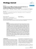
A plasmid (LuIII Lt-Lt) containing two copies of the LuIII
3' termini flanking an E. coli stuffer sequence, was con-
structed (figure 1). In anticipation of the difficulties
expected in manipulating the left end hairpin and to
increase the chances of obtaining the desired construct
two copies of the left end termini were successfully ligated
in vitro, in a tail (nt 278) to head (nt 1) -head to tail orien-
tation, this prior to cloning into pUC19. Sequencing of all
recombinants obtained, with an exception, revealed a sin-
gle copy of the left hairpin of LuIII ligated to E. coli
sequences of ~250 bp. These recombinants all contained
the LuIII hairpin sequence in the same orientation in
pUC19 with respect to the Reverse and Forward Primers,
conserving the LuIII sequence at the 5' end and the E. coli
sequence at the 3' end. Cotmore and Tattersall [22]
reported that the palindromic inserts had a greater ten-
dency for deletions, even in recombination-deficient
strains of E. coli, this probably due to the complex struc-
tures assumed by the inserts. Liu et al. [3] also reported
inherent difficulties in cloning hairpins, resulting in many
incorrect and presumably rearranged clones. The LuIII
sequences may have formed a complex hairpin structure
in vivo, due to its palindromic nature that was removed by
slipped mispairing during replication [23]. Difficulty in
Virology Journal 2005, 2:47 />Page 3 of 11
(page number not for citation purposes)
the sequencing of these clones, particularly with the
Reverse primer (M13R), supports this observation.
LuIII Lt-Lt was cotransfected with pGLu883∆Xba, the
genomic clone of LuIII, by electroporation into HeLa cells.
pGLu883∆Xba provides the trans acting factors necessary
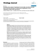
for replication of the minigenome. Southern blot analysis
of the transfection assays are shown in figure 2. The blot
was hybridized with the LuIII Lt-Lt Bam HI fragment
labeled by random primed Digoxigenin-11-dUTP.
Cotransfection of pGLu883∆Xba/LuIII Lt-Lt (lane 2),
resulted in three sets of doublet bands. These doublets
were of ~1.8, ~1.2 and ~.8 kb. These bands do not appear
for the replication of the LuIII genomic clone,
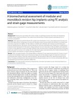
Strategy Used to Construct LuIII Lt-LtFigure 1
Strategy Used to Construct LuIII Lt-Lt. White, grey and dotted regions represent LuIII, pUC19 vector and E. coli
sequences, respectively. Restriction enzyme sites used are indicated. PGLU883 corresponds to the LuIII infectious genomic
clone.
Bam HI
Bam HI / Mlu I
Bam HI (1)
Mlu I (LuIII nt 278)
pGLu883
(7831 bp)
Bam HI
(5139)
Isolation of 278 bp
fragments
T4 DNA Ligation
and Bam HI digestion
T
A
GAG
AG
CTC
TC
Mlu I (278)
Bam HI (1)
LuIII Lt-Lt
(3482 bp)
T4 DNA Ligase
Bam HI
pUC 19
Bam HI
Mlu I
CTC
GAG
GA
T
A
GAG
AG
CTC
TC
CT
A
T
Bam HI
T
A
GAG
AG
CTCTC
Mlu I
CTC
GAG
GA
CT
A
T
Mlu I
Stuffer
Virology Journal 2005, 2:47 />Page 4 of 11
(page number not for citation purposes)
pGLu883∆Xba (lane 1) nor for the transfection of LuIII Lt-
Lt (lane 3) for which only input plasmid was observed
since the plasmid was not capable of replicating in the
absence of helper functions. When DNA samples were
digested with Mlu I (lanes 4–6) pGLu883∆Xba resulted in
a strong band of ~278 bp (lane 4) corresponding to the
left terminus of LuIII. Given the probe used (exclusively
the LuIII Lt-Lt insert) the large fragment corresponding to
nts 279-5135 of the LuIII genome was not observed on
this gel. The presence of this fragment was confirmed by
southern blot analysis using the full length genome of
LuIII (Data not shown). Cotransfection of pGLu883∆Xba/
LuIII Lt-Lt digested with Mlu I (lane 5) resulted in two
bands, one migrating with the ~278 bp band of
pGLu883∆Xba/ Mlu I (lane 4) and a band of greater inten-
sity migrating slightly faster. Digestion of the cotransfec-
tion sample with Mlu I (lane 5) also eliminated the three
sets of doublets observed in the uncut sample (lane 2) of
the cotransfection suggesting that these molecules likely
represent concatemers of a single molecule. Digestion of a
monomer molecule resulting from the replication of LuIII
Lt-Lt with Mlu I is expected to generate two fragments, one
of ~278 bp corresponding to the left hairpin of LuIII and
a band corresponding to the E. coli stuffer sequence which
has a size of ~250 bp; two molecules of the hairpin should
be generated for every copy of the stuffer sequence, there-
with the intensity of the band corresponding to the hair-
pin is expected to be greater than the band corresponding
to the stuffer sequence. Two bands were observed for this
digestion (lane 5); the larger band migrates along side the
band observed for pGLu883∆Xba likely representing the
left end hairpin of LuIII in double stranded form. The
smaller of the two bands, of greater intensity, may repre-
sent the left hairpin with an alternate conformation. A
faint band of similar migration is observed for
pGLu883∆Xba when digested with Mlu I (lane 4). The
band corresponding to the stuffer sequence is not obvi-
ous, this likely due to its similar migration to the LuIII left
end with a different conformation. Lane 6, containing the
transfection sample of only LuIII Lt-Lt shows a band of
~250 bp resulting from the digestion of input plasmid
that was not capable of replicating, this confirms our
assumption that the stuffer sequence observed in lane 6
migrates similar to the left hairpin with an altered confor-
mation hence its greater intensity when compared to the
migration of the double stranded left hairpin.
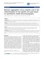
LuIII Lt-Lt was also cotranfected with pCMVNS1, an
expression vector for the MVM nonstructural protein NS1
(figure 3). LuIII Lt-Lt was capable of replicating when only
NS1 was present in trans (lane 7) resulting in the same
banding pattern as observed in figure 2 (lane2). It has
been suggested that the non-structural protein NS1 makes
the excision [4] by introducing a single-stranded nick,
possibly at the 5' end of the viral genome. If the minige-
nome could replicate under these conditions, it contains
all the cis-acting sequences required for excision and DNA
replication. These results suggest that LuIII Lt-Lt was capa-
ble of excision and replication when pGLu883∆Xba or
pCMVNS1 was provided in trans and that only NS1 viral
functions appear to be required for the excision and repli-
cation of LuIII Lt-Lt.
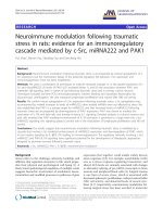
A possible mechanism for the replication of LuIII Lt-Lt is
shown in figure 4. The model proposes a nick at the NS1
nick site present at the left hairpin (step 1); this generates
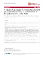
DNA Samples Recovered From Transfection Assays of LuIII Lt-Lt Digested With Mlu IFigure 2
DNA Samples Recovered From Transfection Assays
of LuIII Lt-Lt Digested With Mlu I. Samples correspond
to DNA isolated from transfection assays. Lanes 2 and 5 rep-
resent cotransfections with pGLu883∆Xba and LuIII Lt-Lt.
White lines indicate DNA fragments recovered from the
replication of LuIII Lt-Lt. Sizes of the 2 log ladder (Roche) are
shown. The probe used consisted of the insert of LuIII Lt-Lt
labeled by the DNA random primed labeling method.
Uncut samples Mlu I digested
12 3 4 56
0.8
1.2
1.5
2.0
5.0
(Kb)
278 bp
250 bp
pGlu883∆Xba
Cotransfection
LuIII Lt-Lt
pGlu883∆Xba
Cotransfection
LuIII Lt-Lt
Virology Journal 2005, 2:47 />Page 5 of 11
(page number not for citation purposes)
two origins of replication running in opposite directions
(step 2) that lead to strand displacement. The new hair-
pins assume secondary structures and continue DNA syn-
thesis (step 4), generating a close-end molecule. This step
generates two copies of a molecule estimated to be ~664
nts in length. Both molecules can now generate a mono-
mer length molecule of ~806 bp (step 5). As a result of
replication, the arrangement of the arms in the hairpin
change resulting in hairpins with the GAG triplet present
at the 5'end of the molecules. This forces the molecule to
go through a dimer intermediate (steps 7 and 8) generat-
ing a molecule with a turn around end of ~1192 nts in
length. This dimer is then resolved to generate monomer
length double stranded molecules (step 9). The sizes of
the DNA molecules obtained from this model on the rep-
lication of LuIII Lt-Lt correspond very closely with the
sizes of the products predicted by the model (figure 2 and
3) for the replication of LuIII Lt-Lt.
Parvovirus DNA replication starts when the 3' hydroxyl
group at the left end of the viral genome primes the syn-
thesis of a complementary strand, leading the formation
of a double stranded replicative form known as the cRF. In
vitro studies have shown that the cRF of autonomous par-
vovirus like MVM terminates in closed hairpins at both
ends, making cRF a major, or even obligatory intermedi-
ate of parvovirus replication [8], but only the right-end
hairpin is resolved in the presence of NS1 [8,24]. The cRF
DNA Recovered from Transfection Assays of LuIII Lt-Lt with pCMVNS1Figure 3
DNA Recovered from Transfection Assays of LuIII Lt-Lt with pCMVNS1. DNA samples shown correspond to: 1.
the full length insert isolated from LuIII Lt-Lt, 2. negative control of transfection, 3–7. DNA isolated from transfection assays of
the indicated samples. Arrow heads point to DNA fragments recovered from the replication of LuIII Lt-Lt. Sizes of the 2 log
ladder (Roche) are indicated. The probe used consisted of the insert of LuIII Lt-Lt labeled by the DNA random primed labeling
method.
0.8
1.2
1.5
2.0
5.0
(Kb)
12 345 67
LuIII Lt-Lt / Bam HI
Negative control
pGlu883∆Xba
pGlu883∆Xba / LuIII Lt-Lt
LuIII Lt-Lt
pCMVNS1
pCMVNS1 / LuIII Lt-Lt
Virology Journal 2005, 2:47 />Page 6 of 11
(page number not for citation purposes)
Proposed Model for the Rescue and Replication of LuIII Lt-LtFigure 4
Proposed Model for the Rescue and Replication of LuIII Lt-Lt. Restriction sites and their positions with respect to the
LuIII sequence are indicated. Grey, white and zigzag regions represent pUC19, LuIII left terminus and E. coli sequences respec-
tively. In steps 4 and 5 the molecules generated (a/b and aa/bb) are identical, for this reason only one molecule at each step is
continued throughout the scheme. The estimated sizes of some of the molecules (boxed) are indicated.
CutatbothproposedNS1sites
TC
T
A
A
T
GAG
GAG
CTC
CTC
Mlu I
Mlu I
Stuffer sequence
Bam HI
AG
3’
5’
Bam HI
GA
CT
5’
3’
(1)
(278)
(278)
(1)
Bam HI
Bam HI
TC
T
A
GAG
CTC
AG
3’
5’
(1)
A
T
GAG
GA
(278)
(1)
5’
3’
CT
(278)
Strand displacement and synthesis
3’
5’
TC
T
A
GAG
CTC
AG
A
T
GAG
GA
(278)
5’
3’
CT
(278)
T
AG
CTC
Bam HI
Bam HI
GAG
G
A
G
G
A
T
1.
2.
3.
a
b
4.
a
T
GAG
GA
GAG
T
A
CT
G
A
CTC
5’
3’
GAG
G
A
T
b
Bam HI
is identical to
GA
T
GAG
CTC
T
C
CTC
CT
A
GAG
T
AG
T
GAG
AG
A
TC
CTC
3’
5’
T
A
G
T
GA
GAG
5.
Bam HI
GAG
A
aa
bb
is identical to
A
CTC
CT
TC
GAGAG
CTC
T
CTC
CT
CTC
CT
AG
GAG
T
A
6.
A
A
7.
CTC
CT
CTC
A
CT
AG
GAG
T
GAG
GA
TC
CTC
A
T
Back to
Step 5
8.
9.
GA
T
GAG
CTC
T
C
CTC
CT
A
GAG
T
A
G
A
aa
bb
~806 bp
~664 bp
~806 bp
~1192 bp
T
GAG
AG
GAG
T
A
TC
A
G
CTC
3’
5’
T
A
G
Bam HI
G
A
G
b
a
Virology Journal 2005, 2:47 />Page 7 of 11
(page number not for citation purposes)
is re-opened and copied, producing a right end extended
form (5' eRF) followed by unfolding of the hairpin and
copying of the terminal sequence. This leads to the forma-
tion of dimeric RF (dRF) and higher-order concatamers
that would be resolved into monomeric (mRF) RF DNA.
If the wild type LuIII virus replicates using the mechanism
described for MVM and forms the cRF, the replication of
two copies of the left end such as in LuIII Lt-Lt should
result in a dead molecule that could not be resolved by
NS1. Although the terminal palindromic sequences are
essential for the replication of the APVs genome, the right
and left terminal sequences are not equivalent in function
[25,26]. According to the modified rolling hairpin model
of MVM replication, the right end origin is active in the
covalently closed hairpin configuration and also in the
extended right end telomere [14,24]. In contrast, the
MVM left end inverted repeat does not constitute a repli-
cation origin in the hairpin configuration and needs to be
copied in the form of a left-to-left end bridge to be subse-
quently resolved at the multimeric RF DNA stage
[1,3,8,14,27].
When the dimer bridge origin of MVM is compared to the
left end arrangement in LuIII Lt-Lt (figure 5), it becomes
apparent that the left terminus is an incomplete origin of
replication based on the origin proposed for MVM repli-
cation. A competent replication origin contains, among
other things an NS1 nick site. If like MVM, the left end ter-
minus of LuIII is only processed when present as a bridge
in the dimer RF but not as a hairpin in monomeric repli-
cative form, neither of the left end termini in LuIII Lt-Lt
would be recognized by NS1. As a result, the LuIII insert
would not be excised from the plasmid pUC19, and hence
no replication would be expected to occur. Comparison of
the sequences present in LuIII Lt-Lt with the junction
bridge in the dimer replicative form of MVM [28] (figure
4) illustrates that the A and B arms of the LuIII left end are
organized differently from that proposed for the active
origin of replication for MVM. Unlike the dimer arrange-
ment described for MVM, in LuIII Lt-Lt the CT doublet is
positioned at the 5'end and the CTC triplet is positioned
inboard at the 3'end in both hairpins. In the hairpin
arrangement an NS1 nick site is not present at the 5' end
of the CT bubble as described in the MVM dimer bridge.
Nevertheless, LuIII Lt-Lt was capable of replication sug-
Comparison of the MVM Dimer Bridge (A) with the Hairpin Arrangement in LuIII Lt-Lt (B)Figure 5
Comparison of the MVM Dimer Bridge (A) with the Hairpin Arrangement in LuIII Lt-Lt (B). Hairpins and NS1
recognition nick sites are indicated by dark bold lines and arrows respectively. The grey patterned boxes correspond to
pUC19 sequences.
GAA
GA
CTT
CT
5’
3’
Aarm
Barm
5’ TC CTC GAG GA 3’
3’ AG GAG CTC CT 5’
A. Junction bridge in the dimer replicative form of MVM (28)
B. Hairpin arrangements in Lu III Lt-Lt
Stuffer
Virology Journal 2005, 2:47 />Page 8 of 11
(page number not for citation purposes)
gesting that the left hairpin of LuIII does constitute a rep-
lication origin in the extended double stranded hairpin
configuration.
Given the functionality of the left hairpin of LuIII as an
origin of replication in the extended double stranded
form a replication model of LuIII can be predicted result-
ing in equivalent amounts of plus and minus DNA viral
strands (figure 6). In this model the plus and minus DNA
strands, independently initiate replication from the right
and left hairpins respectively (step 1). The NS1 nick sites
present at the left and right termini in LuIII differ from
each other; there is an insertion of an Adenine residue in
the NS1 nick site present at the 5' terminus of LuIII. This
additional adenine is also not present in the NS1 nick site
described for MVM [29].
This replication model for LuIII predicts flip/flop confor-
mations at both termini. Earlier studies [30] in which the
left and right termini of the minus and plus strands,
respectively, were labeled at the 3' hydroxyl group and
subsequently digested with Hha I suggested that the left
terminus of the LuIII minus strand exists only in the flip
conformation, and the right terminus of the plus strand
Proposed Model for the Replication of Parvovirus LuIIIFigure 6
Proposed Model for the Replication of Parvovirus LuIII. A model for the replication of the (+) and the (-) strand of LuIII
is shown. The NS1 nick site and its complementary sequence (*) are indicated. The unpaired sequences present at the left hair-
pin are shown. The arrows point to NS1 nick sites. A
corresponds to the insertion in the NS1 nick site present at the right ter-
minus of LuIII.
Replication of LuIII using plus strand
1.
2.
3.
4.
5.
Replication of LuIII using minus strand
5’
A
CTC
CT
3’
(+)
A
TC
7
CTC
A
TC
CTC
T
AG
GAG
(-)
(-)
(+)
(+)
(-)
3’
5’
(-)
T
GAG
GA
(ACTATTC)
(GTATAAG) *
A
CTC
CT
(TGATAAG) *
5’
3’
(CATATTC)
(+)
7
T
AG
GAG
A
CTC
CT
(-)
(+)
T
GA
GAG
(+)
(-)
(-)
T
GAG
GA
A
CTC
CT
5’
3’
(+)
T
GAG
GA
3’
5’
(-)
(+)
T
GAG
GA
5’
3’
A
CTC
CT
3’
5’
(-)
7
7
•
•
•
•
•
•
•
•
Virology Journal 2005, 2:47 />Page 9 of 11
(page number not for citation purposes)
exists in both the flip and flop conformations. Numerous
bands were observed when the left terminus of the minus
strand was digested with Hha I yet these were justified as
alternate secondary structures of the hairpin in the flip
conformation. The expected fragments for the digestion of
the flip and flop conformations of the left hairpin are very
similar in size, any slight variation in migration due to the
secondary structures assumed by these fragments could
have impaired the interpretation of the results. The
conformation present at the left end of the plus strand still
remains unknown.
Conclusion
The data presented demonstrates that LuIII Lt-Lt contains
all the cis-acting sequences required for excision and DNA
replication when NS1 viral functions are provided in
trans. These findings suggest that the left hairpin of LuIII
has an active NS1 driven origin of replication at this ter-
minus in the double stranded extended form. This
extended hairpin, capable of acting as an origin of replica-
tion, lacks the arrangement of the specific domains
present in the dimer duplex intermediate of MVM, the
only active form of the left hairpin described for MVM.
This difference between LuIII and MVM has great implica-
tions on the replication of these viruses. The presence of
origins of replication at both the left and right termini can
explain the unique encapsidation pattern observed for
LuIII hinting on the mechanism used by LuIII for the rep-
lication of its viral genome.
Methods
Construction of LuIII Lt-Lt
The LuIII Lt-Lt minigenome (figure 1) has two copies of
the left end palindrome of the autonomous parvovirus
LuIII (nt. 1-278) cloned into the Bam HI site of pUC19
[29] [Genbank L09137
]. The 3' hairpin of LuIII was
obtained from pGLu883 [30], the full-length genomic
clone of LuIII cloned into the pUC19 vector. pGLu883
was digested with both Bam HI (pUC19 nt. 417) and Mlu
I I (LuIII nt. 278) for two hours at 37°C and then electro-
phoresed on a 1.2% agarose gel in 1X TBE buffer at 75 V.
The Bam HI / Mlu I I digestion generated three fragments
of approximately 278, 2686, and 4861 bp. The 278 bp
fragment corresponding to the left end hairpin was
isolated and purified using the Geneclean Spin Kit
®
(QBio-gene, Carlsbad, CA), and then were ligated through
the Mlu I site in an overnight reaction at 4°C using 1 U of
T4 DNA ligase. The ligation was digested with Bam HI
generating a fragment of 568 bp corresponding to the two
copies of the 3' hairpin in a "head to tail-tail to head" con-
formation (nts 1-278, 278-1). The fragment generated was
purified as described and ligated into the Bam HI site of
pUC19 that was previously treated with calf intestinal
alkaline phosphatase (CIAP) (Roche Applied Science,
Indianapolis, IN) for one hour at 37°C.
Preparation of Competent Cells
Two different strains of Escherichia coli were used as com-
petent cells: DH5α [(lacZ.M15. (lacZYA-argF) recA1
endA1 hsdR17 (rkmk+) phoA supE44 thi gyrA96 relA1)]
(ATCC, Rockville, MD) and SURE
®
2 super competent cells
[(e14- (McrA-). (mcrCB-hsd SMR-mrr) 171 endA1 supE44
thi-1 gyrA96 relA1 lac recB recJ sbcC umuC::Tn5 (Kanr)
uvrC (F' proAB lacIqZ.M15 Tn10 (Tetr) Amy Camr)]
(Stratagene, La Jolla, CA). Competent cells were prepared
by the calcium chloride method [31].
Transformation of Competent Cells
The recombinant molecules were transformed in both
DH5α and SURE
®
2 competent cells. Competent cells were
thawed on ice for 15 minutes (min.). The DNA was added
to the cells and incubated on ice for 30 min. Cells were
heat-shocked in a 42°C water bath and subsequently
incubated on ice for 2 min. DH5α and SURE
®
2 competent
cells were heat-shocked for 2 min. and 30 seconds respec-
tively. 100 µL of preheated (42°C) LB broth was added to
both cell samples and incubated at 37°C for 1 hour (h)
with shaking at 225 rpm. DH5α transformed cells were
spread on LB agar plates containing 50 mg/mL ampicillin
and 80 µL of 2% X-gal. SURE
®
2 transformed cells were
spread on LB plates containing 50 mg/mL ampicillin, 100
µL of 2% X-gal and 100 µL of 10 mM IPTG.
Isolation of DNA Recombinants
The resultant plasmids from DH5α and SURE
®
2 trans-
formed cells were purified by the alkaline lysis miniprep
method, described by Ausubel et al. [31] and analyzed
with restriction enzymes. Sequencing was performed at
the New Jersey Medical School, Molecular Resource
Facility.
Tissue Culture
HeLa (ATCC, Rockville, MD) cells were grown in Minimal
Essential Medium (MEM Eagle) (MP Biomedicals, Aurora,
OH) supplemented with 10% fetal bovine serum (FBS)
(HyClone, Logan, UT) and PSG (8 mM Penicillin G, 3
mM Streptomycin Sulfate, 200 mM L-Glutamine). They
were incubated at 37°C in 25 and/or 75 cm
2
plastic tissue
culture flasks. For sub-culturing, the cells were rinsed
twice with Phosphate-Buffered Saline (1X PBS) and incu-
bated in 1X Trypsin (Difco, Detroit, MI) for 5 min. at
37°C. Cells were harvested by centrifugation at 3800 rpm
for 5 min. at 4°C. The resultant pellet was resuspended in
the medium described above and seeded into culture
flasks at a proportion of 1:3.
Transfection Assay
HeLa cells were grown to 100 % confluency in a 75 cm
2
flask. They were washed three times with 1X PBS and then
tripsinized at 37°C for 5 minutes. Cells were harvested by
centrifugation at 3,800 rpm for 5 min. at 4°C and washed
Virology Journal 2005, 2:47 />Page 10 of 11
(page number not for citation purposes)
in 10 ml of PBS. Cells were resuspended and split at a pro-
portion of 1:9. Approximately, 5 µg of pGLu883∆Xba,
LuIII Lt-Lt minigenome and pCMVNS1 were added to the
corresponding tubes and incubated at 37°C for 10 min.
Cells were transferred to sterile cuvettes with a 4-mm gap
width, and electroporated individually at 230 V and 950
µF using a capacitance discharge machine (Gene Pulser,
Bio-Rad Laboratories, Hercules CA). After each pulse, 700
µL of MEM-10% FBS were added to the cuvette and the
cells were resuspended carefully. The electroporated cells
were incubated for 45 min. at 37°C and then transferred
to 25 cm
2
flasks containing 3 mL MEM-10% FBS. After an
overnight incubation at 37°C, the medium was changed,
and the cells were incubated at 37 °C until the low molec-
ular weight DNAs were isolated at five days post-transfec-
tion, as described by Tam and Astell [25]. DNA samples
were resuspended in 30 µL TE (10 mM Tris-HCl, 1 mM
EDTA, pH 8.0).
Southern Blot Analysis
Samples were electrophoresed on a 1.2% agarose gel in 1X
TAE buffer at 80 V, and passively transferred onto a Zeta
Probe nylon membrane (Bio-Rad Laboratories, Hercules,
California) as described by Ausubel et al [31]. Probes were
labelled by the random primed DNA labeling method
with Digoxigenin-11-dUTP (Roche Applied Science, Indi-
anapolis, IN). The blot was hybridized at 50°C and
washed at 55°C. Detection was performed according to
manufacturer's instructions (Roche Applied Science, Indi-
anapolis, IN).
Competing interests
The author(s) declare that they have no competing
interests
Authors' contributions
NDC drafted and revised critically the manuscript, had
the intellectual idea of the study and its design, contrib-
uted significantly in the analysis and interpretation of the
data, proposed the replication models presented and gave
the final approval of the version to be published.
LVP constructed LuIII Lt-Lt, collected the data resulting
from the transfection of LuIII Lt-Lt/pGlu∆Xba, contrib-
uted in the analysis and interpretation of the data, partic-
ipated in the idea and design of the models proposed and
in the drafting and revision of the manuscript.
IDM collected the data resulting from the transfections of
LuIII Lt-Lt/pGlu∆Xba and, LuIII Lt-Lt/pCMVNS1, contrib-
uted in the analysis and interpretation of the data, partic-
ipated in the design of the models proposed and in the
drafting and revision of the manuscript.
All authors read and approved the final manuscript.
Acknowledgements
We thank Dr. David Pintel and Dr. Ian Maxwell for the pCMVMNS1 and
pGLu∆Xba clones respectively and Omayra Rivera-Denizard for her helpful
suggestions in the design of the models.
This work was supported by the Minority Biomedical Research Support,
National Institute of Health Grant SO6GM08103 and the College of Arts
and Sciences, University of Puerto Rico at Mayaguez.
References
1. Astell CR, Chow MB, Ward D: Sequence analysis of the termini
of virion and replicative forms of Minute Virus of Mice DNA
suggests a modified rolling hairpin model for autonomous
parvovirus DNA replication. J Virol 1985, 54:171-177.
2. Cotmore SF, Tattersall P: Parvovirus DNA Replication. In DNA
Replication in Eukaryotic Cells Edited by: Depamphilis ML. New York:
Cold Spring Harbor Lab; 1996:799-813.
3. Liu Q, Yong CB, Astell CR: In vitro resolution of the dimmer
bridge of the Minute Virus of Mice (MVM) genome supports
the modified rolling hairpin model for MVM replication. Virol
1994, 201:251-262.
4. Berns KI: Parvoviridae: the viruses and their replication. In
Fundamental Virology 3rd edition. Edited by: Fields BN, Knipe DM,
Howley PM. Pennsylvania: Lippincott-Raven; 1996:1017-1036.
5. Cotmore SF, Tattersall P: An asymmetric nucleotide in the par-
voviral 3' hairpin directs segregation of a single active origin
of DNA replication. Embo J 1994, 13:4145-4152.
6. Faust EA, Rankin CD: In vitro conversion of MVM virus single-
stranded DNA to the replicative form by DNA polymerase
alpha from Ehrlich ascites tumor cells. Nucl Acids Res 1982,
10:4181-4201.
7. Faisst S, Rommelaere J, (eds): Parvoviruses. From Molecular Biology to
Pathology and Therapeutic Uses. Contrib Microbiol Volume 4. Edited by:
Schmidt A. New York: Karger Press; 2000.
8. Baldauf AQ, Willwand K, Mumtsidu E, Nüesch JP, Rommelaere J:
Specific initiation of replication at the right-end telomere of
the closed species of Minute Virus of Mice replicative-form
DNA. J Virol 1997, 71:971-980.
9. Cotmore SF, Tattersall P: The NS-1 polypeptide of Minute Virus
of Mice is covalently attached to the 5' termini of duplex rep-
licative-form DNA and progeny single strands. J Virol 1988,
62:851-860.
10. Willwand K, Mumtsidu E, Kuntz-Simon G, Rommelaere J: Initiation
of DNA replication at palindromic telomeres is mediated by
a duplex-to-hairpin transition induced by the Minute Virus of
Mice nonstructural protein NS1. J Biol Chem 1998,
273:1165-1174.
11. Kuntz-Simon G, Bashir T, Rommelaere J, Willwand K: Neoplastic
transformation-associated stimulation of the in vitro resolu-
tion of concatemer junction fragments from Minute Virus of
Mice DNA. J Virol 1999, 73:2552-2558.
12. Cotmore SF, Tattersal P: DNA replication in the autonomous
parvoviruses. Semin Virol 1995, 6:271-281.
13. Wilson GM, Hindal HK, Yeung DE, Chen W, Astell CR: Expression
of Minute Virus of Mice major nonstructural protein in insect
cells: Purification and identification of ATPase and helicase
activities. Virol 1991, 185:90-98.
14. Cotmore SF, Nüesch JPF, Tattersall P: Asymmetric resolution of
a parvovirus palindrome in vitro. J Virol 1993, 67:1579-1589.
15. Majaniemi I, Siegl G: Early events in the replication of parvovi-
rus LuIII. Arch Virol 1984, 81:285-302.
16. Christensen J, Cotmore SF, Tattersall P: A novel cellular site-spe-
cific DNA-binding protein cooperates with the viral NS1
polypeptide to initiate parvovirus DNA replication. J Virol
1997, 71:1405-1416.
17. Christensen J, Cotmore SF, Tattersall P: Parvovirus initiation fac-
tor PIF: a novel human DNA-binding factor which coordi-
nately recognizes two ACGT motifs. J Virol 1997, 71:5733-5741.
18. Cotmore SF, Nüesch JP, Tattersall P: In vitro excision and repli-
cation of 5' telomeres of Minute Virus of Mice DNA from
cloned palindromic concatemer junctions. Virol 1992,
190:365-377.
Publish with BioMed Central and every
scientist can read your work free of charge
"BioMed Central will be the most significant development for
disseminating the results of biomedical research in our lifetime."
Sir Paul Nurse, Cancer Research UK
Your research papers will be:
available free of charge to the entire biomedical community
peer reviewed and published immediately upon acceptance
cited in PubMed and archived on PubMed Central
yours — you keep the copyright
Submit your manuscript here:
/>BioMedcentral
Virology Journal 2005, 2:47 />Page 11 of 11
(page number not for citation purposes)
19. Muller DE, Siegl G: Maturation of Parvovirus LuIII in a subcel-
lular system. I. Optimal conditions for in vitro synthesis and
encapsidation of viral DNA. J Gen Virol 1983, 64:1043-1054.
20. Cotmore SF, Tattersall P: High-mobility group 1/2 proteins are
essential for initiating rolling-circle-type DNA replication at
a parvovirus hairpin origin. J Virol 1998, 72:8477-8484.
21. Cotmore SF, Christensen J, Tattersall P: Two widely spaced initi-
ator binding sites create an HMG1-dependent parvovirus
rolling-hairpin replication origin. J Virol 2000, 74:1332-1341.
22. Cotmore SF, Tattersall P: In vivo resolution of circular plasmids
containing concatemer junction fragments from Minute
Virus of Mice DNA and their subsequent replication as linear
molecules. J Virol 1992, 66:420-431.
23. Tam P, Astell CR: Multiple cellular factors bind to cis-regula-
tory elements found inboard of the 5' palindrome of Minute
Virus of Mice. J Virol 1994, 68:2840-2848.
24. Willwand K, Baldauf AQ, Deleu L, Mumtsidu E, Costello E, Beard P,
Rommelaere J: The Minute Virus of Mice (MVM) nonstructural
protein NS1 induces nicking of MVM DNA at a unique site of
the right-end telomere in both hairpin and duplex conforma-
tions in vitro. J Gen Virol 1997, 78:2647-2655.
25. Tam P, Astell CR: Replication of Minute Virus of Mice minige-
nomes: novel replication elements required for MVM DNA
replication. Virol 1993, 193:812-824.
26. Cotmore SF, Tattersall P: Genome packing sense is controlled
by the efficiency of the nick site in the right-end replication
origin of parvoviruses Minute Virus of Mice and LuIII. J Virol
2005, 79:2287-2300.
27. Cotmore SF, Tattersall P: The autonomously replicating parvo-
viruses of vertebrates. Adv Virus Res 1987, 33:91-173.
28. Cotmore SF, Tattersall P: Resolution of parvovirus dimer junc-
tion proceeds through a novel heterocruciform
intermediate. J Virol 2003, 77:6245-6254.
29. Diffoot N, Chen KC, Bates RC, Lederman M: The complete nucle-
otide sequence of parvovirus LuIII and localization of a
unique sequence possibly responsible for its encapsidation
pattern. Virol 1993, 192:339-345.
30. Diffoot N, Shull BC, Chen KC, Stout ER, Lederman M, Bates R: Iden-
tical ends are not required for the equal encapsidation of
plus- and minus- strand parvovirus LuIII DNA. J Virol 1989,
63:3180-3184.
31. Ausubel FM, Roger B, Kingston RE, Moore DD, Seidman JG, Smith JA,
Struhl K: Short protocols in Molecular Biology New York: John Wiley &
Sons; 1999.