báo cáo hóa học: " Neuroimmune modulation following traumatic stress in rats: evidence for an immunoregulatory cascade mediated by c-Src, miRNA222 and PAK1" pptx
Bạn đang xem bản rút gọn của tài liệu. Xem và tải ngay bản đầy đủ của tài liệu tại đây (5.37 MB, 13 trang )
RESEARC H Open Access
Neuroimmune modulation following traumatic
stress in rats: evidence for an immunoregulatory
cascade mediated by c-Src, miRNA222 and PAK1
Hui Zhao
*
, Ranran Yao, Xiaoding Cao and Gencheng Wu
Abstract
Background: Neuroimmune modulation following traumatic stress is accompanied by cortical upregulation of c-
Src expression, but the mechanistic details of the potential regulatory link between c-Src expression and
immunosuppression have not been established.
Methods: We used a combination of techniques to measure temporal changes in: (i) the parallel expression of c-
Src and microRNA222; (ii) levels of PAK1 (p21-activated kinase 1); and (iii) the association between PAK1 and
interleukin 1b signaling, both in cortex of rats following traumatic stress and in primary cortical neurons.
Techniques included real-time PCR, immunoprecipitation, western blotting and subcellular fractionation by
discontinuous centrifugation. We also measured lymphocyte proliferation and natural killer (NK) cell activity.
Results: We confirm robust upregulation of c-Src expression following traumatic stress. c-Src upregulation was
accompanied by marked increases in levels of miRNA222; other studied miRNAs were not affected by stress. We
also established that PAK1 is a primary target for miRNA222, and that increased levels of miRNA222 following
traumatic stress are accompanied by downregulation of PAK1 expression. PAK1 was shown to mediate the
association of IL-1RI with lipid rafts and thereby enhance IL-1 signaling. Detailed analyses in cultured neurons and
glial cells revealed that PAK1-mediated enhancement of IL-1RI activation is governed to a large extent by c-Src/
miRNA222 signaling; this signaling played a central role in the modulation of lymphocyte proliferation and NK cell
activity.
Conclusions: Our results suggest that neuroimmune modulation following trau matic stress is mediated by a
cascade that involves c-Src-mediated enhancement of miRNA222 expression and downregulation of PAK1, which
in turn impairs signaling via IL-1b/IL1-RI, leading to immunosuppression. The regulatory networks involving c-Src/
miRNA222 and PAK1/IL-1RI signaling have significant potential for the development of therapeutic approaches
designed to promote recovery following traumatic injury.
Keywords: c-Src, miRNA222, PAK-1, IL-1b?β?, neuroimmune modulation
Background
Stress refers to the challenge, adversity, hardship, and
affliction that organisms encounter in life, which jeopar-
dize their physical and psychological well being [1]. A
finely tuned spatiotemporal regulation of multiple events
suggests hierarchic involvement of modulatory neuro-
transmitters and modified processes in pathways of gene
expression that together could enable widely diverse
stress responses [2,3]. For example, acetylcholine (ACh)
acts as a stress response-regulatin g transmitter; and
altered ACh levels are variousl y associated with changes
in altern ative splicing of pre-mRNA transcripts in brain
neurons and peripheral blood cells [4]. Surgical trauma
is one form of severe stress, which is associated with
decreased splenocyte proliferation, reduced natural killer
(NK) cell activity, and abnormal levels of several cyto-
kines [5-7]. Importantly, neuroimmune modulation fol-
lowing surgical stress has been ascribed to molecular
* Correspondence:
Department of Integrative Medicine and Neurobiology, State Key Lab of
Medical Neurobiology, Shanghai Medical College, Brain Research Institute,
Fudan University, Shanghai, P. R. China
Zhao et al. Journal of Neuroinflammation 2011, 8:159
/>JOURNAL OF
NEUROINFLAMMATION
© 2011 Zhao et al; licensee BioMed Centra l Ltd. This is an Open Access article distributed under the te rms of the Creative Commons
Attribution License ( which permits unrestricted use, distribution, and reprodu ction in
any medium, pro vided the original work is properly cited.
events taking place in cortical circuits. These can be
separated into two stages - early events of immunosup-
presion operating through an elaborate IL-1b pathway
[8-13], and later progression marked by changes in c-
Src signaling [14]. These dynamic alterations are likely
to take place in distinct cellular compartments control-
ling the activation of different signaling cascades.
c-Src function is crucial for r ecovery from traumatic
stress-mediated immunosuppression [14], but its
mechanistic linkage to infla mmati on onset and progres-
sion remains to be elucidated. c-Src is a member of the
Src family of protein kinases whose members play a cru-
cial role in transducing extracellular signals to cytoplas-
mic and nuclear effectors, and thereby regulate a wide
var iet y of cellular functions, including cell proliferation,
differentiation and stress responses [1 5,16]. Functional
overlap of c-Src and miRNA222 signaling has recently
been demonstrated, and these factors are thought to
play a joint regulatory role in tumor cell migration, ner-
vous system development and neurodegenerative dis-
eases [17]. However, the question of whether such
signaling contributes to neuroimmune modulation in
trauma remains to be clarified.
Of note, many m icroRNAs are involved in the neu-
roimmune pathway, which are named NeurimmiRs.
Both peripheral and central immune i nsults have been
shown to upregulate various NeurimmiRs, either in neu-
rons, in surrounding cells (glia, microglia and infiltrating
leukocytes) or in peripheral leukocytes. Owing to their
physical properties and multiple roles in the nervous
and immune systems, NeurimmiRs may initiate commu-
nication cascades via regulation of expression of numer-
ous genes both in health and disease [18,19]. Besides
reported NeurimmiRs, miRNA222 has been found to
play critical roles in a variety of biological processes in
the central nervous system (CNS), where p21-activated
kinase 1 (PAK1) is one of its targets [20,21]. PAK1 upre-
gulation in hippocampus and cortex is associated with
stroke and neurite outgrowth, whereas downregulation
of PAK1 has been recently reported in depression [22].
Further detailed studies have revealed that precise spa-
tiotempo ral expression of PAK1 proteins is requir ed for
the pleotropic effects of interleukin (IL)-1b [23] that
require appropriate receptor expression and effective
activation of i ntracellular signaling [23,24]. In the CNS,
immune-like processes have been found to underlie
responses not only to immune challenges but also to
physiological and psychological stress. It has become
evident that pro-inflammatory cytokines like IL-1b -
which is produced predominantly by activated cells of
theinnateimmunesystemsuchasmonocytes,macro-
phages, and brain microglia - plays an important role in
neuroendocrine and behavioral responses to variou s
stresses [1,11]. Importantly, further research on IL-1b
signaling has focused on phosphorylation and subcellu-
lar distribution of IL-1 receptor type I (IL- 1RI) in lipid
rafts, where these signaling pathways modulate IL-1b-
induced cellular activation [25,26]. Together, these
observations suggest that PAK1 could be a target for
regulation mediated by c-Src and miRNA222 and
thereby provide a mechanistic link between c-Src signal-
ing and IL-1b activity following traumatic stress.
Notably, the prefrontal cortex (PFC) is known to play
an important role in the integration of affective states
with appropriate modula tion of autonomic and neu-
roendocrine stress regulatory systems. There is evi-
dence for manipulation of prefrontal cortical networks
in conditions involving incorporation of adaptive beha-
vior and prevention of excessive behavioral and physio-
logical stress reactivity [27]. This may be especially
true for traumatic stress-related c-Sr c and IL-1b sig-
naling, which are enriched and initiated within this
region [11,14]. Therefore, in the current study we
sought to characterize molecular aspects of c-Src-
related signaling in PFC which could modulate the
onset or progression of immunosuppression induced
by traumatic stress. It is well established that traumatic
stress in rats leads to constitutive activation of neu-
roimmunomodulatory circuitry [28,29], and we have
investigated the possibility that miRNA222 regulates a
feedback loop that promotes immunosuppression
induced by traumatic stress.
Methods
Traumatic animal model
All animal experiments were carried out in accordance
with the guidelines and regulations for animal experi-
men tation in NIH and Fudan University. SD adult male
rats (Animal Center of Chinese Academy of Sciences,
200-250 g) were used in the current experiment. The
animals were housed in groups (5 per cage) in a con-
trolled environment on a 12 h light-dark cycle, and
allowed to acclimate for a minimum of 5 days before
conducting experiments. Water and food were available
at all times.
Traumatic stress was perfor med as previously
described [11]. Briefly, rats were anesthetized with pen-
tobarbital sodium (35 mg/kg, i.p.), then were incised
longitudinally to a leng th of 6 cm along the dorsal med-
ian line and 5 cm along the abdominal median line.
After surgery, wounds were sutured and animals were
kept warm in single housing, with care taken to keep
sawdust bedding dry and clean. No post-operative infec-
tions occ urre d. The operation was performed 48 h after
implanting a cannula, and tissue samples were taken 1,
3 and 7 days after the operation. Control rats were also
anesthetized an d underwent operation to implant a
cannula.
Zhao et al. Journal of Neuroinflammation 2011, 8:159
/>Page 2 of 13
Intracerebroventricular injection of drugs
Implantation of the cannula w as performed stereotaxi-
cally under anesthesia, a stainless steel guide cannula
(0.5 mm in diameter) with an inserted cannula (0.25
mm in diameter) was implanted into right lateral ventri-
cle (posterior 0.5, lateral 1.5, horizontal 4.5) and fixed
onto the skull with dental cement . IL-1ra (10 units,
Sigma Aldrich, St. Louis, MO), PAK1 antibody (10 μg),
and recombinant adenovirus (5 × 10
9
plaque-forming
units (pfu)) dissolved in sterilized PBS were injected
over 10s via the cannula in a volume of 10 μl. Rats from
the control group were injected with vehicle. At the end
of each procudure, the entire injector system was left in
place for an additional 10 min to minimize reflux. The
position of the cannula was assessed by histological
examination, and data were collected from experiments
in which correct insertion of the cannula was verified.
Animals were operated upon and killed 24 h after IL-
1ra and PAK1 ant ibody injection, or 72 h after recombi-
nant adenovirus injection.
Recombinant adenoviruses
cDNA for dominant-negative (K296R/Y528F, DN c-Src),
or constitutively active (Y528F, CA c-Src) forms
(Upstate Biotechnology, Lake Placid, NY) were cloned
into adenoviral shuttle vector pDE1sp1A (Microb ix Bio-
systems, Inc. Canada). After homologous recombination
in vivo with the backbone vector PJM17, plaques result-
ing from viral cytopathic effects were selected and
expanded in 293 cells. Positive plaques were further pur-
ified and large-scale production of adenovirus was car-
ried out by two sequential CsCl gradients and PD-10
Sephadex chromatography.
Immunofluorescent analysis
Rats were anesthetized with sodium pentobarbital (35
mg/kb, i.p.) and perfused transcardially with fixative (4%
paraformaldehyde). Coronal brain sections (25 μm) were
obtained using a cryostat. Frozen sections were sub-
jected to immunostaining with anti-PAK1 at 1:200 or
anti-c-Src at 1:100 (Upstate Biotechnology, Lake Placid,
NY), then transferred into Alexa594 conjugated anti-
rabbit antiserum (1:1000, Invitrogen, Carlsbad, CA) for
1 h. Data derived from each group were analyzed by
Leika Q500IW image analysis system. Frontal cortex
was chosen for analysis and immunopositive cells were
semi-quantified using photomicrography.
For cell immunofluorescent staining, neuronal or glial
cells were dissociated and plated into covers lips pretreated
with 0.1% polyethylenemine. After 10 days of growth, the
coverslips were subjected to anti-c-Src- and Alexa488-con-
jugated secondary antibodies, and anti-PAK1- and
Alexa594-conj ugated antibodies subsequently. Data were
analyzed using a Leika Q 500IW image analysis system.
Immunological assay
For lymphocyte proliferation, spleens were pressed
through stainless steel mesh and red blood cells were
lysed by treatment with NH
4
Cl solution. Cell w ere sus-
pended at 1 × 10
7
cells/ml in a final volume of 200 μlof
complete tissue culture medium (RPMI 1640 supple-
mented with 10% heat activated fetal calf serum, 2 mM
L-glutamine), and seeded in triplicate in U-bottom 96-
well plates in the presence or absence of concanavalin A
(Con A, 1 mg/L, Sigma Aldrich, St. Louis, MO). Plates
were incubated at 37°C in a 5% CO
2
. After 48 h, cul-
tures were labeled with 0.5 μCi of [
3
H] thymidine
(Amersham Biosciences, Piscataway, NY). Cells were
harvested using a cell harvester 24 h later. Samples wer e
counted in a liquid scintillation counter. Prolifer ation
results are presented as mean cpm ± SD of triplicate
cultures in 5 animals.
For natural kill er cell cytotoxicity, suspensions of
YAC-1 lymphoma cells, with a concentration of 2 ×
10
5
/ml at a final volume of 100 μl, were target ed with
0.5 μCi of [
3
H] thymidine and incubated at 37°C, 5%
CO
2
for 6 h. Then, the spleens were homogenized and
the resultant cell suspensions pooled in the presence or
absence of Con A and seeded in triplicate with effector :
target ratios of 50:1 for 16 h. Cytotoxic a ctivity results
were determined as follows:
Percent response = [(counts in tested well-counts in spontaneous response w ell)/
(counts in maximum response well-counts in spontaneous response w ell)] × 100
IL-1RI Production
IL-1RI expression was measured using an ELISA kit
(R&D systems, Minneapolis, MN). Briefly, frontal cortex
was collected an d suspended i n equal volumes of 50 μl
diluent buffer. Plates were incubated for 2 h at 37°C.
Hybridization reactions were stopped by several washes
and the plates were subsequently incubated with b ioti-
nylated anti-IL-1RI solution for 1 h, streptavidin-HRP
solution for 30 min, and with the stabilized chromogen
for 30 min. Stop solution (100 μl) was added to each
well and t he optical density was measured at 450 nm
using BioRad microreader (Hayward, CA). Data were
normalized, and expressed as mean ± SD from 5 ani-
mals, each performed in triplicate.
TaqMan reverse transcription (RT)-PCR for miRNA
quantification
Total RNA was isolated from frontal cortex (50 mg) or
cortical neurons (1 × 10
6
)withTrizol™ (Invitrogen,
Carlsbad, VA) ac cording to manufacturer’ sprotocol.
MicroRNA 218, 224, 142, 222, 126, 296, 194, 206 quan-
tification was carried out by reverse transcribing total
RNA using Taqman™ microRNA reverse transcription
kit and subjected to real-time PCR using TaqMan™
Zhao et al. Journal of Neuroinflammation 2011, 8:159
/>Page 3 of 13
MicroRNA Assay kit (Applied Biosystems, Carlsbad,
CA). Reactio ns were performed using Stratagene
Mx3000 instrument in triplicate. Real-time PR data was
analyzed using a ΔΔCt calculation. A p value of less
than 0.05, when considering treated animals or cells vs.
control group, was considered significant.
Primary neuron culture and treatment
For primary neuron cultures, rat fetuses were removed
from pregnant rats on embryonic day 18. Cortices were
dissected and collected in Hanks’ balanced salt solution.
Cells were dissociated and plated at a density of 10
6
cells per well into 24-well tissue culture plates pre-
treated with 0.1% polyethylenemine. Cells were main-
tained in serum-free Neurobasal medium containing
B27 supplement (Gibco, Rockville, MD). After 3-4 days
in culture, neurons sent out long processes. By 10 days,
flow cytometry showed that MAP
2
immunopo sitive cells
accounted for more than 95% of cells, and the indica ted
treatments were performed at this same time.
c-Src plasmid, microRNA222 mimetic a nd micro-
RNA222 inhibitor (Dharmacon RNA Technologies,
Lafayette, CO) were transfected into primary neurons
using Lipofectamine 2000 according to the manufac-
turer’s instructions (Qiagen, Valencia, CA). In brief, 1 ×
10
6
neurons were t ransfected with 10 pmol of micro-
RNA222 mimic and microRNA222 inhibitor. Following
transfection, neurons were cultured for another 48 h
prior to experiments.
For experiments using IL-1b (R&D systems, Minnea-
polis, MN. 20 ng/ml, 24 h), IL-1ra (10 ng/ml, 24 h), PP2
(5 μM, 30 min, Tocris Bioscience, Ellisville, MO) was
added to the culture medium for the indicated time per-
iods and followed by analysis.
Detergent-free preparation of lipid rafts
The isolation of lipid rafts in the current study was
adapted from Lisanti’ s lab [30]. Tissues/cells were
homogenized in 2 ml of 500 mM sodium carbonate,
PH11.0. Homogenization was carried out sequentially in
the following order using a loose-fitting Dounce homo-
genizer (10 strokes), three 10 s bursts of a Polytron tis-
sue grinder (Brinkmann Instruments, Inc., Westbury,
NY) at setting 6, followed by one 30 s burst at setting 4
and one 30 s burst at a setting 8 of a sonicator equipped
with a micro-probe (Heat systems-Ultrasonics, Inc.,
Plainview, NY). The homogenate was then adjusted to
45% sucrose by the addition of 2 ml of 90% sucrose pre-
pared in MES-buffered saline (MBS) at pH 6.8 and
placed at the bottom of an ultracentrifuge tube. The
lysate was th en overlaid with 4 ml of 35% sucrose and 4
ml of 5% sucrose, both prepared in MBS containing 250
mM sodium carbonate at pH 11. The discontinuous gra-
dient was centrifuged at 39,000 rpm for 16-20 h in a
SW41 rotor. Light-scattering layers at the 5-35% and
35-45% sucrose interfaces were collected and referred to
as raft (GM-1 positive) and non -raft fractions; proteins
were then analyzed by western blot.
Immunoprecipitation and western blot
Frontal cortex was sonicated with about seven volumes
of protein-extraction buffer containing 20 mM HEPES
(pH 7.5), 10 mM potassium chloride, 1.5 mM magne-
sium chloride, 1 mM ethylenediaminetetraacetic acid, 1
mM EGTA, and 1× Complete Protease Inhibitor (Roche
Applied Science). T he sonicated sample was centrifuged
at 10,000 g for 15 min at 4°C, and the supernatant was
incubated with anti-IL-1RI (1:200; R &D systems, Min-
neapolis, MN) on a rotating platform overnight, f ol-
lowed by incubation with 20 μl protein G agaro se beads
(Pierce Biotechnology) for 2 h at 4°C. The beads were
washed three times in lysis buffer, and pro teins were
extracted and resolved in SDS-polyacrylamide gels, and
transferred to polyvinylidene difluoride membranes
(PVDF, Amersham). The membran es were probed with
anti-PAK1 (1:1000), and subsequent alkaline phospha-
tase-conjugated secondary antibody (1:5000; Amersham
Biosciences, Piscataway, NJ). Bands were detected by
ECF substrate (Amersham Biosciences, Piscataway, NJ)
and were quantified using ImageQquant software.
Statistics
Data are represented as mean ± SD and analyzed with
Prism 5 software. For all data sets, normality and homo-
cedasticity assumptions were reached, validating the
application of one-way ANOVA, followed by Dunnett
testasposthoctesttodocomparisons.Differences
were considered significant for p < 0.05.
Results
Induction of c-Src signaling cascades by traumatic stress
We first examined c-Src expression in the frontal cortex
following traumatic stress. Rats were challenged with
surgical trauma and analysis was performed at days 1, 3
and 7 after trauma-timepoints defined by our previous
observations [11]. Immunofluorescence revealed that c-
Src immunopositivity was increased in frontal cortex,
reaching a maximum at 3 days following trauma. Inter-
estingly, fluorescence progressively decreased thereafter,
returning to control levels at 7 days (Figure 1A, B).
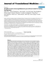
Eight miRNAs have been reported to be regulated by
c-Src [31]. Real-time PCR revealed that levels of
miRNA222 in frontal cortex were robustly increased at
day 3 foll owing trauma, a timepoint corresponding to
maximum upregulation of c-Src. Seven other miRNAs
were also examined: miRNAs 218, 194, 206 showed
weakened signals after traumatic stress whereas there
were no detectable changes in the levels of miRNAs
Zhao et al. Journal of Neuroinflammation 2011, 8:159
/>Page 4 of 13
Figure 1 Induction of c-Src signaling cascades by traumatic stress. Rats were killed 1, 3, or 7 days after traumatic stress (n = 5 for each time
point). Cross sections of frontal cortex were immunostained with anti-c-Src antibody (A), and the density of immunopositive cells was semi-
quantified in three randomly chosen areas (B). Real-time PCR was used to analyze microRNAs in frontal cortex (C). Con: control; T1, 3, 7: 1, 3, 7
days after trauma. Data are presented as percentage of control. Values represent mean ± SD for 3 independent experiments. *P<0.05 vs Con.
Scale bars = 50 μm.
Zhao et al. Journal of Neuroinflammation 2011, 8:159
/>Page 5 of 13
224, 142, 126, or 296. It is therefore possible that
miRNA222 upregulatio n is assoc iated with c-Src signal-
ing and that this could contribute to recovery from
immunosuppression following traumatic stress (Figure
1C).
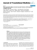
PAK1 is a miRNA222 target
To address whether PAK1 is a target for miRNA222, a
miRNA222 mimetic and/or a miRNA222 inhibitor were
transfected into primary cultured cortical neurons. As
shown in Figure 2A and 2B, the miRNA222 mimetic
decreased mRNA levels for PAK1; conversely, the
miRNA222 inhibitor increased the levels of PAK1
mRNA. Similar effects were observed at the protein
level: expression of PAK1 polypeptide was decreased by
the miRNA222 mimetic whereas the miRNA222 inhib i-
tor increased PAK1 protein levels. We conclude that
PAK1 is negatively regulated by miRNA222.
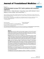
Time-dependent PAK1 expression in response to
traumatic stress
Immunofluorescence using anti-PAK1 antibody on
sections of frontal cortex demonstrated a time-depen-
dent modulation of protein levels following trauma.
Levels were strongly increased by 1 day after trauma,
but decreased progressively at days 3 and 7 (Figure
3A).
Because PAK1 is known to modulate the cellular
effects of IL-1b, we investigated if changes in PAK1
expression are accompanied by parallel changes in
expression of the IL-1 receptor IL-1RI. As shown in Fig-
ure 3B, ELISA assay revealed that IL-1RI expression was
increasedbyover3-fold1dayaftertrauma(354.0±
45.7% control), and gradually decreased thereafter,
returning to control levels at day 7 (Figure 3B). The pat-
tern of PAK1 expression paralleled that previously
reported for IL-1b signaling after trauma [11], suggest-
ing a potential association with neuroimmune modula-
tion in the traumatic rat.
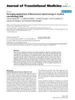
PAK1 and IL-1RI modulation following traumatic stress
It has been previously reported that P AK1 can interact
directly with IL-1RI [25,26]. We therefore investigated
whether the interaction is altered following traumatic
stress. As shown in Figure 4A, B, anti-IL-1RI immuno-
precipitates of rat cortex following trauma were signifi-
cantly enriched in PAK1 material; the binding
interaction was highest at day 1 following trauma and
declined progressively thereafter. This result suggests
that trauma augments the interaction between PAK1
and IL-1R1.
To address whether the increased binding is accompa-
nied by changes in the cellular distribution of IL-1RI,
subcellular fractions from prefrontal cortex were ana-
lyzed by western blotting for IL-1RI. As shown in Figure
4C, monosialoganglioside GM-1, a marker of lipid rafts,
was highly enriched in fractions 4 and 5, indicating that
these represent the lipid-raft membrane microdomain.
In control rats, the IL-1RI immunopositive signal was
generally p resent in the non-raft fract ions. However, at
day 1 following trauma IL-1RI was redistributed, and
immunoreactivity was predominantly associated with
lipid-raft fractions 4 and 5. The proportion of IL-1RI
associated with the raft fraction then declined, and by
days 3 and 7 following trauma IL-1RI immunopositivity
was widely distributed in non-raft fractions (Figure 4C).
IL-1RI activation is known to be accompanied by phos-
phorylation and recruitment into lipid rafts. In addition
to stress induction of IL-1RI expression, our data are
consistent with the possibility that elevated levels of
PAK1 fol lowing traumatic stress also lead to redistribu-
tion and/or activation of receptor, thereby increasing the
cellular effects of IL-1b.
Modulation of PAK-1 signaling in cultured neurons and
glial cells
Neuronal and glial cells cohabit the CNS and both cell
types demonstrate marked changes associated with neu-
roimmune modulation following t raumatic stress [32].
Figure 2 PAK1 is a miRNA222 target. Ra t cortical neurons were transf ected with con trol RNA, microRNA222 mimetic, or microRNA222
inhibitor using Lipofectamine 2000. Two days after transfection, mRNA (A) and protein (B) levels of PAK1 were determined by real-time PCR and
western blot, respectively. Results are normalized against an internal control (b-actin) and further normalized against the results obtained from
cultures transfected with control RNA. The graph depicts percentage expression under the indicated treatments, relative to controls. Data were
analyzed by one-way ANOVA with Dunnett test as a post hoc test for the comparisons.* P<0.05 vs Con, # P<0.05 vs microRNA222 mimetic.
Zhao et al. Journal of Neuroinflammation 2011, 8:159
/>Page 6 of 13
We therefore examined the cellular distribution and
levels of c-Src and PAK1 in neuronal and glial cells in
culture. As shown in Figure 5A, immunostaining for c-
Src (green fluorescence) and PAK1 (red fluorescence)
revealed widespread dual staining in neurons (yellow
coloration), suggesting that c-Src and PAK1 are largely
colocalized in these cells. The same experiment was
repeated for astrocytes and microglia, and overlap of c-
Figure 3 Time-dependent PAK1 expression in response to traumatic stress. Rats were killed 1, 3, or 7 days after traumatic stress (n = 5 for
each time point). Cross sections of frontal cortex were immunostained for anti-PAK1 antibody (A), and the density of immunopositive cells was
semi-quantified in three randomly chosen areas (B). Frontal cortex homogenates were prepared, and IL-1RI expression was determined by ELISA
assay (C). Con: control; T1, 3, 7: 1, 3, 7 days after trauma. Data are presented as percentage of control; each value represents mean ± SD for three
independent experiments. *P<0.05 vs Con. Scale bars = 50 μm.
Zhao et al. Journal of Neuroinflammation 2011, 8:159
/>Page 7 of 13
Src and PAK1 fluorescence was also observed in these
cells (data not presented). This result argues that coex-
pression of the two proteins is likely to be widespread
in the CNS.
We then examined whether c-Src overexpression can
modulate the expression of PAK1. As shown in Figure
5D, E, directed expression of c-Src i n cultured neurons
by transfection with adenovirus expressing con stitutively
active c-Src (CA c-Src) led to a marked reduction in
levels of PAK1 expression and, moreover, upre gulated
levels of miRNA222. Conversely, when endogenous c-
Src activity was blocked by the synthetic inhibitor PP2,
miRNA222 expression was downregulated and PAK1
expression was strengthened (Figure 5B, C). In the
meantime, the association of PAK1 and IL-1RI was
affected by c-Src modulation, which was decreased by
CA c-Src (48 ± 7% control) and elevated by PP2 (291 ±
16% control) (Figure 5D, E).Moreover,whenneurons
were exposed to IL-1b, the association of PAK1 with IL-
1RI was dramatical ly enhanced compared to cells in the
absence of IL-1b, and th is effect was potently and speci-
fically b locked by inhibition of IL-1RI by IL-1ra (Figure
5F, G). Equivalent results were obtained in c ultured
astrocytes and microglia (data not shown).
c-Src signaling in neuroimmune modulation in the
trauma rat
These observ ations together s uggest that c- Src is
strongly upregulated by traumatic stress, and is more-
over a potent regulator of miRNA222 and PAK1: it is
therefore possible that changes in the le vels of PAK1
following stress could in turn could be responsible for
alterations in IL-1RI receptor activation following
trauma. We therefore addressed whether m odulation of
c-Src ac tivity in vivo would impact upon the expression
of miRNA222 and PAK1 and on the PAK1 interaction
with IL-1RI.
Accordingly, at day 3 following trauma rats were
injected icv with adenovirus expressing the dominant-
negative (DN) form of c-Src, and changes in levels of
miRNA222 and PAK1 were measured 72 hour later. As
shown in Figure 6A, B, DN-c-Src resulted in a dramatic
reduction in levels of miRNA222 and an equally robust
incre ase in levels of PAK1 expression. We also explored
the effects of administering the equivalent form of con-
stitutively active (CA) c-Src. CA-c-Src administration
resulted in an inverse effect, leading to increased
miRNA222 levels and decreased PAK1 expression.
Furthermore, the association of PAK1 and IL-1RI was
also similarly modulat ed by administration of DN-c-SRc
or CA-c-Src (Figure 6C, D).
These data argue that c-Src is a positive regulator of
miRNA222. Because PAK1 is a target for miRNA222, it
is possible that c-Src modulates the PAK1-IL-1RI inter-
action by upregulating miRNA222 and inhibiting the
expression of PAK1. Given that c-Src is strongly upre-
gulated by traumati c stress, miR NA222 is a strong con-
tender for the mechanistic link between c-Src activation
and neuroimmune modulation following traumatic
stress. To address this possibility we studied the effects
Figure 4 PAK1 and IL-1RI modulation following traumatic stress. Rats were killed 1, 3, or 7 days after traumatic stress (n = 5 for each time
point), and frontal cortex homogenates were prepared. Immunoprecipitation was used to analyze alterations of PAK1 and IL-RI interaction. The
immunoprecipitation antibody was anti-IL-1RI and the immunoblotting antibody was anti-PAK1 (A). Panel B depicts quantitative analysis of A.
Data are presented as percentage of control, with the density of PAK1 in the control group (without operation) set at 100%. Values represent
mean ± SD for 3 independent experiments. *P<0.05 vs Con. A lipid raft preparation was prepared to determine subcellular distribution of IL-1RI.
Western blot analysis was used to detect IL-1RI expression in fractions 3-11, and GM-1 immunopositive fractions were identified as lipid raft
fractions (C). Con: control; T1, 3: 1, 3 days after trauma.
Zhao et al. Journal of Neuroinflammation 2011, 8:159
/>Page 8 of 13
Figure 5 Modulation of PAK-1 signaling in cultured neurons. Rat cortical neurons were grown on coverslips for 10 days. Neurons were then
immunostained using anti-c-Src and anti-PAK1 antibodies, and double-labeled cells were identified using a Leika Q500IW image analysis system
(A). Neurons were treated with vehicle or PP2 for 30 min, and then assessed for microRNA222 (B) and PAK 1 (C) expression using real-time PCR
and western blot, respectively. (D) Directed expression of c-Src in cultured neurons by transfection with adenovirus expressing active c-Src (CA c-
Src). Endogenous c-Src activity was blocked using the synthetic inhibitor PP2, and association of PAK1 with IL-1RI was determined by
immunoprecipitation assay. The immunoprecipitation antibody was anti-IL-1RI and the immunoblotting antibody was anti-PAK1. (E) The graph
depicts expressions as percentages of controls. (F) Neurons were exposed to IL-1b and IL-1ra as described in Methods, and immunoprecipitation
was used to analyze the association between PAK1 and IL-RI. The immunoprecipitation antibody was anti-IL-1RI and the immunoblotting
antibody was anti-PAK1. The graph depicts expressions as percentages of controls (G). The results were normalized against an internal control
and further normalized against the results obtained from control cultures. Data were analyzed by one-way ANOVA with Dunnett test as a post
hoc test to assess comparisons.* P<0.05 vs Con, # P<0.05 vs c-Src, or IL-1b. Scale bars = 50 μm.
Zhao et al. Journal of Neuroinflammation 2011, 8:159
/>Page 9 of 13
ofc-Srcmodulationonthesuppressionoflymphocyte
proliferation and NK cell activity following traumatic
stress. This revealed that inhibition of c-Src by DN c-
Src led to a significant reduction of both lymphocyte
proliferations and NK cell activity, [
3
H] incorporation
for lymphocyte proliferation was 71 ± 8 and 71 ± 9% of
control at day 3 after trauma and DN c-Src injection
respectively. For NK cell activity, they were 70 ± 9 and
79 ± 7% of control, whereas, conversely, c-Src activation
promoted the recovery from immunosuppression (Fig-
ure 7A).
To investigate the potential involvement of PAK1 in
this process, antibody against PAK1 was injected icv. As
shown in Figure 7B, abrogation of PAK1 activity with
anti-PAK1 decreased lymphocyte proliferation and NK
cell activity. We attribute this effect to inhibition of
PAK1 enhancement of IL-1RI receptor expression and
activation. To c onfirm that IL-1RI plays a role in this
system, we investigated the effects of administering IL-
1ra. As also shown in Figure 7B, IL-1ra exerted a similar
progressive effect on PAK1 in the traumatic rat.
Discussion
Recently, it has been reported that c-Src is likely to play
a regulatory role in immunosuppression induced by
trauma in rats [14]. In the present paper we have shown
that activation of c-Src is accompanied by strong upre-
gulation of expression of miRNA222, an inhibitor of the
immunoregulator PAK1. We therefore postulate that
miRNA222 provides a mechanistic link between c-Src
and immunosuppression following traumatic stress.
Members of the Src family of protein tyrosine kinases
are known to mediate a signaling cascade that relays
information from the cell surface to the nucleus, pro-
moting an array of cellular responses [14,33]. Tyrosine-
phosphorylated signaling molecules have been directly
implicated in neurite outgrowth that is thought to
reflect an early step in neuronal regeneration [34-36].
Figure 6 PAK1 signaling modulation in traumatiz ed rats. Rats were subjected to surgical trauma, and 3 days later some of these rats were
injected icv with adenovirus expressing either the dominant-negative (DN) form of c-Src or the equivalent form of constitutively active (CA) c-
Src. Thus, 4 groups of rats were created: Controls (rats with no trauma), T3 (rats killed 3 days after trauma), T3+DN c-Src (rats treated with DN c-
Src 3 days after trauma and killed 72 hours later), and T3+CA c-Src (rats treated with CA c-Src 3 days after trauma and killed 72 hours later) (n =
5 for each group). Homogenates of frontal cortex were prepared and assessed for microRNA222 (A) and PAK1 (B) expression using real-time PCR
and western blot, respectively. The interaction of PAK1 and IL-RI was assessed by immunoprecipitation (C, D). Data are presented as percentage
of control. Values represent mean ± SD for 3 independent experiments. *P<0.05 vs Con. Con: control; T3: 3 days after trauma.
Zhao et al. Journal of Neuroinflammation 2011, 8:159
/>Page 10 of 13
Recent studies have revealed that c-Src overlaps func-
tionally with miRNA222, and their combined effects
modula te not only cancer cell proliferation [37] but also
the fine tuning of gene expression during cell differen-
tiation and brain development [38]. The interaction
between c-Sr c and miRNA222 expression reported here
isthereforelikelytoplayaroleinorchestratingneu-
roimmune changes following traumatic stress.
miRNAs are small noncoding RNAs that typically bind
to the 3’-untranslated regions of protein-coding genes,
repressing their expression by translational inhibition
and/or promoting mRNA degradation [39]. PAK1 has
been identified as one of the targets of miRNA222
[40,41], a finding confirmed here. Extending this work,
we demonstrate that, following traumatic stress,
miRNA222 and PAK1 expression in frontal cortex are
inversely modulated. Specifically, PAK1 expression is
strongly upregulated at day 1 following trauma, and
thereafter progressively decreases to control levels at day
7. These observations suggest the hypothesis that
Figure 7 c-Src signaling in neuroimmune modulation in traumatized rats. Four groups of rats were prepared as described in Fig. 6: Con, T3,
T3+DN c-Src, T3+CA c-Src (n = 5 for each group). Homogenates of spleen were prepared, and lymphocyte proliferation and NK cell activity were
assayed by [
3
H] incorporation (A). Rats were subjected to surgical trauma, and 1 day later some of these rats were treated with PAK-1 Ab or IL-
1ra. Thus, 4 groups of rats were created: Controls (Con; no trauma), T1 (rats killed 1 day after surgical trauma), T1+PAK-1 Ab (icv administration of
antibody against PAK1, 1 day after trauma and killed 24 hours later), and T1+IL-1ra (icv administration of antibody against IL-1ra 1 day after
trauma and killed 24 hours later) (n = 5 for each group). Homogenates of spleen were collected, and lymphocyte proliferation and NK cell
activity were assayed by [
3
H] incorporation (B). Data are presented as percentage of control. Values represent mean ± SD for 3 independent
experiments. *p < 0.05 vs Con, #p < 0.05 vs traumatized.
Zhao et al. Journal of Neuroinflammation 2011, 8:159
/>Page 11 of 13
increased miRNA levels induced by traumatic stress
exert their cellular effect s by inhibiting PAK1 expres-
sion, and thereby downregulating PAK1-mediated facili-
tation of IL-1 signaling and, potentially, mediating
recovery from immunosuppression following trauma.
IL-1b was the first cytokine to be associated with
modulation of neuroendocrine systems, particularly the
hypothalamic pituitary-adrenal axis (HPA) and the
hypothalamic-pituitary-gonadal axis, in the 1980s. To
date, IL-1b has remained the most studied inflammatory
cytokine in the mediation of immunological and psycho-
logical stress responses. IL-1b signaling is mediated by a
complex system, and IL-1 receptor type I (IL-1RI)
appears to mediate all of the known biological functions
of IL-1 [1]. In regard to traumatic stress, we have found
that elevated IL-1b expression is a primary response to
traumatic stress [11], and that activation of IL-1b signal-
ing is postulated to mediate immunosuppression in the
traumatic rat [11]. In the present study we report that
IL-1RI expression is dramatically increased at day 1 fol-
lowing trauma. Importantly, upregulation was accompa-
nied by both increased PAK1 binding and accumulation
of IL-1RI in lipid-raft fractions, a marker of receptor
activation. Detailed examination of the role of PAK1
indicated that activated PAK1 is enriched primarily in
postmitotic neurons and major axonal tracts [42,43].
PAK1 activation reflects its recruitment t o the plasma
membrane, and this has been demonstrated both in
post-traumatic disorders and in progressive neurodegen-
erative conditions [44]. It was also recently reported that
PAK1 activation could b e respon sible for changes in IL-
1b/IL-RI activation i n the CNS following stress [45-48].
It is therefore possible that PAK1 modulates IL-1RI acti-
vation and translocation into lipid rafts, thereby fine-
tuning IL-1b signaling in the onset and progression of
immunosuppression in the traumatic rat.
It is noteworthy that these signaling cascades al so
appear to operate in cultured neuron and glial cells.
PAK1 is a target for miRNA222, and c-Src reproducibly
produced up- or downregulation of miRNA222 and
PAK1. PAK1 could function as a conveying molecule to
enhance phosphorylation and lipid-raft association of
IL-1RI, thereby facilitating IL-1b signaling. Importantly,
c-Src activation is likely to modulate the association of
PAK1 an d IL-1RI. We therefore propose that c-Src
upregulation following traumatic stress exerts its neu-
roimmunomodulatory effects by upregulating
miRNA222, thereby reducing PAK1 expressi on and
downregulating IL1-b/IL1-RI signaling.
Certai nly, there ar e many NeurimmiRs, which primar-
ily enable modulation of both immune and stress
responses through direct or indirect alterations of neu-
ron-glia signaling. Based on the present observations,
miRNA222 may ac t as a “negotiato r” between c-Src and
IL-1b signaling compartments [18]. Also, miRNA222 is
likely to have other targets in addition to PAK1, and the
impact of c-Src modulation of miRNA222 expression
may not be restricted to PAK1. However, the observa-
tion that the time course of miRNA222 overexpression
following traumatic stress matches both the depression
of PAK1 expression and immunosuppression supports
the contention that PAK1 is a primary target for
miRNA222 and contributes to neuroimmune modula-
tion following traumatic stress.
Conclusions
In summary, we report that c-Src activation following
traumatic stress leads to a robust increa se in lev els of
miRNA222 and a corresponding decrease in expression
of the neuromodulator PAK1, a confirmed target for
miRNA222. PAK1 is a key regulator of IL-1b signaling
through its association with IL-1RI and, moreover, mod-
ulates the subcellular distribution of IL-1RI, enhancing
its association with lipid rafts and its signaling activity.
Together our data suggest that the pronounced immu-
nosuppression that occurs in the CNS following trau-
matic stress [47,48] is mediated by a regulatory cascade
involving c-Src, miRNA22, and PAK1 that then l eads to
abrogation of IL-1RI signaling.
Abbreviations
Icv: intracerebraventricular injection; IL-1β: interleukin-1β; IL-1RI: type 1 IL-1
receptor; MBS: MES-buffered saline (MES: 2-[morpholino]ethanesulfonic acid);
NK cell: natural killer cell; PAK1: Rac1/p21-activated kinase 1; PP2: 4-amino-5-
(4-chlorophenyl)-7-(t-butyl)pyrazolo[3,4-d]pyrimidine).
Acknowledgements
The study is supported by the National Key Basic Research program of China
(2007CB512502).
Authors’ contributions
HZ produced the hypothesis for this study. XC and GW gave extensive
advice on the study. RY was responsible for the animal and primary neuron
and glial cell culture study. All authors read the manuscript, studied it
critically for its intellectual content and approved the final draft.
Competing interests
The authors declare that they have no competing interests.
Received: 5 September 2011 Accepted: 14 November 2011
Published: 14 November 2011
References
1. Goshen I, Yirmiya R: Interleukin-1 (IL-1): A central regulator of stress
responses. Front Neuroendocrinol 2009, 30:30-45.
2. Stahel PF, Kariya K, Shohami E, Barnum SR, Eugster HP, Trentz O,
Kossmann T, Morganti-Kossmann MC: Intracerebral complement C5a
receptor (CD88) expression is regulated by TNF and lymphotoxin-a
following closed head injury in mice. J Neuroimmunol 2000, 109:164-172.
3. Ginis I, Hallenbeck JM, Liu J, Spatz M, Jaiswal R, Shohami E: Tumor necrosis
factor and reactive oxygen species cooperative cytotoxicity is mediated
via inhibition of NF-κB. Mol Med 2000, 6:1028-1041.
4. Shaked I, Meerson A, Wolf Y, Avni R, Greenberg D, Gilboa-Geffen A,
Soreq H: MicroRNA-132 Potentiates Cholinergic Anti-Inflammatory
Signaling by Targeting Acetylcholinesterase. Immunity 2009, 31:965-973.
Zhao et al. Journal of Neuroinflammation 2011, 8:159
/>Page 12 of 13
5. Zhao H, Du LN, Jiang JW, Wu GC, Cao XD: Modulatory effect of orphanin
FQ on the IL-1beta transcripts in hippocampus. Sheng Li Xue Bao 2001,
53:209-214.
6. John GR, Lee SC, Song X, Rivieccio M, Brosnan CF: IL-1-regulated
responses in astrocytes: relevance to injury and recovery. Glia 2005,
49:161-176.
7. Agay D, Andriollo-Sanchez M, Claeyssen R, Touvard L, Denis J, Roussel AM,
Chancerelle Y: Interleukin-6, TNF-alpha and interleukin-1 beta levels in
blood and tissue in severely burned rats. Eur Cytokine Netw 2008, 19:1-7.
8. Murray CA, McGahon B, McBennett S, Lynch MA: Interleukin-1 beta
inhibits glutamate release in hippocampus of young, but not aged, rats.
Neurobiol Aging 1997, 18:343-348.
9. Konsman JP, Tridon V, Dantzer R: Diffusion and action of
intracerebroventricularly injected interleukin-1 in the CNS. Neuroscience
2000, 101:957-67.
10. Li Y, Liu L, Kang J, Sheng JG, Barger SW, Mrak RE, Griffin WS: Neuronal-glial
interactions mediated by interleukin-1 enhance neuronal
acetylcholinesterase activity and mRNA expression. J Neurosci 2000,
20:149-55.
11. Zhao H, Huang HW, Wu GC, Cao XD: Effect of orphanin FQ on
interleukin-1 beta mRNA transcripts in the rat CNS. Neuroscience 2002,
114:1019-1031.
12. Oprica M, Hjorth E, Spulber S, Popescu BO, Ankarcrona M, Winblad B,
Schultzberg M: Studies on brain volume, Alzheimer-related proteins and
cytokines in mice with chronic overexpression of IL-1 receptor
antagonist. J Cell Mol Med 2007, 11:810-825.
13. Smith DE, Lipsky BP, Russell C, Ketchem RR, Kirchner J, Hensley K, Huang Y,
Friedman WJ, Boissonneault V, Plante MM, Rivest S, Sims JE: A central
nervous system-restricted isoform of the interleukin-1 receptor
accessory protein modulates neuronal responses to interleukin-1.
Immunity 2009, 30:817-831.
14. Xiao S, Wang J, Jiang JW, Cao XD, Wu GC, Zhao H: Characterization of Fyn
signaling on the age-dependent immunomodulation on traumatic rats.
Brain Res 2009, 1255:162-169.
15. Basuroy S, Dunagan M, Sheth P, Seth A, Rao RK: Hydrogen peroxide
activates focal adhesion kinase and c-Src by a phosphatidylinositol 3
kinase-dependent mechanism and promotes cell migration in Caco-2
cell monolayers. Am J Physiol Gastrointest Liver Physiol 2010, 299:G186-95.
16. Takino T, Tsuge H, Ozawa T, Sato H: MT1-MMP promotes cell growth and
ERK activation through c-Src and paxillin in three-dimensional collagen
matrix. Biochem Biophys Res Commun 2010, 396:1042-7.
17. Li X, Shen Y, Ichikawa H, Antes T, Goldberg GS: Regulation of miRNA
expression by Src and contact normalization: effects on nonanchored
cell growth and migration. Oncogene 2009, 28:4272-83.
18. Meerson A, Cacheaux L, Goosens KA, Sapolsky RM, Soreq H, Kaufer D:
Changes in brain microRNAs contribute to cholinergic stress reactions. J
Mol Neurosci
2010, 40:47-55.
19. Soreq H, Wolf Y: NeurimmiRs: microRNAs in the neuroimmune interface.
Trends Mol Med 2011, 17:548-555.
20. Zhang J, Han L, Ge Y, Zhou X, Zhang A, Zhang C, Zhong Y, You Y, Pu P,
Kang C: miR-221/222 promote malignant progression of glioma through
activation of the Akt pathway. Int J Oncol 2010, 36:913-20.
21. Cardinali B, Castellani L, Fasanaro P, Basso A, Alemà S, Martelli F, Falcone G:
Microrna-221 and microrna-222 modulate differentiation and maturation
of skeletal muscle cells. PLoS One 2009, 4:e7607.
22. Asrar S, Meng Y, Zhou Z, Todorovski Z, Huang WW, Jia Z: Regulation of
hippocampal long-term potentiation by p21-activated protein kinase 1
(PAK1). Neuropharmacology 2009, 56:73-80.
23. Viviani B, Bartesaghi S, Gardoni F, Vezzani A, Behrens MM, Bartfai T,
Binaglia M, Corsini E, Di Luca M, Galli CL, Marinovich M: Interleukin-1beta
enhances NMDA receptor-mediated intracellular calcium increase
through activation of the Src family of kinases. J Neurosci 2003,
23:8692-8700.
24. Bergamaschi A, Corsi M, Garnier MJ: Synergistic effects of cAMP-
dependent signalling pathways and IL-1 on IL-6 production by H19-7/
IGF-IR neuronal cells. Cell Signal 2006, 18:1679-1684.
25. Walaas SI, Greengard P: Protein phosphorylation and neuronal function.
Pharmacological Reviews 1991, 43:299-349.
26. Zhou L, Yan C, Gieling RG, Kida Y, Garner W, Li W, Han YP: Tumor necrosis
factor-alpha induced expression of matrix metalloproteinase-9 through
p21-activated Kinase-1. BMC Immunol 2009, 10:15.
27. Robbins TW: Shifting and stopping: fronto-striatal substrates,
neurochemical modulation and clinical implications. Philos Trans R Soc
Lond B Biol Sci 2007, 362:917-932.
28. Hopkins SJ: Central nervous system recognition of peripheral
inflammation: a neural, hormonal collaboration. Acta Biomed 2007,
78(Suppl 1):231-247.
29. Tsirpanlis G: Cellular senescence and inflammation: a noteworthy link.
Blood Purif 2009, 28:12-14.
30. Song KS, Li SW, Okamoto T, Quilliam LA, Sargiacomo M, Lisanti MP: Co-
purification and direct interaction of Ras with caveolin, an integral
membrane protein of caveolae microdomains. A detergent-free
purification of caveolae microdomains. J Biol Chem 1996, 271:9690-9697.
31. Li X, Shen Y, Ichikawa H, Antes T, Goldberg GS: Regulation of miRNA
expression by Src and contact normalization: effects on nonanchored
cell growth and migration. Oncogene 2009, 28:4272-83.
32. Zhao H, Xiao S, Kong X, Wang J, Cao XD, Wu GC, Loh HH, Law PY: Neuron-
glial cell communication in the traumatic stress-induced
immunomodulation. Synapse
2011, 65:433-440.
33. Cooper JA, Qian H: A mechanism for SRC kinase-dependent signaling by
noncatalytic receptors. Biochemistry 2008, 47:5681-5688.
34. Endo M, Yamashita T: Inactivation of Ras by p120GAP via focal adhesion
kinase dephosphorylation mediates RGMa-induced growth cone
collapse. J Neurosci 2009, 29:6649-62.
35. Martinez-Rico C, Pincet F, Thiery JP, Dufour S: Integrins stimulate E-
cadherin-mediated intercellular adhesion by regulating Src-kinase
activation and actomyosin contractility. J Cell Sci 2010, 123:712-22.
36. Messa M, Congia S, Defranchi E, Valtorta F, Fassio A, Onofri F, Benfenati F:
Tyrosine phosphorylation of synapsin I by Src regulates synaptic-vesicle
trafficking. J Cell Sci 2010, 123:2256-65.
37. Khanna S, Roy S: Micromanaging vascular biology: tiny microRNAs play
big band. Sen CK, Gordillo GM, J Vasc Res 2009, 46:527-40.
38. Hussein K, von Neuhoff N, Büsche G, Buhr T, Kreipe H, Bock O: Opposite
expression pattern of Src kinase Lyn in acute and chronic
haematological malignancies. Ann Hematol 2009, 88:1059-67.
39. Newman MA, Hammond SM: Emerging paradigms of regulated
microRNA processing. Genes Dev 2010, 24:1086-92.
40. Reddy SD, Ohshiro K, Rayala SK, Kumar R: MicroRNA-7, a homeobox D10
target, inhibits p21-activated kinase 1 and regulates its functions. Cancer
Res 2008, 68:8195-200.
41. Siomi H, Siomi MC: Posttranscriptional regulation of microRNA biogenesis
in animals. Mol Cell 2010, 38:323-32.
42. Hayashi K, Ohshima T, Hashimoto M, Mikoshiba K: Pak1 regulates dendritic
branching and spine formation. Dev Neurobiol 2007, 67:655-69.
43. Causeret F, Terao M, Jacobs T, Nishimura YV, Yanagawa Y, Obata K,
Hoshino M, Nikolic M: The p21-activated kinase is required for neuronal
migration in the cerebral cortex. Cereb Cortex 2009, 19:861-75.
44. Jacobs T, Causeret F, Nishimura YV, Terao M, Norman A, Hoshino M,
Nikolić M: Localized activation of p21-activated kinase controls neuronal
polarity and morphology. J Neurosci 2007, 27:8604-15.
45. Clerk A, Sugden PH: Activation of p21-activated protein kinase alpha
(alpha PAK) by hyperosmotic shock in neonatal ventricular myocytes.
FEBS Lett 1997, 403:23-5.
46. Basak C, Pathak SK, Bhattacharyya A, Mandal D, Pathak S, Kundu M: NF-
kappaB- and C/EBPbeta-driven interleukin-1beta gene expression and
PAK1-mediated caspase-1 activation play essential roles in interleukin-
1beta release from Helicobacter pylori lipopolysaccharide-stimulated
macrophages. J Biol Chem 2005, 280:4279-88.
47. Demyanenko GP, Halberstadt AI, Rao RS, Maness PF: CHL1 cooperates with
PAK1-3 to regulate morphological differentiation of embryonic cortical
neurons. Neuroscience 2010, 165:107-15.
48. Kreis P, Barnier JV: PAK signalling in neuronal physiology. Cell Signal 2009,
21:384-93.
doi:10.1186/1742-2094-8-159
Cite this article as: Zhao et al.: Neuroimmune modulation following
traumatic stress in rats: evidence for an immunoregulatory cascade
mediated by c-Src, miRNA222 and PAK1. Journal of Neuroinflammation
2011 8:159.
Zhao et al. Journal of Neuroinflammation 2011, 8:159
/>Page 13 of 13