báo cáo hóa học:" Genetic lesions within the 3a gene of SARS-CoV" pdf
Bạn đang xem bản rút gọn của tài liệu. Xem và tải ngay bản đầy đủ của tài liệu tại đây (517.8 KB, 4 trang )
BioMed Central
Page 1 of 4
(page number not for citation purposes)
Virology Journal
Open Access
Short report
Genetic lesions within the 3a gene of SARS-CoV
Timothy HP Tan*
1
, Timothy Barkham
2
, Burtram C Fielding
1
, Chih-
Fong Chou
1
, Shuo Shen
1
, Seng Gee Lim
1
, Wanjin Hong
1
and Yee-Joo Tan
1
Address:
1
Institute of Molecular and Cell Biology, 61 Biopolis Drive, 138673 Singapore and
2
Tan Tock Seng Hospital, 11 Jalan Tan Tock Seng,
308433 Singapore
Email: Timothy HP Tan* - ; Timothy Barkham - ;
Burtram C Fielding - ; Chih-Fong Chou - ; Shuo Shen - ;
Seng Gee Lim - ; Wanjin Hong - ; Yee-Joo Tan -
* Corresponding author
Abstract
A series of frameshift mutations within the 3a gene has been observed in culture-derived severe
acute respiratory syndrome coronavirus (SARS-CoV). We report here that viral RNA from clinical
samples obtained from SARS-CoV infected patients also contains a heterogeneous population of
wild-type and mutant 3a transcripts.
Findings
Introduction
Numerous isolates of the severe acute respiratory syn-
drome coronavirus (SARS-CoV) have been completely
sequenced [1-4]. In most cases, only synonymous or non-
synonymous substitutions have been reported within the
major viral genes, which include replicase 1a/1b gene
products, spike, membrane, nucleocapsid and envelope
[1-4]. On the other hand, large insertions or deletions
have been found in the part of viral genome that encodes
the SARS-CoV accessory proteins which have no viral
homologues. The ORF 8a/8b region appears to be partic-
ularly prone to mutations as deletions of up to 415 bp
have been observed in some isolates [2]. Although these
mutations do not appear to have any adverse effect on the
survival of the virus, it is conceivable that these mutations
may have effects on viral pathogenesis in vivo, as have
been observed for other coronaviruses [5].
We have previously reported that a frameshift mutation
occurs within the 3a gene of culture-derived SARS-CoV,
which results in a protein with a distinctively shorter N-
terminus than the wild-type form [6]. Protein 3a is one of
the SARS-CoV accessory proteins and the expression of the
3a protein has been demonstrated during both in vitro and
in vivo infection [5]. To determine if the mutation arises
from repeated passages of the virus or if the mutation
exists in the virus that is replicating in SARS-CoV infected
patients, we analyzed viral RNA isolated directly from 8
clinical samples and determined the sequence of the 3a
gene. Interestingly, we have found evidence of a heteroge-
neous population of subgenomic RNA 3 (sgRNA3) tran-
scripts in patients with acute SARS-CoV infection
containing copies of wild-type and mutant 3a genes.
The Study
Total RNA was extracted from 8 patients confirmed with
SARS-CoV infection, as defined by WHO guidelines. The
use of clinical samples for this study was approved by the
Tan Tock Seng Hospital ethics committee. Reverse tran-
scription (RT, Superscript II RT, Invitrogen) was per-
formed on all samples, according to the manufacturer's
protocol, and was followed up by a nested polymerase
chain reaction (PCR). The PCR conditions and
Published: 20 June 2005
Virology Journal 2005, 2:51 doi:10.1186/1743-422X-2-51
Received: 09 May 2005
Accepted: 20 June 2005
This article is available from: />© 2005 Tan et al; licensee BioMed Central Ltd.
This is an Open Access article distributed under the terms of the Creative Commons Attribution License ( />),
which permits unrestricted use, distribution, and reproduction in any medium, provided the original work is properly cited.
Virology Journal 2005, 2:51 />Page 2 of 4
(page number not for citation purposes)
subsequent cloning steps have been described elsewhere
[6]. Essentially, 15 independent clones from each of the
eight SARS patient samples were sequenced. As a polymer-
ase fidelity control of the RT and PCR system, full-length
3a RNA was in vitro transcribed from pXJ40-3a, a cDNA
construct for expressing 3a in mammalian cells [6], and
subjected to an identical follow-up PCR and cloning
protocol.
For the fluorescence-activated cell sorting (FACS) and
Western blot analysis, Vero E6 cells were transiently trans-
fected with pXJmyc-GST, pXJmyc-3a or pXJmyc-3amut1 as
previously described (3a and 3amut1 are also known as
U274 and U274mut1, respectively, in ref. 6). All these
constructs were tagged with the c-myc epitope at the N-ter-
minus. All these experiments were performed as previ-
ously described [6].
Results and Conclusions
We have previously identified an oligo(T) tract within the
3a gene, located 16 bp after the first ATG initiation codon,
which is prone to insertional mutations [6]. According to
several analyses of about 100 genomic sequences of SARS-
CoV isolates (in total) obtained from human and animal
populations and that have been deposited with Genbank,
there has been no report of mutations in this region [1-
4]]. It is possible that direct sequencing results do not
show this mutation if it is present in only a minority pop-
ulation. There was a previous report on a frameshift muta-
tion in the 3a gene but the identity of this isolate was not
mentioned [7]. The 3a gene from culture-derived SARS-
CoV isolates contained heterogeneous extensions at this
internal oligo(T) tract. The nature of this extension was
such that up to three additional T's were added to the 6T's
tract. Any change in the number of T's in this oligo(T)
tract, other than in multiples of three, would result in a
frameshift mutation and premature translation termina-
tion of 3a.
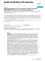
We analyzed this region of the 3a gene from eight patients
confirmed with SARS-CoV infection and have detected the
presence of sgRNA3 transcripts, carrying 6T's to 10T's
tract, in these patients (Figure 1). As our polymerase fidel-
ity controls have confirmed that it is highly unlikely that
sequencing and PCR errors are the source of these nucle-
otide aberrations (data not shown), and these results
showed that different variants of the 3a gene exist in the
viruses that were replicating in these patients. The percent-
age of the different mutant transcripts varied considerably
from patient to patient. However, in 6 out of the 8
patients, more than 50 % of the sgRNA3 transcripts con-
tains either 6T's or 9T's, which means that the full-length
3a (or with 1 additional amino acid) will be expressed. In
patient D, less than 10 % of the transcripts are in-frame,
while in patient E, none of the transcripts is in-frame,
indicating that the full-length 3a protein will be expressed
at a low level (Pat D) or not expressed at all (Pat E). In
comparison, about 27 % of the transcripts from a culture-
derived virus are in-frame [6]. Overall, these results
showed that the frameshift mutations of the 3a gene are
not specific to culture-derived SARS-CoV and that they
point towards the existence of quasispecies within a given
population of SARS-CoVs. As deduced from their study of
sequence variation of the spike gene from viral isolates,
another group has also suggested that SARS-CoV quasis-
pecies exists in vivo [8].
Translation of a frameshifted 3a gene would terminate
shortly after 18 residues or so. Nonetheless, there are two
alternative initiation codons located downstream. Trans-
lation from these would result in two possible smaller 3a
gene products. We have shown that at least one of these
truncated forms of 3a, named as 3amut1, can be detected
in the lysate of infected cells [6]. It has been shown that
the 3a is localized to the cell surface of SARS-CoV infected
cells [6,9]. FACS analysis showed that while the myc-3a
protein (full-length 3a with a c-myc epitope at the N-ter-
minus) was transported to the cell surface in transiently
transfected Vero E6 cells, the myc-3amut1 (truncated 3a
with the same c-myc epitope) could not be detected on cell
surface (Figure 2A). Western blot analysis was also per-
formed to ensure that the expressions of the full-length
and truncated 3a proteins in the transfected cells were
comparable (Figure 2B). As 3amut1 corresponds to 101 to
274 amino acids of 3a and lacks the first two transmem-
brane domains [6], our results showed that these two
transmembrane domains are essential for the expression
of 3a on the cell surface.
It is possible that the aberrant nature of the oligo(T) tract
in the 3a gene is akin to that of the nucleotide insertion
event in the oligo(A) tract of the 3b gene of infectious
bronchitis virus (IBV), another coronavirus [10]. The
insertion of a single adenylate nucleotide into this region
of 3b resulted in a C-terminally truncated gene product.
The corresponding mutant 3b was also localized differ-
ently within the cell. In view of the fact that 3a has also
been shown to interact with the spike protein [6,7] and
that both proteins have a tendency to co-mutate [7], it
would be interesting to know whether these serial
frameshift mutations can also be correlated with a recog-
nizable mutation pattern of the spike gene.
In other RNA viruses, such as the foot-and-mouth disease
virus (FMDV) and human respiratory syncytial virus,
internal poly(A) extensions have been identified before as
a hot spot for mutations [11,12]. Similarly, these exten-
sions also create frameshift mutations in the affected
genes. FMDV populations with longer poly(A) extensions
seem to have a lower fitness value as compared to those
Virology Journal 2005, 2:51 />Page 3 of 4
(page number not for citation purposes)
Distribution of 3a sequence variants within a population of SARS-CoV obtained from clinical and culture-derived samplesFigure 1
Distribution of 3a sequence variants within a population of SARS-CoV obtained from clinical and culture-
derived samples. Pat A to H: patients confirmed with acute SARS-CoV infection. Lab: Vero E6 cells infected with SARS-CoV.
The value above each column represents the percentage of in-frame 3a genes within the population.
Detection of myc-3amut1 in transfected Vero E6 cells by FACS and Western blot analysisFigure 2
Detection of myc-3amut1 in transfected Vero E6 cells by FACS and Western blot analysis. (A) FACS analysis of
live cells transfected with myc-3a, myc-3amut1 and myc-GST. Cells were initially probed with anti-myc monoclonal antibodies
followed by the corresponding FITC-conjugated antibody. (B) Expression levels of the myc-tagged proteins in cells used for the
FACS experiment were determined by Western blot analysis. Probing was done with anti-myc polyclonal antibody.
Publish with BioMed Central and every
scientist can read your work free of charge
"BioMed Central will be the most significant development for
disseminating the results of biomedical research in our lifetime."
Sir Paul Nurse, Cancer Research UK
Your research papers will be:
available free of charge to the entire biomedical community
peer reviewed and published immediately upon acceptance
cited in PubMed and archived on PubMed Central
yours — you keep the copyright
Submit your manuscript here:
/>BioMedcentral
Virology Journal 2005, 2:51 />Page 4 of 4
(page number not for citation purposes)
with shorter extensions [11]. This is despite the fact that
both the wild-type and mutant forms of the affected
FMDV protein, the L protease, have equal functionality
[13]. In addition, viral genomes possessing different
lengths of the poly(A) tract could be recognized even from
just a single PFU of FMDV [11].
It is intriguing to find that unlike 3a, the 3amut1 is not
transported to the cell surface as the cell surface expression
(and endocytotic properties) of 3a may be involved in
modulating the trafficking properties of the spike protein
[14]. Our results showed that the viruses in some of the
patients appear to encode only for the truncated form(s)
of 3a and not the full-length 3a protein (Figure 1) and
indicated that the functionality of full-length 3a is not
essential for virus replication. However, it is also conceiv-
able that the different variants of 3a have different stabili-
ties and/or functions, and hence would contribute
differently to viral pathogenesis in vivo. In addition, it was
recently reported that 3a is a structural protein and at least
2 truncated forms of 3a were dominantly present in the
virion [9]. Further studies will reveal if the truncated
forms of 3a, which results from frameshift mutations in
the viral genome, can be incorporated in the virion and if
there are phenotypic effects of a truncated 3a during the
infection cycle. With respect to viral viability in the natural
host, does full-length 3a confer a fitness gain over trun-
cated 3a? If so, the possibility remains that under selective
pressure, the distribution of viral genotypes could tip in
favor to those which carry the wild-type 3a gene. A similar
genotypic reversion event has been documented for
FMDV [11]. Further studies on a larger cohort of patients
will be necessary to establish if there is a relationship
between the mutations observed in the 3a transcripts and
the severity of the clinical symptoms in individual
patients.
Competing interests
The author(s) declare that they have no competing
interests.
Authors' contributions
THPT carried out all experimental work and drafted the
manuscript. YJT conceived of the study and helped to draft
the manuscript. TB provided the clinical samples. BCF,
CFC, SS, SGL and WH also assisted THPT and YJT in draft-
ing the manuscript. All authors read and approved the
final manuscript.
Acknowledgements
This work was supported by grants from the Agency for Science, Technol-
ogy and Research (A*STAR), Singapore.
References
1. Vega VB, Ruan Y, Liu J, Lee WH, Wei CL, Se-Thoe SY, Tang KF, Zhang
T, Kolatkar PR, Ooi EE, et al.: Mutational dynamics of the SARS
coronavirus in cell culture and human populations isolated in
2003. BMC Infect Dis 2004, 4:32.
2. Chinese SARS Molecular Epidemiology Consortium: Molecular evo-
lution of the SARS coronavirus during the course of the
SARS epidemic in China. Science 2004, 303:1666-1669.
3. Zhu Y, Liu M, Zhao W, Zhang J, Zhang X, Wang K, Gu C, Wu K, Li
Y, Zheng C, et al.: Isolation of virus from a SARS patient and
genome-wide analysis of genetic mutations related to patho-
genesis and epidemiology from 47 SARS-CoV isolates. Virus
Genes 2005, 30:93-102.
4. Yeh SH, Wang HY, Tsai CY, Kao CL, Yang JY, Liu HW, Su IJ, Tsai SF,
Chen DS, Chen PJ: Characterization of severe acute respira-
tory syndrome coronavirus genomes in Taiwan: molecular
epidemiology and genome evolution. Proc Natl Acad Sci U S A
2004, 101:2542-2547.
5. Tan YJ, Lim SG, Hong W: Characterization of viral proteins
encoded by the SARS-coronavirus genome. Antiviral Res 2005,
65:69-78.
6. Tan YJ, Teng E, Shen S, Tan TH, Goh PY, Fielding BC, Ooi EE, Tan
HC, Lim SG, Hong W: A novel severe acute respiratory syn-
drome coronavirus protein, U274, is transported to the cell
surface and undergoes endocytosis. J Virol 2004, 78:6723-6734.
7. Zeng R, Yang RF, Shi MD, Jiang MR, Xie YH, Ruan HQ, Jiang XS, Shi
L, Zhou H, Zhang L, et al.: Characterization of the 3a protein of
SARS-associated coronavirus in infected vero E6 cells and
SARS patients. J Mol Biol 2004, 341:271-279.
8. Xu D, Zhang Z, Wang FS: SARS-associated coronavirus quasis-
pecies in individual patients. N Engl J Med 2004, 350:1366-1367.
9. Ito N, Mossel EC, Narayanan K, Popov VL, Huang C, Inoue T, Peters
CJ, Makino S: Severe acute respiratory syndrome coronavirus
3a protein is a viral structural protein. J Virol 2005,
79:3182-3186.
10. Shen S, Wen ZL, Liu DX: Emergence of a coronavirus infectious
bronchitis virus mutant with a truncated 3b gene: functional
characterization of the 3b protein in pathogenesis and
replication. Virology 2003, 311:16-27.
11. Escarmis C, Davila M, Charpentier N, Bracho A, Moya A, Domingo E:
Genetic lesions associated with Muller's ratchet in an RNA
virus. J Mol Biol 1996, 264:255-267.
12. Garcia-Barreno B, Delgado T, Melero JA: Oligo(A) sequences of
human respiratory syncytial virus G protein gene: assess-
ment of their genetic stability in frameshift mutants. J Virol
1994, 68:5460-5468.
13. Medina M, Domingo E, Brangwyn JK, Belsham GJ: The two species
of the foot-and-mouth disease virus leader protein,
expressed individually, exhibit the same activities. Virology
1993, 194:355-359.
14. Tan YJ: The Severe Acute Respiratory Syndrome (SARS)-
coronavirus 3a protein may function as a modulator of the
trafficking properties of the spike protein. Virol J 2005, 2:5.