báo cáo hóa học:" Strategically examining the full-genome of dengue virus type 3 in clinical isolates reveals its mutation spectra" doc
Bạn đang xem bản rút gọn của tài liệu. Xem và tải ngay bản đầy đủ của tài liệu tại đây (319.72 KB, 10 trang )
BioMed Central
Page 1 of 10
(page number not for citation purposes)
Virology Journal
Open Access
Research
Strategically examining the full-genome of dengue virus type 3 in
clinical isolates reveals its mutation spectra
Day-Yu Chao*
1
, Chwan-Chuen King
1
, Wei-Kung Wang
2
, Wei-June Chen
3
,
Hui-Lin Wu
4
and Gwong-Jen J Chang
5
Address:
1
Institute of Epidemiology, College of Public Health, National Taiwan University (NTU), Taipei, Taiwan (100), Republic of China
(R.O.C.),
2
Institute of Microbiology, College of Medicine, NTU, Taipei, Taiwan (100), Republic of China (R.O.C.),
3
Dept. of Parasitology, Chang
Gung College of Medicine and Technology, Kwei-San, Tao-Yuan, Taiwan (100), Republic of China (R.O.C.),
4
Hepatitis Research Center, NTU
Hospital, Taipei, Taiwan (100), Republic of China (R.O.C.) and
5
Division of Vector-Borne Infectious Diseases, National Center for Infectious
Diseases, Centers for Disease Control and Prevention (CDC), Fort Collins, USA
Email: Day-Yu Chao* - ; Chwan-Chuen King - ; Wei-Kung Wang - ; Wei-
June Chen - ; Hui-Lin Wu - ; Gwong-Jen J Chang -
* Corresponding author
Quasispeciesmutation spectramicro-evolution of dengue virus serotype 3dengue hemorrhagic fever (DHF)sequence diversityTaiwan
Abstract
Background: Previous studies presented the quasispecies spectrum of the envelope region of
dengue virus type 3 (DENV-3) from either clinical specimens or field-caught mosquitoes. However,
the extent of sequence variation among full genomic sequences of DENV within infected individuals
remains largely unknown.
Results: Instead of arbitrarily choosing one genomic region in this study, the full genomic
consensus sequences of six DENV-3 isolates were used to locate four genomic regions that had a
higher potential of sequence heterogeneity at capsid-premembrane (C-prM), envelope (E),
nonstructural protein 3 (NS3), and NS5. The extentof sequence heterogeneity revealed by clonal
sequencing was genomic region-dependent, whereas the NS3 and NS5 had lower sequence
heterogeneity than C-prM and E. Interestingly, the Phylogenetic Analysis by Maximum Likelihood
program (PAML) analysis supported that the domain III of E region, the most heterogeneous region
analyzed, was under the influence of positive selection.
Conclusion: This study confirmed previous reports that the most heterogeneous region of the
dengue viral genome resided at the envelope region, of which the domain III was under positive
selection pressure. Further studies will need to address the influence of these mutations on the
overall fitness in different hosts (i.e., mosquito and human) during dengue viral transmission.
Background
Dengue viruses (DENV), which consisted of four antigen-
ically distinct serotypes (DENV-1, 2, 3 and 4), are the
most important arthropod-borne viruses affecting
humans. After infection, it may result in dengue fever
(DF), dengue haemorrhagic fever (DHF), dengue shock
syndrome (DSS) or death [1,2]. It is estimated that close
to 50–100 million cases of DF and 30,000 fatal cases of
Published: 24 August 2005
Virology Journal 2005, 2:72 doi:10.1186/1743-422X-2-72
Received: 29 June 2005
Accepted: 24 August 2005
This article is available from: />© 2005 Chao et al; licensee BioMed Central Ltd.
This is an Open Access article distributed under the terms of the Creative Commons Attribution License ( />),
which permits unrestricted use, distribution, and reproduction in any medium, provided the original work is properly cited.
Virology Journal 2005, 2:72 />Page 2 of 10
(page number not for citation purposes)
DHF/DSS occur annually in tropical and subtropical
regions. With the increased numbers of dengue patients, it
is indicated the global expansion of epidemic areas, and
increased frequencies of severe DHF/DSS and case fatality
[3]. Considerable efforts have been devoted to developing
vaccines to prevent dengue, but the success of the vaccines
will be dependent on the vaccine strain chosen to direct
against the diversity and evolution of DENV genome.
DENV belongs to the genus Flavivirus, family Flaviviridae,
possessing a positive-sense, single-stranded RNA genome,
which is approximately 10,700 bases in length and con-
tains a single open reading frame [4]. A single polyprotein
translated from the viral RNA is cleaved into 3 structural
proteins [capsid (C), premembrane (prM) and envelope
(E) protein] and 7 nonstructural proteins (NS), with the
gene order as 5'-C-prM/M-E-NS1-NS2A-NS2B-NS3-NS4A-
NS4B-NS5-3'. Like many RNA viruses, the genomic
sequence of a single DENV isolate exists in nature as a col-
lection of highly similar but not identical variants known
as quasispecies due to its high average mutation rate of 10
-
3
to 10
-5
substitution per nucleotide copied and per round
of replication [5,6]. Previous studies using a clonal
sequencing approach amplified viral RNA directly from
DENV-3 infected patients' plasma and the extent of
sequence heterogeneity in the envelope region with mean
pairwise difference ranging from 0.21 to 1.67% have been
observed [7]. There are obvious reasons for selecting the E
gene region for this study, mainly due to its important
biological functions such as receptor-mediated endocyto-
sis, virus-induced cellular tropism and eliciting neutraliza-
tion antibodies. However, one cannot exclude the
biological significance of the sequence heterogeneity in
other genomic regions including non-structural (NS) pro-
teins, 5' and/or 3' non-coding regions (NCR). The well-
studied example of hepatitis C virus (HCV) demonstrated
that the quasispecies dynamics and composition of the
NS5A region may play a role in disease prognosis and in
response to interferon and ribavirin therapy [8]. Although
the previous attempt to correlate the sequence heteroge-
neity of the capsid gene with NS protein 2B gene region of
DENV-3 has observed very similar sequence heterogeneity
with mean pairwise p-distance 0.12–1.2% [9], the extent
of sequence variation among full genomic sequences of
DENV within infected individuals remains largely
unknown. Thus, it is important to address whether the
evidence of different evolutionary processes, such as adap-
tive evolution, shape the population genetics of DENV at
specific genomic regions other than the E region.
An outbreak of DHF, attributed to genotype 2 of DENV-3,
resulted in 111 DF and 23 DHF cases in Tainan (southern
Taiwan) from October 1998 to January of 1999 [10].
DENV-3 was the only serotype isolated during this out-
break, and the seroepidemological study clearly demon-
strated that DHF cases were not associated with secondary
DENV infection [10]. Here we report the selection of the
most prominent variable regions identified by the full-
genomic sequencing of DENV isolates from six clinical
patients during this outbreak. The application of the
clonal sequencing of those variable regions enabled us to
study quasispecies structure of DENV isolates and to pro-
vide a better understanding of the changes in mutation
spectrum at the clonal level and virus evolution.
Results
Heterogeneous regions identified at full genomic scale of
DENV-3
To identify the potential heterogeneous regions of DENV-
3 in the whole-genomic scale, acute-phase plasma sam-
ples were obtained from six dengue patients, including
three DF (designated 1F, 2F and 3F) and three DHF
patients (designated 1H, 2H and 3H). The sequencing
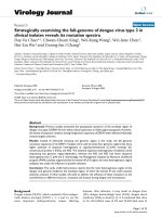
strategy is depicted in Fig 1. These patients' plasma sam-
ples were used to infect the C6/36 mosquito cell line to
obtain sufficient viral genomic RNA for full-genomic con-
sensus sequencing for the identification of regions with
sequence heterogeneity for follow-up clonal sequencing.
The consensus sequence similarity of these six viruses was
as high as 99.73%. The 2H and 3H virus each had two
silent changes at nucleotide positions of 808 (G to A),
9979 (T to C), 4204 (C to T) and 8785 (T to C), respec-
tively (Table 1). There were no consistent nucleotide
changes that might correlate with disease severity among
paired viruses using this consensus sequencing approach.
However, the potential heterogeneous sequence regions
were clearly observed and identified by close examination
of the overlapping chromatogram files using the SeqMan
program in the Lasergene software package (DNASTAR
inc., Madison, WI). Special attention was paid to identify
the regions which consistently presented mixed-chroma-
tographic peaks in the respective trace files obtained from
at least two independent sequencing primers. These
potential heterogeneous regions, located at C/PrM, E, NS3
and NS5 genes (Table 1), were selected for the clonal
sequencing analysis. Five genomic fragments were ampli-
fied directly from six patients' viremic plasma by five
flanking primer pairs (Table 4) at nucleotide position of
1–764 (5'NCR/C/prM), 1259–2550 (E/NS1), 5443–6337
(NS3) and 8501–10316 (NS5/3'NCR) using Titan™ one
tube RT-PCR System (Boehringer Mannheim). After
excluding the primer sequences, the C/PrM region was
752 nucleotides in length with 225 amino acids in the
coding region; the E/NS1 region was 1239 nucleotides in
length covering 413 amino acids which included 40
amino acids at the N terminal end of NS1 protein, the NS3
region was 866 nucleotides in length covering 288 amino
acids, and the NS5 region was 1791 nucleotides in length
with 586 amino acids in viral coding sequences.
Virology Journal 2005, 2:72 />Page 3 of 10
(page number not for citation purposes)
Clonal sequencing of the heterogeneous regions among
dengue viral genomes
The pCRII-TOPO™ T/A cloning kit (Invitrogen, San Diego,
CA) was used to clone PCR products representing hetero-
geneous sequence regions identified by the consensus
sequencing as described previously [7]. At least 20 to 30
clones containing the PCR amplicons from four heteroge-
neous gene regions (C-PrM, E, NS3 and NS5) were
sequenced, aligned, and analyzed using the program GCG
and MEGA v3.0 [11]. In general, the transitional substitu-
tions were higher than transversional substitutions for all
samples analyzed. The transitional changes (A to G or T to
C) constituted overall substitution rate of 72.8 ± 5.1%,
73.7 ± 11% at C/PrM, E of structural proteins, and the NS3
and NS5 regions had relatively lower such changes of 63.2
± 7.5%, 44.7 ± 18%. The lowest nucleotide mutation fre-
quencies were observed in NS3 region with a mean ±
standard deviation (SD) of 0.6 ± 0.3 × 10
-3
for all clones
analyzed, followed by C/PrM (1.2 ± 0.15 × 10
-3
), NS5 (1.5
± 0.4 × 10
-3
) and envelope region (1.8 ± 0.8 × 10
-3
), which
was statistically significant (p < 0.01). Similarly, the sub-
stitution frequencies of amino acids were also variable
among the viral genome regions with the lowest fre-
quency observed in NS3 (1.3 ± 0.4 × 10
-3
), followed by C/
PrM (2.3 ± 0.3 × 10
-3
), E (3.1 ± 1.8 × 10
-3
) and NS5 region
(3.3 ± 0.8 × 10
-3
) (Table 2).
The mean pairwise p-distance as described in the previous
study [7] was employed to compare the extent of sequence
variation among different viral genome regions. Consist-
ently, NS3 had the lowest pairwise p-distance among NS5,
C/PrM or E protein. The average mean p-distance in nucle-
otides and SD for NS3, C-PrM, NS5, and E were 1.4 ± 0.6
× 10
-3
, 2.3 ± 0.3 × 10
-3
, 3.1 ± 0.9 × 10
-3
, and 3.7 ± 0.7 × 10
-
3
, respectively. At the amino acid level, NS3 also had the
lowest mean p-distance (3 ± 1.4 × 10
-3
) and E proteins had
the highest variability (6 ± 1.2 × 10
-3
) (Table 2). The dif-
ference of mean p-distance in nucleotides or amino acids
among different genes was statistically significant (p <
0.01). No consistent correlation between any two differ-
ent genes from the same human isolates with the extent of
the nucleotide heterogeneity could be made. This would
suggest that different genes are governed by different
mutation rates, which resulted in different sequence (qua-
sispecies) spaces/sizes in different gene regions.
Different selection pressures on different domains of E
gene of DENV-3
Our previous analysis of the E gene of DENV-3 covered
only 131 amino acids [7]. The PCR amplification by
primer pair p1259A and cdc2503B in this study covered
1239 nucleotides encoding 413 amino acids, including
portion of domain I and II, 3 hinge regions, and complete
domain III to the end of the stem-anchor region [12].
However, genetic instability was observed when the PCR
product was cloned into the T/A vector and propagated in
E. coli. The genetic truncation occurred consistently at the
location following amino acid position 412 of the enve-
lope gene (E412). This truncation was observed in 29
clones (21.5%) out of 135 clones sequenced. In order to
increase the sample size and to investigate the extent of
amino acid substitution in the E protein, the deduced
amino acid sequences of all 135 clonally obtained
sequences from patients' viremic plasma were aligned and
trimmed so that it contained 293 amino acids, ranging
from E118 to E412, which include portions of domain I
Strategy in clonal-sequencing the whole genome of genomic RNA of DENV-3Figure 1
Strategy in clonal-sequencing the whole genome of genomic
RNA of DENV-3.
Plasma samples collected from
dengue patients
C6/36 mosquito
cell line
propagation
Extract viral RNA
from C6/36-
passage one virus
RT-PCR to
amplify complete
genomic region in
six overlapping
fragments
PCR direct
sequencing and
identification of
heterogeneous
regions from trace
file
Extract viral RNA
from viremic
plasma
RT-PCR to amplify
the selected
heterogeneous
region
Clonal sequencing
and
characterization of
the mutation
spectra of the
regions
Virology Journal 2005, 2:72 />Page 4 of 10
(page number not for citation purposes)
and II, the complete domain III and a portion of stem-
anchor region for analysis (Table 3). Consistently, there
was a higher mean amino acid p-distance in dengue hem-
orrhagic patients (1H: 0.008 ± 0.002, 2H: 0.012 ± 0.002,
3H: 0.009 ± 0.002) than in dengue fever patients (1F:
0.006 ± 0.001, 2F: 0.007 ± 0.002, 3F: 0.007 ± 0.001) with
statistical significance (p < 0.05).
Since the E protein is the major determinant of viral entry,
cellular tropism and the target of both humoral and cellu-
lar immune selection [13,14], amino acid changes associ-
ated with particular site changes were further investigated
using Phylogenetic Analysis by Maximum Likelihood pro-
gram (PMAL) [15]. The non-synonymous (d
N
) to synony-
mous (d
S
) substitution ratio, referred to as parameter
omega (ω) in the model, was calculated with the
CODEML program from the PAML package, which ana-
lyzed and compared the ω ratios codon-by-codon using
the maximum likelihood ratio test among three domains
[16]. In this study, the M3 model of codon evolution was
used since it often provides the best evidence for positive
selection. Although some variations were observed, in
general, domains III and I were influenced by positive
selection as indicated by the d
N
/d
S
ratio larger than 1, but
domain II was influenced by neutral selection, as the d
N
/
d
S
ratio was smaller than 1. The average value of d
N
/d
S
was
the highest in Domain III (21.52), followed by Domain I
(2.45), then Domain II (0.92) (Table 3).
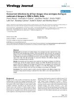
Phylogenetic analysis of DEN-3 Virus
To determine the evolutionary history of the DENV-3
viruses found in Taiwan in 1998, the nucleotide sequence
of their partial E protein genes were compared with those
from all previous published DENV-3 E gene sequence
available in the GenBank. The phylogenetic tree analysis
for 141 clonal sequences from six virus isolates of this
study and 24 global DENV-3 sequences separated the
viruses into five main subgroups, which had been previ-
ously defined as five different genotypes. As in previous
studies of DENV-3 diversity, the 1963 Puerto Rico strain
formed a distinct outlier, which served as the outgroup for
the phylogenetic tree. The tree topology was very similar
based on either neighbor-joining (NJ) or parsimony
(PAR) method. Based on the phylogenetic tree, the virus
isolates from Taiwan in 1998 formed a tight cluster with
strong bootstrap support, which fell closer to the isolates
from Thailand as they belong to DEN-3 genotype II,
according to the classification of Lanciotti et al (20)(Fig
2). Most of the population from the clonal sequences
formed a tightly cluster, which represented the highly
homogeneous nucleotide sequences during the same epi-
demic. Interestingly, some clones from different individ-
ual isolates appeared to form the different subgroups
under the Thailand genotype, with 50–100% bootstrap
support. This indicated viral evolution did occur during
the epidemic period, probably under selection pressure.
Discussion and Conclusion
To the best of our knowledge, this was the first systematic
attempt to understand the sequence spectrum of the entire
genome of DENV-3. Previous studies, focused on certain
genomic regions such as the envelope gene, the capsid
gene or the NS2B gene, have revealed the presence of qua-
sispecies structure as indicated by the simultaneous pres-
ence of multiple variant genomic sequences of the dengue
virus isolates from either the clinical samples or field-
caught mosquitoes [7,9,17-19]. Instead of arbitrarily
choosing one genomic region in this study, the full
genomic consensus sequences of six DENV-3 isolates were
used to locate the four most prominent heterogeneous
regions, the C/PrM and E in the structure, and NS3 and
NS5 in the nonstructural regions.
Table 1: Identification of the positions of potential heterogeneity nucleotide sequence by the full genome consensus sequence of
DENV-3
a
viruses isolated during 1998–1999 dengue outbreak in Taiwan.
Virus ID Disease Status
c
Nucleotide Changes at Positions Indicated
b
320–322 444–445 808 1693 1716 4204 5322 6045 6079 8785 9076 9979 10105 10128
1F DF RRR TG (CA) G G(C) C(T) C T A C T T T C C
1H DHF GGG TG G C C(T) C T A C T T T C C
2H DHF GGG TG A G C(T) C T A C T T(A) C T C
2F DF GGG TG G G C(T) C T A C T T T C C
3H DHF GGG TG G G(C) C(T) T T(C) A(C) C(T) C T T C C
3F DF GGG TG G G C(T) C T A C T T T C C(T)
a
Nucleotide in parentheses indicated "mix nucleotide sequence" based on mix chromatographic signals in the sequencing trace file and nucleotide
position number referred to the reference strain H87 of DENV-3 (genebank accession number: M93130)
b
DENV-3 viruses were isolated from the plasma of six dengue patients by one passage in the C6/36 mosquito cell culture.
c
Disease status was classified based on WHO criteria [30]. DF: dengue fever; DHF: dengue hemorrhagic fever.
Virology Journal 2005, 2:72 />Page 5 of 10
(page number not for citation purposes)
Table 2: Sequence diversity (mean p-distance) among different genomic regions of DENV-3
Nucleotide Amino acid
Virus ID
No.
Region No of
Clones
No of
change/
total
Mutation
frequency
a
(10
-3
)
A→G or
U→C
b
(%)
Mean p-
distance
c
(10
-3
)
Range (10
-
3
)
No of
change/
total
Mutation
frequency
a
(10
-3
)
Mean p-
distance
c
(10
-3
)
Range (10
-
3
)
1F C/PrM 23 24/17733 1.4 66.7 2.67 0–6.7 13/5175 2.5 5 0–8.9
1H C/PrM 29 29/22359 1.3 69 2.45 0–6.7 17/6525 2.6 5.12 0–22.2
2H C/PrM 21 16/16191 1.0 81.3 2.03 0–6.7 9/4725 1.9 3.81 0–13.3
2F C/PrM 22 18/16962 1.1 72.2 2.17 0–6.7 10/4950 2.0 4 0–13.3
3H C/PrM 13 11/10023 1.1 72.7 1.81 0–4 7/2925 2.4 4.7 0–17.8
3F C/PrM 24 24/18504 1.3 75 2.55 0–8 13/5400 2.4 4.8 0–17.8
Mean ± std C/PrM 22 1.2 ± 0.15 72.8 ± 5.1 2.28 ± 0.3 2.3 ± 0.3 4.6 ± 0.5
1F E/NS1 26 40/32240 1.2 72.5 2.23 0–6.5 21/10192 2.1 4.3 0–10.2
1H E/NS1 13 47/16250 2.9 61.7 3.82 0–6.4 17/5096 3.3 6.6 0–7.6
2H E/NS1 25 33/16250 2.0 93.9 3.88 0–5.6 34/9800 3.5 6.4 0–12.6
2F E/NS1 13 15/22103 0.7 66.7 3.18 0–2.8 4/5096 0.8 7.6 0–9
3H E/NS1 23 44/20240 2.2 72.7 3.83 0–4.8 55/9016 6.1 6.2 0–12.6
3F E/NS1 20 39/24960 1.6 74.4 2.56 0–6.8 20/7840 2.6 4.8 0–21.7
Mean ± std E/NS1 20 1.8 ± 0.8 73.7 ± 11 3.7 ± 0.7 3.1 ± 1.8 6 ± 1.2
1F NS3 19 5/17005 0.3 60 0.9 0–3.5 8/4978 1.6 3.3 0–11.4
1H NS3 27 16/24165 0.7 56.3 1.2 0–4.6 11/7074 1.6 3.1 0–11.4
2H NS3 26 9/23270 0.4 33.3 1.2 0–4.6 6/6812 0.9 3.2 0–11.5
2F NS3 25 16/22375 0.7 40 2.4 0–5.8 12/6550 1.8 5.7 0–15.3
3H NS3 23 25/20585 1.2 16 1.6 0–4.6 6/6026 1.0 2.0 0–11.4
3F NS3 18 8/16110 0.5 62.5 1.0 0–4.6 4/4716 0.8 1.6 0–11.4
Mean ± std 23 0.6 ± 0.3 44.7 ± 18 1.4 ± 0.6 1.3 ± 0.4 3 ± 1.4
1F NS5 13 29/23335 1.2 69 1.7 0–4.5 17/7748 2.2 4.1 0–8.4
1H NS5 17 32/30515 1.0 71.9 2.4 0–5.6 31/10132 3.1 3.5 0–8.4
2H NS5 18 51/32310 1.6 64.7 3.0 0–8.9 33/10728 3.1 6.1 0–18.5
2F NS5 25 60/44875 1.3 65 2.5 0–5.6 47/14900 3.2 3.6 0–8.4
3H NS5 16 64/28720 2.2 56.3 4.3 0–7.3 46/9536 4.8 6.8 0–15.2
3F NS5 26 69/46670 1.5 52.2 3.7 0–6.7 52/15496 3.4 6.5 0–11.8
Mean ± std 19 1.5 ± 0.4 63.2 ± 7.5 3.1 ± 0.9 3.3 ± 0.8 5.1 ± 1.5
a
Mutation frequency is defined as the proportion of mutations relative to the consensus nucleotide or amino acid sequence for each patient and
calculated by dividing the number of mutations relative to consensus by the total number of nucleotides or amino acid sequenced in each sample.
b
Percentage of A→G or U→C transitional mutations
c
p-distance is calculated by pairwise comparison of nucleotide or amino acid sequences between clones by the program MEGA d Indicated the virus
derived from mosquito inoculation by C6/36-passaged one virus of patient ID#1H.
Table 3: Mean p-distance and ratio of dN to dS per site of amino acid among different domains of the E protein in DENV-3 infected
patients
Virus ID No of
sequences
Envelope (293aa) Domain I (70aa) Domain II (106aa) Domain III (100aa)
Mean p-
distance
dN/dS Mean p-
distance
dN/dS Mean p-
distance
dN/dS Mean p-distance dN/dS
1F 26 0.006 ± 0.001 2.04 0.009 ± 0.003 1.82 0.005 ± 0.002 1.72 0.005 ± 0.002 2.8
1H 21 0.008 ± 0.002 1.11 0.008 ± 0.004 2.53 0.007 ± 0.003 0.59 0.007 ± 0.002 1.2
2F 18 0.007 ± 0.002 1.38 0.009 ± 0.004 2.52 0.006 ± 0.003 1.04 0.007 ± 0.003 2.57
2H 25 0.012 ± 0.002 1.06 0.013 ± 0.004 2.68 0.012 ± 0.004 1.08 0.011 ± 0.003 0.77
3F 23 0.007 ± 0.001 0.81 0.012 ± 0.004 0.96 0.005 ± 0.002 0.45 0.003 ± 0.002 1.76
3H 23 0.009 ± 0.002 1.46 0.014 ± 0.005 4.19 0.006 ± 0.003 0.62 0.005 ± 0.002 120.03
Average 22.7 0.008 ± 0.002 1.31 0.011 ± 0.002 2.45 0.007 ± 0.002 0.92 0.006 ± 0.002 21.52
Virology Journal 2005, 2:72 />Page 6 of 10
(page number not for citation purposes)
Use of clonal sequencing to study the mutation spectrum
needs to ensure that sequencing artifacts due to RT-PCR
amplification are reduced to minimum. In this study, we
used viral RNAs extracted from patients' viremic plasma
directly. In addition, a thermostable polymerase with
proof-reading function was incorporated in the RT-PCR,
which has been shown to be a simple and valuable
method for characterization of mutant spectra of virus
quasispecies [20]. The nucleotide changes from four
sequenced-viral genomic regions (range of 4.4–11.6 × 10
-
5
changes/nucleotide/cycle of PCR) were greater than
those predicted based on reverse transcriptase (10
-4
) and
proof-reading DNA polymerase (Pfu, 10
-6
error/site/cycle)
combined [21]. Based on the experimental data, Arias et
al pointed out that the biological and molecular clones
were statistically indistinguishable when defining the
mutation spectrum with regard to the types and distribu-
tions of mutations, mutational hot-spots and mutation
frequencies [20]. Similarly, we believe that the full-
genomic characterization followed by clonal sequencing
procedure employed in this study is a reasonable and
justifiable approach for the characterization of mutation
spectra (quasispecies dynamic) of DENV-3 viruses.
DENV, like other RNA viruses, exists as quasispecies with
the sequence diversity of the envelope gene in the DENV-
3 virus population from 6 clinical isolates, ranging from
0.22–0.39% of mean p-distances in this study. These val-
ues are within the range calculated by other studies (0.12
to 0.84%) for different portions of the E protein genes of
DENV-3 viruses from either the clinical or field-caught
mosquito isolates [7,9,17-19]. Our study confirmed use
of the structural protein, especially the E gene with higher
sequence heterogeneity to study the viral quasispecies,
instead of NS protein and 5' and 3' NCR. The extent of
sequence variation observed in this study was similar to or
lower than what has been reported for acute infection of
HIV-1 or HCV [8,22-25]. A study of sequence variation of
HIV-1 after sexual transmission revealed that the nucle-
otide mean diversity of the E gene (gp120) was 0.24% and
that of the gag gene (p17) was 0.5% [22]. Similar results
by studying variants of hepatitis C virus (HCV) from a sin-
gle infected blood donor and 13 viraemic recipients were
traced to examine the sequence diversity in hypervariable
region 1 with sequence p-distance ranged from 0.3% to
6.2% [23]. These data might support an important con-
cept in the evolution of arthropod-borne RNA viruses
(arboviruses) which evolve more slowly than RNA viruses
transmitted by other routes due to intrinsic constraints
associated with dual replication in mammalian and inver-
tebrate hosts [26]. Consistent with this interpretation was
that the lower sequence diversity was observed at the same
E protein gene from the field-caught mosquito DENV-3
isolates [19] or after inoculation of clinical serum of
DENV-3 into mosquitoes (data not shown).
Table 4: The Oligonucleotide primers and conditions used for RT-PCR of full-length genome of DENV-3
PCR Primer
a
Sequence (5' → 3') Genome Position
b
Size(nt)
c
P1A AGT TGT TAG TCT RCG TGG 1–18 1181
CP1181B TCC ARG CAC CTT CAG ATG 1181–1199
DC530A AAC AWR TGC ACC CTC 540–555 1164
CDC1694B TGC ATK GCT CCT TCT TGR 1694–1712
P1259A GGC AAG GGA AGC TTG GTG ACA TGC GC 1259–1285 1244
CDC2503B GGG AGT CTG CTT GGA ATT 2503–2521
DC2171A GCC ATT CTR GGW GAC ACC GCY TGG GA 2171–2197 1246
CDC3417B TCT CTT CTT TGT CMT TCA 3417–3435
D3-3142A CCA AAG AGT CTA GCT GGT CC 3142–3162 1535
D3-4677B CAT TGT GCG TCA ACA CTG CC 4677–4697
d3NS2B1A AGC TGG CCA CTG AAT GAG G 4124–4143 1562
D35686B CAA AAG TCT TCC TAC TAA GTT G 5686–5708
D3-5443A GCC GCA ATT TTC ATG ACA 5443–5461 2034
D3-7477B AAC AGC TAT CGT GGT GTT CC 7477–7497
d37246A AAG AAT CCA ACG GTG GAT GG 7246–7266 1454
d38750B TCC CTT GTG CAT AAT CTG GG 8750–8770
d38501A CAG GCT CAG CCT CCT CC 8501–8518 1654
d310316B GCT TCT TCC GTA CTG TGG C 10316–10335
d39991A CTT ACT GTC TGG AAC AGG G 9991–10010 648
d310688B GTT GAT TCA ACA GCA CCA TTC 10688–10709
a Primer names with A in the end indicate a viral-sense orientation; names with B in the end indicate a complementary sense orientation
b Genome positions are given according to the published sequence of strain H87 of dengue virus serotype 3
c nt indicated nucleotide
Virology Journal 2005, 2:72 />Page 7 of 10
(page number not for citation purposes)
Phylegenetic tree showing the evolutionary relationships of the E gene among 54 sequences from 30 clonal sequences of 6 DEN-3 clinical isolates and 24 global isolatesFigure 2
Phylegenetic tree showing the evolutionary relationships of the E gene among 54 sequences from 30 clonal sequences of 6
DEN-3 clinical isolates and 24 global isolates. Bootstrap support values presented as percentage are given for key nodes only
and the genotype designations are given. The horizontal branch length of the trees was drawn to scale. GenBank accession
numbers of the global DEN-3 strains used in this analysis are as follows: Fiji92 (L11422
), India84 (L11424), Indonesia73
(L11425
), Indonesia78 (L11426), Indonesia85 (L11428), Malaysia74 (L11429), Malaysia81 (L11427), Mozambique85 (L11430),
H87 (L11423
), Philippines83 (L11432), Puerto Rico77 (L11434), Puerto Rico63 (L11433), Samoa86 (L11435), SriLanka81
(L11431
), Sri Lanka85 (L11436), Sri Lanka89 (L11437), Tahiti65 (L11439), Tahiti89 (L11619), Thailand62 (L11440), Thailand73
(L11620
), Thailand87 (L11442).
PHY LIP_1
100
Pu Ric o63
PuRic o 7 7
Tahiti65
H87
Fiji92
Tahiti89
Malay 81
Indon73
Indon85
Indon78
P hili83
Ma la y 7 4
Thai land62
Moz ambique
India8 4
Samoa86
SriLank81
SriLank85
SriLank91
SriLank89
T hai l and73
T hailand86
Thai 87
1Hclone27
2Hcl one19
2Fclone17
1Hclone26
1Hcl one13
1F clone18
3F clone9
1F clone26
2Hclone28
3Fclone3
1Fclone12
2Hclone27
2Hclone29
3H clone16
1Hcl one25
3Hclone15
1F clone7
2F clone29
2H clone24
1Fclone6
3Hcl one30
3F clone13
3Hcl one12
2Fclone15
2Fclone14
3Hclone1
3F clone15
1Hcl one29
2Fclone30
3F clone26
1 Substitution/site
GenotypeII
GenotypeIII
GenotypeI
100
50
100
100
74
98
51
GenotypeIV
Virology Journal 2005, 2:72 />Page 8 of 10
(page number not for citation purposes)
The larger mutation spectra in structural proteins than
non-structural proteins probably imply less genetic con-
straint on the structure proteins to maintain proper func-
tion than non-structural proteins. However, the
mutations in the structural or non-structural proteins did
not accumulate randomly during replication. The muta-
tion rates vary in different functional/structural domains.
Even within the envelope structure protein, where
domain III, the proposed receptor-binding and neutraliz-
ing antibody-binding sites [13] had highest sequence
heterogeneity than Domain I or II. The detail analysis in
our study further indicated that the different selection
pressure was exerted on different domain of the E gene of
DENV-3. Domain III and domain I were under the influ-
ence of positive selection (d
N
/d
S
:21.52, 2.45) and domain
II was under the influence of neutral selection (d
N
/
d
S
:0.92). The particularly higher d
N
/d
S
ratio in domain III
of viral isolates 3H was caused by the value of 0 of d
S
at the
denominator. The positive selection on the domain III is
not surprising since domain III contains the receptor-
binding domain and major type-specific neutralization
epitopes [12]. However, the complete E gene sequence
may be required to clarify the evolutionary selection on
domain I and II due to incomplete sequence obtained in
this study.
In contrast to other studies which suggested the strong
purifying selection in the E gene of dengue virus evolu-
tion, the consensus sequences used for analysis repre-
sented dengue viral gene conservation during long-term
evolution [27]. The clonal sequences obtained from our
study represented the selection pressure imposed on viral
populations during the short term of evolution, which
might explain the substantially different d
N
/d
S
value
within hosts and among genotypes. The majority of the
nonsynonymous mutations that arise within each host
occurred as singletons with relatively low frequency in the
population; thus are likely to be deleterious. Such hetero-
geneous gene pool may give rise to various viruses able to
occupy new ecological niches or to adapt to sudden selec-
tion pressures on the cycle of replication. It is evident that
certain nonsynonymous nucleotide mutations at specific
sites repeatedly occurred among different virus isolates as
well as after mosquito inoculation in our study (data not
shown), which has been proposed as quasispecies mem-
ory in another study [28]. Further studies are needed to
address the influence of these mutations on the overall fit-
ness in different hosts (i.e., mosquito and human) during
dengue viral transmission.
Materials and methods
Study subjects and virus isolation
Six dengue patients were identified by RT-PCR to be
DENV-3 positive during the 1998 epidemic and their
acute-phase viremic plasma samples were collected within
seven days following the onset of fever. These plasma
samples were used to infect C6/36 Aedes albopictus mos-
quito cell lines as described previously [29]. The study
protocol was approved by the College of Public Health
Research Ethics Review Committee at the National Tai-
wan University with the informed consent obtained from
six dengue patients. Six adult dengue cases between 38
and 63 years of age, including one DF (F) and one DHF
(H) cases, whose disease status were classified based on
WHO criteria [30], were represented as 1F, 1H, 2F, 2H, 3F
and 3H, respectively.
DENV-3 was confirmed by indirect immuno-fluorescent
antibody (IFA) tests using serotype-specific monoclonal
antibodies (DENV-1:H47, DENV-2:H46, DENV-3:H49,
DENV-4:H48) [31]. The C6/36-passage one viral stock
was used for full genomic consensus sequencing to iden-
tify regions with sequence heterogeneity for clonal
sequencing as described later.
Preparation of viral RNA, RT-PCR amplification and
consensus sequencing of PCR products
Viral RNA was extracted either from viremic plasma spec-
imens or from the C6/36-passaged one cell culture fluids
using QIAamp viral RNA mini kit (Qiagen, Germany) by
following the manufacturer's protocol. The eluted RNA
was used as the template and overlapping regions of
DENV-3 genome amplified by Titan™ one tube RT-PCR
System (Boehringer Mannheim, Germany) following the
manufacturer's suggestions. The oligonucleotide primer
pairs were designed based on published full-length
DENV-3 sequence data for the strains of H87 and 80-2
(GenBank Accession number M93130
and AF317645)
and the unpublished DENV-3 sequences (Chang, G-J. per-
sonal communication). Ten overlapping fragments were
generated which spanned genomic regions of DENV-3 at
the following nucleotide (nt) positions: 1 to 1199, 540 to
1712, 1259 to 2521, 2171 to 3435, 3142 to 4697, 4124 to
5708, 5443 to 7497, 7246 to 8770, 8501 to 10335, 9991
to 10709. Primer sequences used for PCR amplification
were summarized in Table 4. The obtained PCR products
were sequenced by using the Big Dye Terminator Sequenc-
ing kit (Perkin-Elmer, Applied Biosystems, Foster City,
CA) and analyzed by the 3100 automate sequencer (Per-
kin-Elmer, Applied Biosystems) with a short capillary.
Preparation of plasmid templates for clonal sequencing
We used pCRII-TOPO™ T/A cloning kit (Invitrogen, San
Diego, CA) to clone PCR products representing heteroge-
neity sequence regions identified by the consensus
sequencing protocol at the previous section. The T/A vec-
tor ligated PCR product was used to transform Escherichia
coli TOP10 competent cells (Invitrogen) and at least 30
white colonies were picked, to grow in 3 ml LB broth at
37°C overnight. Plasmid DNAs were extracted by the
Virology Journal 2005, 2:72 />Page 9 of 10
(page number not for citation purposes)
QIAprep Spin Miniprep kit (Qiagen), and each plasmid
DNA with the desired inserts was completely sequenced
using insert flanking primers, T7 and cSP6.
Nucleotide and Amino acid sequence analysis
Overlapping chromatogram files retrieved from the auto-
mate sequencer were analyzed and edited using the Seq-
Man program in the Lasergene software package
(DNASTAR inc., Madison, WI). The derived consensus
sequences after excluding the sequences of amplifying
primers were aligned using GCG package (Genetic Com-
puter Group, WI). For full-length genomic sequences we
paid special attention to identify the regions which con-
sistently presented mixed-chromatographic peaks in the
trace file obtained from at least two independent sequenc-
ing primers. These regions were selected for the follow-up
clonal sequence analysis. Pairwise comparisons of both
nucleotide and amino acid sequences between isolates
and clonal sequences were performed using the program
MEGA v3.0 (Molecular Evolutionary Genetics Analysis,
Pennsylvania State University, PA) to determine the num-
bers of transition and transversion changes, and the mean
and proportion of difference, Hamming distance and p-
distance, as described previously [8,32,33]. Synonymous
(d
S
) and nonsynonymous (d
N
) distances relative to the
consensus sequences were calculated within each isolate
by maximum likelihood ratio method in the CODEML
program from the PAML package [15]. Instead of assum-
ing that all sites are under the same selection pressure with
the same underlying d
N
/d
S
ratio, it allows variable selec-
tion intensity to vary among amino acid sites [34,35]. In
this study, M3 model of codon evolution was applied for
which often provides the best evidence for positive selec-
tion [16]. An excess of nonsynonymous substitutions over
synonymous substitutions (ie. the ratio of d
N
/d
S
> 1) is an
indicator of positive natural selection at the molecular
level.
The results were expressed as the mean ± standard devia-
tion (SD). T-tests were performed on two-sampled tests
and a one-way ANOVA was performed to compare data
from different genomic regions, family clusters or
different domains in envelope region. In all tests, a p-
value less than 0.05 was considered statistically
significant.
Evolutionary analysis
The nucleotide sequences generated in this study were
combined with those of all other DENV-3 E protein gene
sequences available on GenBank, which resulted in a total
data set of 154 sequences. Phylogenetic trees were esti-
mated using parsimony method available in the Phylip
v3.6 package [36]. Bootstrap resampling analysis of 500
replicates was generated with the SEQBOOT program to
prove the stability of the trees. Phylogenetic trees were
delineated using the TreeView (v.1.6.6) program by using
Puerto Rico 1963 isolate as the outgroup. For better pres-
entation of the phylogenetic tree, only 30 clonal
sequences from six different clinical isolates and 24 global
isolates were shown in Fig 2.
Nucleotide sequences accession numbers
The sequences from four heterogeneous regions of dengue
viruses from the six patients studied here have all been
submitted to GenBank, and their accession numbers are
from DQ109039
to DQ109173 for the E region, from
DQ109174
to DQ109305 for the capsid/prM region,
from DQ109306
to DQ109405 for the NS5 region and
from DQ109406
to DQ109524 for the NS3 region.
Competing interests
The author(s) declare that they have no competing
interests.
Authors' contributions
DYC designed and performed all the experiments and
helped drafted this manuscript. CCK helped with collect-
ing field isolates and instructed the experiments, together
with WKW and HLW. WJC helped for the mosquito injec-
tion experiments and GJC formulated the idea for this
study and also provided critical comments regarding this
manuscript.
Financial support
The study was supported by the grants from the National
Health Research Institute (NHRI), Taipei, Taiwan (grant
number: NHRI#DD01-861X-CR-501P and NHRI#CN-
CL8903P) and International Society of Infectious Disease
(ISID).
Acknowledgements
We sincerely thank Shih-Ting Ho at the Sin-Lau Christian Hospital, Chien-
Ming Li at the Chi-Mei Foundation Medical Center and Shih-Chung Lin at
the Kuo General Hospital for their enthusiasm for kindly providing the clin-
ical samples. This study was supported by the grants from the National
Health Research Institute (NHRI), Taipei, Taiwan (NHRI#DD01-861X-CR-
501P and NHRI#CN-CL8903P) and the training grant to D Y. Chao from
International Society of Infectious Disease (ISID).
References
1. Gubler DJ: Dengue and dengue hemorrhagic fever. Clin Micro-
biol Rev 1998, 11(3):480-496.
2. Halstead SB: The XXth century dengue pandemic: need for
surveillance and research. Wld Hlth Statist Quart 1992,
45:292-296.
3. McBride WJH, Bielefeldr-Ohmann H: Dengue viral infections;
pathogenesis and epidemiology. Microbes Infect 2000,
2:1041-1050.
4. Chambers TJ, Hahn CS, Galler R, Rice CM: Flavivirus genome:
organization, expression and replication. Annu Rev Microbiol
1990, 44:649-688.
5. Twiddy SS, Holmes EC, Rambaut A: Inferring the rate and time-
scale of dengue virus evolution. Mol Biol Evol 2003,
20(1):122-129.
Publish with BioMed Central and every
scientist can read your work free of charge
"BioMed Central will be the most significant development for
disseminating the results of biomedical research in our lifetime."
Sir Paul Nurse, Cancer Research UK
Your research papers will be:
available free of charge to the entire biomedical community
peer reviewed and published immediately upon acceptance
cited in PubMed and archived on PubMed Central
yours — you keep the copyright
Submit your manuscript here:
/>BioMedcentral
Virology Journal 2005, 2:72 />Page 10 of 10
(page number not for citation purposes)
6. Domingo E: Quasispecies theory in virology. J Virol 2002,
76(1):463-465.
7. Wang WK, Lin SR, Lee CM, King CC, Chang SC: Dengue type 3
virus in plasma is a population of closely related
genomes:quasispecies. J Virol 2002, 76(9):4662-4665.
8. Farci P, Shimoda A, Coiana A, Diaz G, Peddis G, Melpolder JC,
Strazzera A, Chien DY, Munoz SJ, Balestrieri A, Purcell RH, Alter HJ:
The outcome of acute hepatitis C predicted by the evolution
of the viral quasispecies. Science 2000, 288:339-344.
9. Wang WK, Sung TL, Lee CN, Lin TY, King CC: Sequence diversity
of the capsid gene and the nonstructural gene NS2B of den-
gue-3 virus in vivo. Virology 2002, 303:181-191.
10. Chao DY, Lin TH, Hwang KP, Huang JH, Liu CC, King CC: 1998 den-
gue hemorrhagic fever epidemic in Taiwan. Emerg Infect Dis
2004, 10(3):552-554.
11. Kumar S, Tamura K, Nei N: MEGA3: Integrated software for
Molecular Evolutionary Genetics Analysis and sequence
alignment. Brief Bioinform 2004, 5(2):150-163.
12. Modis Y, Ogata S, Clements D, Harrison SC: Variable surface
epitopes in the crystal structure of dengue virus type 3 enve-
lope glycoprotein. J Virol 2005, 79(2):1223-1231.
13. Rey FA, Heinz FX, Mandl C, Kunz C, Harrison SC: The envelope
glycoprotein from tick-borne encephalitis virus at 2 A
resolution. Nature 1995, 375:291-298.
14. Modis Y, Ogata S, Clements D, Harrison SC: A ligand-binding
pocket in the dengue virus envelope glycoprotein. Proc Natl
Acad Sci 2003, 100(12):6986-6991.
15. Yang Z: PAML: a program package for phylogenetic analysis
by maximum likelihood. Comput Appl Biosci 1997, 13(5):555-556.
16. Yang Z, Nielsen R, Goldman N, Pedersen AMK: Codon-substitu-
tion models for heterogeneous selection pressure at amino
acid sites. Genetics 2000, 155:431-449.
17. Craig S, Thu HM, Lowry K, Wang X, Holmes EC, Aaskov J: Diverse
dengue type 2 virus populations contain recombinant and
both parental viruses in a single mosquito host. J Virol 2003,
77(7):4463-4467.
18. Wittke V, Robb TE, Thu HM, Nisalak A, Nimmannitya S, Kalayanrooj
S, Vaughn DW, Endy TP, Holmes EC, Aaskov J: Extinction and
rapid emergence of strains of dengue 3 virus during an
interepidemic period. Virology 2002, 301:148-156.
19. Lin SR, Hsieh SC, Yueh YY, TH L, Chao DY, Chen WJ, King CC,
Wang WK: Study of sequence variation of dengue type 3 virus
in naturally infected mosquitoes and human hosts: implica-
tions for transmission and evolution. J Virol 2004,
78(22):12717-12721.
20. Arias A, Lazaro E, Escarmis C, Domingo E: Molecular intermedi-
ates of fitness gain of an RNA virus: characterization of a
mutant spectrum by biological and molecular cloning. J Gen
Virol 2001, 82:1049-1060.
21. Mullan B, Sheehy P, Shanahan F, Fanning L: Do Taq-generated RT-
PCR products from RNA viruses accurately reflect viral
genetic heterogeneity? J Viral Hepat 2004, 11:108-114.
22. Zhu T, Mo H, N W, D.S. N, Cao Y, Koup RA, Ho DD: Genotypic
and phenotypic characterization of HIV-1 in patients with
primary infection. Science 1993, 261:1173-1181.
23. Love A, Molnegren V, Mansson AS, Smaradottir A, Thorsteinsson SB,
Widell A: Evolution of hepatitis C virus variants following
blood transfusion from one infected donor to several recipi-
ents: a long-term follow-up. J Gen Virol 2004, 85(2):441-450.
24. Curran R, Jameson CL, Craggs JK, Grabowska AM, Thomson BJ, rob-
ins A, Irving WL, Ball JK: Evolutionary trends of the first hyper-
variable region of the hepatitis C virus E2 protein in
individuals with differing liver disease severity. J Gen Virol 2002,
83:11-23.
25. Delwart E, Magierowska M, Royz M, Foley B, Peddada L, Smith R,
Heldebrant C, Conrad A, Busch M: Homogeneous quasispecies
in 16 out of 17 individuals during very early HIV-1 primary
infection. AIDS 2002, 16:189-195.
26. Domingo E, Biebricher CK, Eigen M, Holland JJ: Quasispecies and
RNA virus evolutioon:principles and consequences. Landes
Bioscience, Texas, USA 2001:82-108.
27. Holmes EC: Patterns of intra- and interhost nonsynonymous
variation reveal strong purifying selection in dengue virus. J
Virol 2003, 77(20):11296-11298.
28. Domingo E, Ruiz-Jarabo CM, Sierra S, Arias A, Pariente N, Bar-
anowski E, Escarmis C: Emergence and selection of RNA virus
variants: memory and extinction. Virus Res 2002, 82:39-44.
29. Gubler DJ, Kuno G, Sather GE, Velez M, Oliver A: Mosquito cell
cultures and specific monoclonal antibodies in surveillance
for dengue viruses. Am J Trop Med Hyg 1984, 33:158-165.
30. World Health Organization: Dengue haemorrhagic fever: diag-
nosis, treatment, prevention and control. 2nd edition.
Geneva. Wld Hth Org 1997:12-23.
31. Kao CL, Wu MC, Chiu YH, Lin JL, Wu YC, Yueh YY, Chen LK, Shaio
MF, King CC: Flow cytometry compared with indirect immun-
ofluorescence for rapid detection of dengue virus type 1
after amplification in tissue culture. J Clin Microbiol 2001,
39(10):3672-3677.
32. Kumar S, Tamura K, Nei M: MEGA: Molecular Evolutionary
Genetics Analysis (Pennsylvania State Univ., University
Park). 1993.
33. Nei M, Gojobori T: Simple methods for estimating the num-
bers of synonymous and nonsynonymous nucleotide
substitutions. Mol Biol Evol 1986, 3:418-426.
34. Yang Z, Bielawski JP: Statistical methods for detecting molecu-
lar adaptation. Trends Ecol Evol 2000, 15:496-503.
35. Nielsen R, Yang Z: Likelihood models for detecting positively
selected amino acid sites and applications to the HIV-1 enve-
lope gene. Genetics 1998, 148:929-936.
36. Felsenstein J: PHYLIP: Phylogeny Inference Package (Univ. of
Wahsington, Seattle), Version 3.5c. 1993.