báo cáo hóa học:" An inexpensive and rapid diagnostic method of Koi Herpesvirus (KHV) infection by loop-mediated isothermal amplification" docx
Bạn đang xem bản rút gọn của tài liệu. Xem và tải ngay bản đầy đủ của tài liệu tại đây (1.16 MB, 8 trang )
BioMed Central
Page 1 of 8
(page number not for citation purposes)
Virology Journal
Open Access
Methodology
An inexpensive and rapid diagnostic method of Koi Herpesvirus
(KHV) infection by loop-mediated isothermal amplification
Hatem Soliman and Mansour El-Matbouli*
Address: Institute of Zoology, Fish Biology and Fish Diseases, Faculty of Veterinary Medicine, University of Munich, Germany
Email: Hatem Soliman - ; Mansour El-Matbouli* -
* Corresponding author
Abstract
Background: Koi Herpesvirus (KHV) affects both juvenile and adult common carp and koi, and is
especially lethal to fry. The high mortalities caused by the disease have had a negative impact on the
international koi trade. Different diagnostic techniques have been used to detect KHV, including:
isolation of the virus in cell culture, electron microscopy, several PCR tests, ELISA and in situ
hybridisation. All of these methods are time consuming, laborious and require specialised
equipment.
Results: A rapid field diagnosis of KHV in common and koi carp was developed using loop-
mediated isothermal amplification (LAMP). The LAMP reaction rapidly amplified nucleic acid with
high specificity and efficiency under isothermal conditions using a simple water bath. Two methods
of extracting DNA from host tissue were compared: extraction by boiling and by using a
commercial extraction kit. A set of six primers – two inner primers, two outer primers and two
loop primers – was designed from a KHV amplicon. The reaction conditions were optimised for
detection of KHV in 60 min at 65°C using Bst (Bacillus stearothermophilus) DNA polymerase. When
visualised by gel electrophoresis, the products of the KHV LAMP assay appeared as a ladder
pattern, with many bands of different sizes from 50 base-pairs (bp) up to the loading well. The KHV
LAMP product could also be simply detected visually by adding SYBR Green I to the reaction tube
and observing a colour change from orange to green. All samples positive for KHV by visual
detection were confirmed positive by gel electrophoresis. The KHV LAMP had the same sensitivity
as a standard PCR assay for the detection of KHV.
Conclusion: This paper describes an accelerated LAMP assay for diagnosis of KHV. The entire
procedure took only 90 minutes to produce a result: 15 minutes for DNA extraction; 60 min for
the LAMP reaction; 2 min for visual detection using SYBR Green I. The test can be used under field
conditions because the only equipment it requires is a water bath.
Background
Koi Herpesvirus (KHV) is a highly contagious viral disease
which causes significant morbidity and mortality in com-
mon carp (Cyprinus carpio) and its ornamental domesti-
cated form, koi carp [1]. Although the virus is currently
regarded as a DNA-virus belonging to family Herpesviri-
dae [1], some reports have disputed this classification and
have renamed the virus as Carp Nephritis and Gill Necro-
sis Virus, CNGV [2]. More recently, reports based on
Published: 17 October 2005
Virology Journal 2005, 2:83 doi:10.1186/1743-422X-2-83
Received: 27 May 2005
Accepted: 17 October 2005
This article is available from: />© 2005 Soliman and El-Matbouli; licensee BioMed Central Ltd.
This is an Open Access article distributed under the terms of the Creative Commons Attribution License ( />),
which permits unrestricted use, distribution, and reproduction in any medium, provided the original work is properly cited.
Virology Journal 2005, 2:83 />Page 2 of 8
(page number not for citation purposes)
morphology and genetics have demonstrated strong evi-
dence that KHV is indeed a herpesvirus [3].
The international trade in live fish is arguably the most
effective dispersal pathway of fish diseases through inci-
dental movement of pathogenic organisms [4].With
respect to koi, exhibitions and national and international
trading have facilitated the rapid global spread of KHV.
The disease struck koi population in the USA and Israel in
1998 and spread rapidly [5]; it has been reported in Ger-
many [6], Korea [7,8], Indonesia [9], Japan [10], South
Africa, and Thailand (unpublished data).
Clinical signs of KHV are often non-specific and mortality
may occur rapidly. Discoloration and severe necrosis of
the gills is the most consistent sign of infection, with dis-
orientation and erratically swimming prior to death,
which can occur within 24–48 hours after the onset of
clinical signs [11,12]. KHV has caused considerable eco-
nomic losses in both the koi and carp culture industries:
to fish breeders, retailers and hobbyists impacted by the
cumulative mortalities associated with outbreaks [4,2].
There is a clear need for a reliable, rapid diagnostic proce-
dure for the detection of KHV infection.
Rapid virological diagnosis through isolation of the virus
has proven difficult and time consuming. A far more effi-
cient approach is nucleic acid amplification; one of the
most valuable tools in virtually all life science fields [13].
One of the most widely used techniques is the polymerase
chain reaction (PCR) which uses heat denaturation of
double-stranded DNA products to promote the next
round of DNA synthesis [14,15]. A widely used PCR assay
for KHV was developed [16], and a second PCR assay for
KHV has been described [12]. A real-time TaqMan PCR
assay for KHV has also been developed to detect and
quantify KHV DNA in infected tissues [17]. While these
PCR techniques have significantly increased our ability to
detect KHV infection in koi and common carp, their
requirement for a high precision thermacycler has pre-
vented their widespread use in private clinics, for exam-
ple, as a routine diagnostic tool.
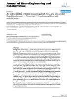
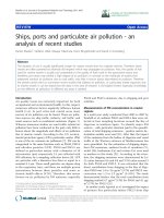
Nucleotide sequence of the KHV amplicon (GenBank acces-sion number AF411803) used for construction of the inner and outer primersFigure 1
Nucleotide sequence of the KHV amplicon (GenBank acces-
sion number AF411803) used for construction of the inner
and outer primers. The primer sequences are indicated in
bold letters. Inner primers FIP and BIP comprise sequences
within the amplicon; FIP is the complementary sequence of
F1 and F2, BIP is B1 plus the complementary sequence of B2.
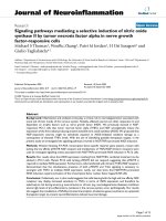
Agarose gel showing the effect of time on amplification of KHV DNA by LAMP assay, using four primers (FIP, BIP, F3, B3), carried out at 65°C for durations of 10–60 minFigure 2
Agarose gel showing the effect of time on amplification of
KHV DNA by LAMP assay, using four primers (FIP, BIP, F3,
B3), carried out at 65°C for durations of 10–60 min. Lane
mar = 100 bp DNA molecular weight standard, lane -ve =
negative control. The LAMP assay detected KHV after 60
min.
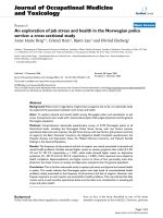
Agarose gel showing the effect of time on amplification of KHV DNA by LAMP assay using six primers (FIP, BIP, F3, B3, loopF, loopB), carried out at 65°C for durations of 10–60 minFigure 3
Agarose gel showing the effect of time on amplification of
KHV DNA by LAMP assay using six primers (FIP, BIP, F3, B3,
loopF, loopB), carried out at 65°C for durations of 10–60
min. Lane mar = 100 bp DNA molecular weight standard,
lane -veco = negative control. The LAMP assay detected
KHV as early as 30 min.
Virology Journal 2005, 2:83 />Page 3 of 8
(page number not for citation purposes)
A novel nucleic acid amplification method, loop-medi-
ated isothermal amplification (LAMP), has been devel-
oped that does not require a theramcycler. LAMP relies
instead on autocycling strand displacement DNA synthe-
sis by a Bst DNA polymerase, to amplify DNA with high
specificity, efficiency, and speed under isothermal condi-
tions [13,18,19]. LAMP requires two specially designed
inner and two outer primers to improve specificity
[20,21]; if two additional 'loop' primers are added, the
reaction time can be halved [20]. The amplification prod-
ucts are stem-loop DNA structures with several inverted
repeats of the target, and cauliflower-like structures com-
prising multiple loops [22]. In the present study, we used
a LAMP technique for diagnosis of KHV, and evaluated its
sensitivity, specificity, and applicability.
Results
Optimisation of the KHV LAMP reaction
The LAMP reaction was performed using purified KHV
genomic DNA as a template to determine the optimal
primer combination and duration of reaction. The ampli-
con was formed using either 4 or 6 primers. With 4 prim-
ers, a LAMP product was detected after 60 min at 65°C
(Fig. 2) while with 6 primers the amplification product
was detected as early as 30 min (Fig. 3). KHV DNA
extracted either by kit or by boiling gave rise to a typical
ladder pattern: many bands of different size up to the
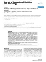
loading well as shown in Figures 2, 3 and 5. After addition
of 1 µl of diluted SYBR Green I to the reaction tube, posi-
tive reactions (amplified products) turned green, whereas
all negative controls remained orange, the starting colour
of SYBR Green (Fig. 4). The optimal primer concentration
is stated in Methods.
Specificity of the KHV LAMP primers and assay
Reaction products were detected only when KHV DNA
was present, giving rise to a typical ladder-like pattern.
There were no amplification products detected with Her-
pesvirus cyprini (CHV), channel catfish virus (CCV) or koi
fish genomic DNA (Fig. 6).
Sensitivity of the LAMP reaction in detection of KHV
The reaction was tested using 10-fold serial dilutions of
KHV DNA from both purified viral DNA and from DNA
extracted from positive clinical samples, and compared
against results from the commonly used PCR assay. The
detection limit of both the LAMP and PCR assays using
Visual detection of KHV LAMP products using SYBR Green I stainFigure 4
Visual detection of KHV LAMP products using SYBR Green I
stain. 1: negative LAMP reaction remained orange. 2: positive
LAMP reaction turned green.
Agarose gel showing LAMP products of KHV DNA extracted by boilingFigure 5
Agarose gel showing LAMP products of KHV DNA extracted
by boiling. The reaction was carried out at 65°C using the 6
primer set. Lanes: mar = 100 bp molecular weight marker; 1
= KHV DNA extracted by boiling; 2 = negative fish tissue; 3
= negative control.
Virology Journal 2005, 2:83 />Page 4 of 8
(page number not for citation purposes)
purified KHV viral DNA was 10
-7
(Fig. 7, 9). The detection
limit of both assays was 10
-5
for clinical samples (Fig. 8,
10). These were the limits for the KHV LAMP reaction
under optimal conditions: using 6 primers at 65°C for 60
min. If the reaction was run for 30 min, the detection limit
of the LAMP assay was 10
-3
for the purified KHV viral DNA
and 10
-1
for clinical samples. Increasing the primer con-
centrations did not affect these detection limits (data not
shown).
Applicability of the KHV LAMP reaction
50 clinical cases with suspected KHV infections were sub-
mitted to our laboratory and were investigated both with
the LAMP assay and standard PCR. 37 out of 50 tested
positive with both the PCR and LAMP; 13 were negative.
No sample that was negative with the LAMP assay tested
positive with the PCR, and vice versa.
Discussion
The most extensively used diagnostic methods for KHV
are cell culture and PCR. These techniques, however,
require a relatively long time to produce results or are not
practical for commercial producers, retailers, and regula-
tors because of the equipment and expertise needed to
conduct the assays. Moreover, the Taq DNA polymerase
used in the PCR assay is easily inactivated by tissue- and
blood-derived inhibitors such as myoglobin, hem-blood
protein complex and immunoglobulin G [25-28]. Loop-
mediated isothermal amplification (LAMP) is a novel
method that facilitates rapid nucleic acid amplification
using only simple equipment [13]. In the first step of the
LAMP reaction, Bst polymerase synthesises new DNA
between the F3 and B3 primers; this is the same reaction
Agarose gel illustrating the specificity of the designed primers to KHV DNAFigure 6
Agarose gel illustrating the specificity of the designed primers
to KHV DNA. The reaction was carried out at 65°C using
the 6 primer set for 1 hr. Lanes: 1 = KHV DNA; 2 = Herpes-
virus cyprini (CHV) DNA showing no amplification; 3 = chan-
nel catfish virus (CCV) showing no amplification; 4 =
uninfected koi tissue; mar = 100 bp DNA molecular weight
marker.
Agarose gel illustrating the sensitivity of the LAMP assay using 10-fold serial dilutions of purified KHV viral DNAFigure 7
Agarose gel illustrating the sensitivity of the LAMP assay
using 10-fold serial dilutions of purified KHV viral DNA. The
amplification shows a ladder-like pattern, and detected puri-
fied KHV viral DNA down to a dilution of 10
7
. Lanes: -1 =
dilution of 10
-1
; -2 = 10
-2
and so on; mar = 100 bp DNA
molecular weight standard. -veco = negative control.
Agarose gel demonstrating the sensitivity of the LAMP assay using 10-fold serial dilutions of KHV DNA extracted from a clinical sampleFigure 8
Agarose gel demonstrating the sensitivity of the LAMP assay
using 10-fold serial dilutions of KHV DNA extracted from a
clinical sample. The amplification shows a ladder-like pattern,
and detected KHV DNA in a clinical sample at a dilution of
10
-5
. Lanes: 0 = undiluted KHV DNA; -1 = dilution of 10
-1
; -2
= 10
-2
and so on; mar = 100 bp DNA molecular weight
standard. -veco = negative control.
Virology Journal 2005, 2:83 />Page 5 of 8
(page number not for citation purposes)
as standard PCR and requires homology between the
primers and the template DNA. In the next step, the newly
synthesised strands are recognised by the inner primers
FIP and BIP to start loop mediated autocycling amplifica-
tion [29] to produce stem-loop DNA structures with sev-
eral inverted repeats of the target and cauliflower-like
structures with multiple loops [22]. Amplification is spe-
cific and rapid when template which includes sequences
that the loop primers recognise is present [20]. To acceler-
ate the LAMP reaction 6 primers were used instead of 4.
The two additional primers hybridised to the stem-loops
(except for those loops that had been hybridized by the
inner primers) [20].
KHV DNA extraction was performed by boiling fish tis-
sues in a buffer solution; a simple and rapid technique
[31-33] AL buffer was used to inactivate DNase and to
elute DNA from tissues. Immediately after boiling, both
undiluted and diluted DNA samples were trialled as tem-
plates for the LAMP reaction. No amplification products
were detected for the undiluted DNA; addition of 800 µl
TE buffer was necessary to dilute reaction inhibitors which
were present in the boiled solution [30]. The LAMP assay
was sensitive enough to detect KHV DNA at this (1:4)
dilution. A specific type of DNA polymerase was required
for the LAMP reaction, Bst DNA polymerase, which has
two distinct activities: linear target isothermal multimeri-
sation and amplification, and cascade rolling-circle
amplification [34]. The mechanism of loop mediated
isothermal amplification is similar to cascade rolling cir-
cle amplification. Occasionally, a different LAMP amplifi-
cation pattern appeared as a result of linear target
isothermal multimerisation and amplification, as LAMP
primers and target DNA seem to randomly multimerize
[29]. Betaine was used in the LAMP reaction mixture to
reduce base stacking [35-37] and to increase not only the
overall rate of reaction but also target selectivity by signif-
icantly reducing amplification of irrelevant sequences
[13]. Use of SYBR Green I for visual inspection of LAMP
amplification products was a simple and superior tech-
nique, with no gel electrophoresis and staining with
ethidium bromide required. Only 1 µl of diluted SYBR
Green I added to the reaction mixture was enough to see
a result: if the reaction mix turned from orange to green it
was judged as positive. This visualisation technique is
effective due to the high specificity and amplification effi-
ciency of LAMP [22].
The detection limit of the KHV LAMP reaction was deter-
mined through amplification of 10-fold serial dilutions of
both purified KHV viral DNA and DNA from positive clin-
ical samples (containing both fish and KHV DNA). The
LAMP reaction was performed at 65°C for 30 and 60 min,
and compared with the results of the standard PCR assay.
There was no difference between the detection limit of the
LAMP reaction and the PCR reaction at 60 min: both were
positive at 10
-7
dilution of purified virus DNA, and at 10
-
5
from the clinical samples. However, at 30 min the LAMP
detected down to only 10
-3
dilution of viral DNA and 10
-
1
dilution DNA from clinical samples. Hence the optimal
LAMP conditions were determined to be 65°C for 60 min
to detect KHV virus down to a concentration of 0.1 pg.
Although the LAMP reaction had equivalent sensitivity to
the PCR test, it is considered superior because it is a sim-
pler technique which can be carried out in most situations
where a rapid diagnostic method is required: under field
conditions, in private clinics, and at quarantine inspec-
tion stations. A water bath is the only equipment needed,
Agarose gel illustrating the sensitivity of the PCR assay using 10-fold serial dilutions of the purified KHV viral DNAFigure 9
Agarose gel illustrating the sensitivity of the PCR assay using
10-fold serial dilutions of the purified KHV viral DNA. The
PCR shows a 484 bp amplification product, and detected
purified KHV viral DNA down to a dilution of 10
7
. Lanes: 0 =
undiluted KHV DNA; -1 = dilution of 10
-1
; -2 = 10
-2
and so
on; mar = 100 bp DNA molecular weight standard; -veco =
negative control without target DNA.
Agarose gel showing the sensitivity of the PCR assay using 10-fold serial dilutions of the KHV DNA extracted from a clinical sampleFigure 10
Agarose gel showing the sensitivity of the PCR assay using
10-fold serial dilutions of the KHV DNA extracted from a
clinical sample. The PCR reveals a 484 bp amplification prod-
uct, and detected KHV in a clinical sample at a dilution of 10
-
5
. Lanes: 0 = undiluted KHV DNA; -1 = dilution of 10
-1
; -2 =
10
-2
and so on; mar = 100 bp DNA molecular weight stand-
ard; -ve = negative control without target DNA.
Virology Journal 2005, 2:83 />Page 6 of 8
(page number not for citation purposes)
and is used for both the DNA extraction and nucleic acid
amplification.
Although the application of LAMP for the detection of
KHV has been reported previously [38], these authors use
only 4 primers which target the KHV tk gene. In the cur-
rent study, 6 primers which recognise 8 distinct regions on
the KHV DNA were used, thereby enhancing the specifi-
city of the reaction and eliminating false positive results
[20]. Also, DNA extraction by boiling prior to the LAMP
test and visualisation of reaction products using SYBR
Green I DNA stain were employed to reduce the time
needed to perform the KHV test and to simplify the
procedure.
In conclusion, the KHV LAMP reaction is a highly sensi-
tive, rapid, and reliable method that can be used under
field condition for diagnosis of the KHV infection.
Methods
DNA oligonucleotides
Six primers were designed from a KHV amplicon (Gen-
bank Accession number AF411803), which recognise
eight distinct regions of the target DNA. Forward inner
primer (FIP) comprised the antisense sequence of F1
(23nt), a TTTT linker and a sense sequence of F2 (23nt):
5'- CAACAATGCTTCTTGTGATTACA-TTTT-GAACCCG
AGGGGACTGCTCGCTT-3'. Backward inner primer (BIP)
consisted of the sense sequence of B1 (23nt), a TTTT linker
and the antisense sequence of B2 (23nt): 5'- CC GAT-
GGAGTGAAACTGGAACTG-TTTT-CGTCATGCTCTC-
CGAGGCCAGCG-3'. The outer primers were F3 (19nt):
5'- GAGGAAGCGCAAAAAGAAC-3', and B3 (19nt): 5'-
TTCAGTCTGTTCCTCAACC-3'. The loop primers were,
loop F (20nt): 5'-ATTATTATAC AACAACAATA-3'; and
loop B (20nt): 5'-TGAGCGTGGGGTCAAAGTT G-3'. (Fig.
1). Primers used in the PCR assay were constructed
according to Gilad et al. (2002). Forward primer- KHV9/
5F: 5'- GACGACGCCGGAGACCTTGTG-3', and reverse
primer- KHV9/5R: 5'-
CACAAGTTCAGTCTGTTCCTCAAC-3'. This primer set
amplified a 484 bp segment of the KHV template.
DNA extraction
Gills, kidney, spleen, and brain were sampled from fish
sent to our laboratory with suspected KHV infections.
DNA extraction was performed using both a commercial
kit and a tissue boiling method. For the QIAamp DNA
mini kit (QIAGEN GmbH, Hilden Germany), one gram of
each organ was ground thoroughly in liquid nitrogen
using a mortar and pestle. 20 mg of tissue powder was
placed in a 2 ml microfuge tube, 180 µl of lysis buffer and
20 µl of proteinase K were added, then incubated at 56°C
in a water bath until the tissues were completely lysed (1–
3 h). DNA extraction was then completed according to the
manufacturer's instructions, with final elution of DNA in
100 µl elution buffer, and storage at -20°C.
The second method of DNA extraction was by boiling: 20
mg of each tissue were placed in 2 ml microfuge tubes
with 200 µl AL buffer (QIAGEN GmbH, Hilden, Ger-
many), and placed in boiling water for 15 min. 800 µl of
Tris- EDTA buffer (TE: 10 mM Tris-HCl, 0.1 mM EDTA,
pH 8.0) was then added to the tube, mixed well, and
centrifuged at 14,000 rpm for 3 min. The supernatant con-
tained the DNA was used immediately in the KHV assays.
LAMP reaction
The 25 µl reaction mixture comprised: 20 mM Tris-HCl
(pH 8.8), 10 mM KCl, 6 mM MgSO
4
, 10 mM (NH
4
)
2
SO
4
,
0.1% Triton X-100, 1.6 M betaine, deoxynucleotide
triphosphates 2.8 mM each, 1.6 µM each FIP and BIP, 0.8
µM each loop-F and loop-B, 0.2 µM each F3 and B3 prim-
ers, 8 U Bst DNA polymerase (New England BioLabs,
GmbH, Frankfurt, Germany), 2 µl template DNA, distilled
water to 25 µl. As a negative control, template DNA was
omitted from the reaction. The mix was incubated at 65°C
for 60 min and then heated at 80°C for 2 min to terminate
the reaction.
Analysis of LAMP products
1 µl of 1:10 diluted SYBR Green I Nucleic acid gel stain,
10,000× concentration in DEMSO (Cambrex Bio Science,
Rockland, Inc, ME USA) was added directly to the reaction
tube and any colour change observed. The solution turned
green if LAMP reaction products were present, otherwise it
remained orange. Reaction products were also analysed
by gel electrophoresis: 5 µl aliquots were analysed on a
2% agarose gel and subsequently stained with ethidium
bromide; a DNA molecular weight marker, 100 bp DNA
Ladder, (Cambrex Bio Science, Inc, Rockland, ME USA)
was used to determine the size of the products.
PCR assay
Amplification was performed according to Gilad et al.
(2002) in a standard reaction volume of 50 µl comprising
3 µl template DNA and 47 µl 1.1× ReaddyMix PCR Master
mix: 75 mM Tris-HCl (pH 8.8), 20 mM (NH
4
)
2
SO
4
, 1.5
mM MgCl
2
, 0.01% Tween20, 0.2 mM each of dATP, dCTP,
dGTP, dTTP, 1.25 U Taq DNA polymerase and red dye for
electrophoresis (ABgene, Hamburg, Germany) and for-
ward and reverse primers (20 pmol each). The reaction
mixture was subjected to 39 amplification cycles under
the following conditions: denaturation at 94°C for 1 min,
annealing at 68°C for 1 min, extension at 72°C for 30s.
The amplification cycles were preceded by a denaturation
step at 94°C for 5 min and followed by an extended elon-
gation step at 72°C for 7 min.
Virology Journal 2005, 2:83 />Page 7 of 8
(page number not for citation purposes)
Detection of PCR products
products were analysed by electrophoresis on 1.5% agar-
ose gels stained with ethidium bromide. 100 bp DNA
Ladder (Cambrex Bio Science, Inc, Rockland, ME USA)
was used to determine the size of the PCR products.
Optimisation of KHV LAMP reaction conditions
varying concentrations of the FIP, BIP, F3, B3, loop-F,
loop-B primers were trialled, as well as use of only 4
primers (excluding loop-F and loop-B). Time of reaction
was varied in 5 minute increments from 10–60 min to
determine detection time of KHV genomic DNA.
Specificity of the KHV LAMP assay
The reaction was tested using DNA from Herpesvirus
cyprini (CHV), channel catfish virus (CCV) and koi
genomic DNA.
Sensitivity of the KHV LAMP reaction
The detection limits of the KHV LAMP assay were evalu-
ated using 10-fold serial dilutions of purified KHV DNA
and DNA extracted from positive clinical samples. The
reaction was performed at 65°C for both 30 and 60 min,
and compared with the PCR assay results.
Applicability of the KHV LAMP reaction
After the initial validation studies, the KHV LAMP reaction
was used to test 50 suspected clinical cases submitted to
our laboratory and the results compared with the PCR
assay results of those 50 cases.
Author's contributions
ME conceived and supervised the study and drafted the
manuscript. HS carried out all the experimental work and
data acquisition.
References
1. Hedrick RP, Gilad O, Yun S, Spangenberg JV, Marty GD, Nordhausen
RW, Kebus MJ, Bercovier H, Eldar A: A herpesvirus associated
with mass mortality of juvenile and adult koi, a strain of com-
mon carp. J Aquat Anim Health 2000, 12:44-57.
2. Ronen A, Perelberg A, Abramowitz J, Hutoran M, Tinman S, Bejerano
I, Steinitz M, kotler M: Efficient vaccine against the virus causing
a lethal disease in cultured Cyprinus carpio. Vaccine 2003,
21:4677-4684.
3. Waltzek TB, Kelley GO, Yun SC, McDowell TS, Hedrick RP: Rela-
tionships of Koi Herpesvirus (KHV) to Herpes-like Viruses of
Fish and Amphibians. Proceedings, 35th Annual Conference Interna-
tional Association for Aquatic Animal Medicine, Galveston, TX 2004:16-17.
4. Hedrick RP: Movement of pathogens with the international
trade of live fish; Problems and solutions. Rev Sci Tech 1996,
15:523-531.
5. Gilad O, Yun S, Adkison M, Way K, Willits N, Bercovier H, Hedrick
RP: Molecular comparison of isolates of an emerging fish
pathogen, koi Herpesvirus, and the effect of water tempera-
ture on mortality of experimentally infected koi. J Gen Virol
2003, 84:2661-2668.
6. Bretzinger A, Fischer-Scherl T, Oumouna M, Hoffmann R, Truyen U:
Mass mortalities in koi, Cyprinus carpio, associated with gill
and skin disease. Bull Eur Ass Fish Pathol 1999, 19:182-185.
7. Oh MJ, Jung SJ, Choi TJ, Kim HR, Rajendran KV, Kim YJ, Park MA,
Chun SK: A viral disease occurring in cultured carp Cyprinus
carpio in Korea. Fish pathol 2001, 36:147-151.
8. Choi D, Sohn S, Bang J, Do J, Park M: Ultrastructural identifica-
tion of a herpes-like virus infection in common carp Cyprinus
carpio in Korea. Dis Aquat Org 2004, 61:165-168.
9. Rukyani A: Koi Herpesvirus infection in Indonesia. ProMed Jun
30 2002 [
]. Archive No. 20020630.4639
10. Miyazaki T, Okamoto H, Kageyama T, Kobayashi T: Viremia associ-
ated ana-aki-byo, a new viral disease in colour carp Cyprinus
carpio in Japan. Dis Aquat Org 2003, 39:183-192.
11. Hartman KH, Yanong RP, Petty BD, Francis-Floyd R, Riggs AC: Koi
Herpes Virus (KHV) Disease. Fact sheet VM-149, Extension service,
Institute of Food and Agricultural Sciences, University of Florida 2004 [http:/
/edis.ifas.ufl.edu].
12. Gray WL, Mullis L, LaPatra SE, Groff JM, Goodwin A: Detection of
Koi herpesvirus DNA in tissues of infected fish. J Fish Dis 2002,
25:171-178.
13. Notomi T, Okayama H, Masubuchi H, Yonekawa T, Watanabe K,
Amino N, Hase T: Loop-mediated isothermal amplification of
DNA. Nuc Acids Res 2000, 28:e63.
14. Saiki RK, Scharf S, Faloona F, Mullis KB, Horn GT, Erlich HA, Arnheim
N: Enzymatic amplification of beta-globin genomic
sequences and restriction site analysis for diagnosis of sickle
cell anaemia. Science 1985, 230:1350-1354.
15. Saiki RK, Gelfand DH, Stoffel S, Scharf S, Higuchi R, Horn GT, Mullis
KB, Erlich HA: Primer-directed enzymatic amplification of
DNA with a thermostable DNA polymerase. Science 1988,
239:487-491.
16. Gilad O, Yun S, Andree KB, Adkison MA, Zlotkin A, Bercovier H,
Eldar A, Hedrick RP: Initial characteristics of koi Herpesvirus
and development of a polymerase chain reaction assay to
detect the virus in koi, Cyprinus carpio koi. Dis Aquat Org 2002,
48:101-108.
17. Gilad O, Yun S, Zagmutt-Vergara FJ, Leutenegger CM, Bercovier H,
Hedrick RP: Concentrations of a Koi herpesvirus (KHV) in tis-
sues of experimentally-infected Cyprinus carpio koi as
assessed by real-time TaqMan PCR. Dis Aquat Org 2004,
60:179-187.
18. Nagamine K, Watanabe K, Ohtsuka K, Hase H, Notomi T: Loop-
mediated isothermal amplification reaction using a nonde-
natured template. Clin Chem 2001, 47:1742-1743.
19. Mori Y, Nagamine K, Tomita N, Notomi T: Detection of loop-
mediated isothermal amplification reaction by turbidity
derived from magnesium pyrophosphate formation. Biochem
Biophys Res Commun 2001, 289:150-154.
20. Nagamine K, Hase T, Notomi T: Accelerated reaction by loop-
mediated isothermal amplification using loop primers. Mol
Cell Probes 2002, 16:223-229.
21. Ihira M, Yoshikawa T, Enomoto Y, Akimoto S, Ohashi M, Suga S,
Nishimura N, Ozaki T, Nishiyama N, Ozaki T, Nishiyama Y, Notomi
T, Ohta Y, Asano Y: Rapid diagnosis of Human Herpesvirus 6
infection by a novel DNA amplification method, loop-medi-
ated isothermal amplification. J Clin Microbiol 2004, 42:140-145.
22. Iwamoto T, Sonobe T, Hayashi K: Loop-mediated isothermal
amplification for direct detection of Mycobacterium tubercu-
losis complex, M. avium, and M. intracellulare in sputum
samples. J Clin Microbiol 2003, 41:2616-2622.
23. Waltzek TB, Hedrick RB: Koi Herpesvirus update. California
Veterinarian . 2004 July-August
24. Way K, LeDeuff RM, Stone DM, Denham KL, St-Hilaire S: Koi Her-
pesvirus diagnosis and research at CEFAS Weymouth labo-
ratory 2000–2003. International workshop on koi Herpesvirus 2004
[ />].
25. Belec L, Authier J, Eliezer-Vanerot M, Piedouillet C, Mohamed A,
Gherardi R: Myoglobin as a polymerase chain reaction (PCR)
inhibitor: a limitation for PCR from skeletal muscle tissue
avoided by the use of Thermus thermophilus polymerase. Mus-
cle and Nerve 1998, 21:1064-1067.
26. Akane A, Matsubara K, Nakamura H, Takahashi S, Kimura K: Identi-
fication of the heme compound copurified with deoxyribo-
nucleic acid (DNA) from bloodstains, a major inhibitor of
polymerase chain reaction (PCR) amplification. J Forensic Sci
1994, 39:362-372.
Publish with BioMed Central and every
scientist can read your work free of charge
"BioMed Central will be the most significant development for
disseminating the results of biomedical research in our lifetime."
Sir Paul Nurse, Cancer Research UK
Your research papers will be:
available free of charge to the entire biomedical community
peer reviewed and published immediately upon acceptance
cited in PubMed and archived on PubMed Central
yours — you keep the copyright
Submit your manuscript here:
/>BioMedcentral
Virology Journal 2005, 2:83 />Page 8 of 8
(page number not for citation purposes)
27. Al-Soud W, Önsson LJ, Radström P: Identification and character-
ization of immunoglobulin G in blood as a major inhibitor of
diagnostic PCR. J Clin Microbiol 2000, 38:345-350.
28. Johnson S, Martin D, Cammarata C, Morse S: Alterations in sam-
ple preparation increase sensitivity of PCR assay for diagno-
sis of chancroid. J Clin Microbiol 1995, 33:1036-1038.
29. Kuboki N, Inoue N, Sakurai T, Di Cello F, Grab DJ, Suzuki H, Sugim-
oto C, Igarashi I: Loop-mediated isothermal amplification for
detection of African trypanosomes. J Clin Microbiol 2003,
41:5517-5524.
30. Ikeda N, Bautista N, Yamada T, Kamijima O, Ishii T: Ultra-simple
DNA extraction method for marker-assisted selection using
microsatellite markers in rice. Plant Mol Biol Rep 2001, 19:27-32.
31. Valsecchi E: Tissue boiling: a short-cut in DNA extraction for
large-scale population screening. Mol Ecol 1998, 7:1243-1245.
32. Sepp R, Szabo I, Uda H, Sakamoto H: Rapid techniques for DNA
extraction from routinely processed archival tissue for use in
PCR. J Clin Pathol 1994, 47:318-323.
33. Afghani B, Stutman HR: Polymerase chain reaction for diagnosis
of M. tuberculosis: comparison of simple boiling and conven-
tional methods for DNA extraction. Biochem Mol Med 1996,
57:14-18.
34. Hafner GJ, Yang IC, Wolter LC, Stafford MR, Giffard PM: Isothermal
amplification and multimerization of DNA by Bst DNA
polymerase. BioTechniques 2001, 30:852-867.
35. Rees WA, Yager TD, Korte J, Von Hippel PH: Betaine can elimi-
nate the base pair composition dependence of DNA melting.
Biochemistry 1993, 32:137-144.
36. Baskaran N, Kandpal RP, Bhargava AK, Glynn MW, Bale A, Weissman
SM: Uniform amplification of a mixture of deoxyribonucleic
acids with varying GC content. Genome Res 1996, 6:633-638.
37. Rajendrakumar CS, Suryanarayana T, Reddy AR: DNA helix desta-
bilization by proline and betaine: possible role in the salinity
tolerance process. FEBS Letters 1997, 410:201-205.
38. Gunimaladevi I, Kono T, Venugopal MN, Sakai M: Detection of koi
Herpesvirus in common carp, Cyprinus carpio L., by loop-
mediated isothermal amplification. J Fish Dis 2004, 27:583-589.