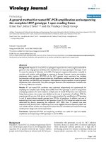
báo cáo hóa học:" A general method for nested RT-PCR amplification and sequencing the complete HCV genotype 1 open reading frame" pdf
Bạn đang xem bản rút gọn của tài liệu. Xem và tải ngay bản đầy đủ của tài liệu tại đây (542.35 KB, 9 trang )
BioMed Central
Page 1 of 9
(page number not for citation purposes)
Virology Journal
Open Access
Methodology
A general method for nested RT-PCR amplification and sequencing
the complete HCV genotype 1 open reading frame
Ermei Yao
1
, John E Tavis*
1,2
and the Virahep-C Study Group
Address:
1
Department of Molecular Microbiology and Immunology, Saint Louis University School of Medicine, Saint Louis, Missouri 63104, USA
and
2
Saint Louis University Liver Center, Saint Louis University School of Medicine, Saint Louis, Missouri 63104, USA
Email: Ermei Yao - ; John E Tavis* - ; the Virahep-C Study Group -
* Corresponding author
Abstract
Background: Hepatitis C virus (HCV) is a pathogenic hepatic flavivirus with a single stranded RNA
genome. It has a high genetic variability and is classified into six major genotypes. Genotype 1a and
1b cause the majority of infections in the USA. Viral genomic sequence information is needed to
correlate viral variation with pathology or response to therapy. However, reverse transcription-
polymerase chain reaction (RT-PCR) of the HCV genome must overcome low template
concentration and high target sequence diversity. Amplification conditions must hence have both
high sensitivity and specificity yet recognize a heterogeneous target population to permit general
amplification with minimal bias. This places divergent demands of the amplification conditions that
can be very difficult to reconcile.
Results: RT and nested PCR conditions were optimized independently and systematically for
amplifying the complete open reading frame (ORF) from HCV genotype 1a and 1b using several
overlapping amplicons. For each amplicon, multiple pairs of nested PCR primers were optimized.
Using these primers, the success rate (defined as the rate of production of sufficient DNA for
sequencing with any one of the primer pairs for a given amplicon) for amplification of 72 genotype
1a and 1b patient plasma samples averaged over 95% for all amplicons. In addition, two sets of
sequencing primers were optimized for each genotype 1a and 1b. Viral consensus sequences were
determined by directly sequencing the amplicons. HCV ORFs from 72 patients have been
sequenced using these primers. Sequencing errors were negligible because sequencing depth was
over 4-fold and both strands were sequenced. Primer bias was controlled and monitored through
careful primer design and control experiments.
Conclusion: Optimized RT-PCR and sequencing conditions are useful for rapid and reliable
amplification and sequencing of HCV genotype 1a and 1b ORFs.
Background
Hepatitis C virus (HCV) is a human hepatotropic flavivi-
rus. It is the major cause of non-A, non-B hepatitis, infect-
ing about 3% of people world-wide [1]. Nearly 4 million
people in the United States are infected with HCV [2], pre-
dominantly with genotypes 1a and 1b. HCV infection
becomes chronic in about 80% of infected individuals.
These chronically infected patients are at high risk of
developing serious liver disease, including cirrhosis and
hepatocellular carcinoma [3]. No effective vaccine has
Published: 01 December 2005
Virology Journal 2005, 2:88 doi:10.1186/1743-422X-2-88
Received: 17 June 2005
Accepted: 01 December 2005
This article is available from: />© 2005 Yao et al; licensee BioMed Central Ltd.
This is an Open Access article distributed under the terms of the Creative Commons Attribution License ( />),
which permits unrestricted use, distribution, and reproduction in any medium, provided the original work is properly cited.
Virology Journal 2005, 2:88 />Page 2 of 9
(page number not for citation purposes)
been developed to prevent HCV infection. The best avail-
able therapy for HCV infection is a combination of
pegylated interferon α and ribavirin, an oral guanosine
analogue [4]. The response rate to therapy varies depend-
ing on HCV genotype, viral load, patient sex, patient age,
and the stage of liver fibrosis [5].
The HCV genome is a positive polarity, single-stranded
RNA about 9600 nucleotides long. It contains one long
ORF flanked by 5' and 3' untranslated regions (UTR). The
genome is highly variable due to the poor fidelity of the
viral RNA dependent RNA polymerase (RdRp) and the
lack of genome repair mechanisms. HCV genomic varia-
bility is not uniform throughout the genome. The 5'UTR
and the terminal 98 nucleotides of the 3'UTR are con-
served, but the region of the 3'UTR immediately down-
stream of the open reading frame and the adjacent U-rich
sequence are highly variable [6]. Significant sequence var-
iation is also present in the ORF at both the nucleotide
and the amino acid level, especially in hypervariable
regions (HVR1 and HVR2) within the E2 region [7,8].
Analysis of the NS5B region encoding the viral RNA
polymerase from a wide range of HCV isolates led to the
classification of HCV into six major genotypes and a series
of subtypes [9,10]. Genotypes share less than 72% nucle-
otide homology. Within genotypes, subtypes have homol-
ogies of 75%–86%.
HCV sequences within an infected individual exist as a
group of related but distinct variants [11,12]. This distri-
bution of sequences is common among RNA viruses and
is referred to as "quasispecies". Quasispecies variation can
lead to significant amino acid variation of the encoded
proteins [11,13]. The distribution of sequences in a qua-
sispecies clusters around a master sequence, and the
"center" of the genetic distribution can be described either
as the dominant quasispecies (the single most common
sequence in the viral population) or as the consensus
sequence (an "average" sequence comprised of the pre-
dominant sequence at each nucleotide position). This
protocol is designed to yield the consensus sequence.
The high genomic heterogeneity of HCV may contribute
to viral immune evasion [9], promote chronicity [14], and
may influence the outcome of interferon α therapy in
HCV-infected individuals [11,15,16]. Therefore, system-
atic examination of HCV sequence variation has impor-
tant implications in understanding HCV biology and
could open novel avenues for anti-viral therapy.
HCV viremia is relatively low compared to many other
viruses, rarely exceeding 10
6
–10
7
genomes per milliliter.
Therefore, reverse transcription-polymerase chain reac-
tion (RT-PCR) of the HCV genome must overcome not
only high target sequence diversity, but also low template
HCV genotype 1b ampliconsFigure 1
HCV genotype 1b amplicons. Amplicons are numbered sequentially as amplicon 1 to 4 starting from 5' of the genome.
Amplicon 1 and 4 are divided into halves named 1x, 1y and 4x, 4y. The amplicon boundaries indicate the 5' ends of the inner-
most amplification primers against the genome of strain J4.
Amplicon 1 Amplicon 3
Amplicon 2 Amplicon 4
4x
4y
5’UTR
3’UTR
ORF
57 4995 7698
2407 5026 7283 8373
8288 9524
1x
1y
1364
1313 2566
Virology Journal 2005, 2:88 />Page 3 of 9
(page number not for citation purposes)
concentration. Hence, the amplification conditions must
have high sensitivity and specificity yet recognize a heter-
ogeneous target population. These divergent demands are
difficult to reconcile. In this paper, we report a general
method to amplify and sequence the whole ORF of HCV
genotypes 1a and 1b. We systematically optimized all
steps in the process, including isolation of HCV RNA from
patient plasma or serum, RT, PCR primer sequences, PCR
conditions, template preparation, sequencing and assem-
bly. We have a success rate of over 95% in RT-PCR ampli-
fication and have successfully sequenced HCV ORFs from
over 72 patients using this system.
Results and discussion
Amplification strategy
The HCV ORF is over 9 kb long, so long range PCR was
initially attempted to amplify partial or full HCV ORFs. Its
success frequency was inadequate for large-scale HCV
genome sequencing projects, so this approach was aban-
doned. Efficient amplification with regular PCR is limited
to 3 kb. Therefore, to maximize PCR sensitivity, we
divided the genome into four amplicons that were num-
bered sequentially as amplicon 1 to 4 starting from the 5'
end of the genome, with each amplicon being less than 3
kb and overlapping with the adjacent amplicon(s). This
strategy was effective for amplifying all amplicons except
for amplicon 4 for both genotype 1a and 1b and amplicon
1 for genotype 1b. To increase the sensitivity of amplifica-
tion for these regions, they were subdivided, which
resulted in efficient amplification. The HCV ORF was
therefore partitioned into amplicons 1, 2, 3, 4x and 4y for
genotype 1a and amplicons 1x, 1y, 2, 3, 4x and 4y for gen-
otype 1b. Figure 1 shows the amplicon partition for geno-
type 1b.
Optimization of RT conditions for genotype 1b amplicon 2Figure 2
Optimization of RT conditions for genotype 1b amplicon 2. R2V1 and R2V2 were RNAs isolated from the same aliq-
uot of a patient plasma; R2V1 employed guanidine thiocyanate and phenol/chloroform extraction and R2V2 employed the Viral
RNA Mini Kit. RT primer B4R1 is a specific primer targeted to the 3'UTR. Rndm, random hexamers; M-MLV, Murine Leukemia
Virus Reverse Transcriptase; AMV, Enhanced Avian Reverse Transcriptase. Different PCR primers were used for odd or even
numbered lanes. Lanes 11 and 12 are negative controls in which template RNA was omitted.
Reverse
transcriptase
RT primer
RT template
1 2
B4R1 B4R1
Lane
B4R1Rndm Rndm Rndm B4R1
M-MLV M-MLV M-MLV AMV AMV AMV M-MLV
R2V1 R2V2 H
2
O
λ
Hi
n
d
I
I
I
Hy
p
e
r
l
a
d
d
e
r
I
3.0kb
1.0kb
1.5kb
3 4 5 6 7 8 9 10 11 12 13 14
Virology Journal 2005, 2:88 />Page 4 of 9
(page number not for citation purposes)
Optimization of RNA extraction
RNA isolation must be suitable for extracting HCV RNA
from both patient plasma and serum because these are
common sources of HCV. RNA isolation must be efficient
to yield adequate amounts of high purity template due to
the limited amount of patient plasma or serum that is
often available and the relatively low titer of the virus. We
tried three RNA isolation protocols to isolate RNA from
plasma/serum samples including guanidine thiocyanate
denaturation plus phenol/chloroform extraction, the ZR
Viral RNA Kit (ZYMO Research) and the QIAamp Viral
RNA Mini Kit (Qiagen). The QIAamp Viral RNA Mini Kit
(Qiagen) worked best. The manufacturer's protocol was
followed without modification. Processing 140 µl plasma
sample routinely yielded about 60 µl viral RNA solution,
of which 15 µl was sufficient for an RT reaction. RNA iso-
lation was equally efficient using this kit with either serum
or plasma.
Optimization of cDNA synthesis
The reverse transcriptases tested include Cloned AMV
Reverse Transcriptase (Invitrogen), AMV Reverse Tran-
scriptase (Promega), Moloney Murine Leukemia Virus
Reverse Transcriptase (M-MLV RT; Promega) and
Enhanced Avian Reverse Transcriptase (AMV-RT; Sigma),
an enhanced avian myeloblastosis virus reverse tran-
scriptase. Reactions were assembled per manufacturer's
instructions employing a constant amount of HCV RNA
(15 µl for a 50 µl reaction). Because HCV RNA has a rela-
tively high GC percentage and has many secondary struc-
tures that may interfere with RT, incubation temperatures
between 30°C – 50°C were tested at 5°C intervals for
each enzyme. After RT, nested PCR was performed to test
the RT efficiency. Figure 2 shows part of the optimization
of RT conditions for genotype 1b amplicon 2. Different
sets of PCR primers were used for odd and even numbered
lanes. RNA isolated by the Viral RNA Mini Kit (R2V2) was
much more efficient than RNA processed through guani-
dine thiocyanate and phenol/chloroform extraction
(R2V1) (compare lanes 1 and 2 versus 3 and 4). Random
hexamers (Rndm) were more efficient than B4R1, a
primer specific to the 3'-UTR (lanes 3 and 4 versus 5 and
6, or lanes 7 and 8 versus 9 and 10). For amplicon 2, AMV-
RT and M-MLV RT worked equally well (lanes 3 and 4 ver-
sus 7 and 8, or lanes 5 and 6 versus 9 and 10). Lanes 11
and 12 are negative controls in which template RNA was
omitted.
M-MLV RT and AMV-RT both worked very well for ampli-
cons 1, 2, 3 and 4x. For amplicon 4y, AMV-RT worked
much better, especially if the enzyme was stored at -75°C
or lower (data not shown). RT reactions were suitable for
amplicons 1, 2, 3 and 4x when stored at -20°C for several
months, but for amplicon 4y, fresh RT reactions worked
much better.
Optimization of nested PCR
We optimized nested PCR conditions for each amplicon
independently. The process is summarized in Figure 3.
First, we designed primers for nested PCR. Because viral
genetic heterogeneity will prevent a given primer from
working well on all isolates, we optimized three sense and
three anti-sense primers for each amplicon as shown in
Figure 4. The three anti-sense primers must reside 3' to all
three sense primers for the downstream amplicon to pre-
vent gaps between the amplicons. Primers were targeted
to relatively conserved regions of the genome to maximize
the number of isolates they recognize. We employed
Oligo Explorer 1.2 [17] to guide primer design. The soft-
ware considers melting temperature and length of primers
while avoiding sequences prone to dimer or hairpin for-
mation or self-complementary primers. To use the pro-
gram, a reference sequence must be provided. We used
consensus sequences generated by aligning all full length
HCV 1a or 1b genome sequences available in Genbank
because these consensus sequences represent "average" 1a
or 1b isolates. We first chose the rough boundaries of the
amplicons, and then designed primers within 500 nucle-
otides at both ends of each amplicon. Candidate primers
of 20–25 nucleotides were designed and compared to the
1a or 1b alignment from which the reference sequence
was generated. For positions with unavoidable variability
Amplification optimization processFigure 3
Amplification optimization process.
Design primers
Optimize primer concentration,
[Mg++], annealing temperature and
permutation on cloned HCV DNA
Test primers on cDNA from serum samples
> 80% success rate <80%success rate
Research primer problem
Keep primers
Reject primer
s
Virology Journal 2005, 2:88 />Page 5 of 9
(page number not for citation purposes)
within the primer, degenerated bases were used. Generally
no more than 5 mixed bases per primer were employed
because we found that primers with more mixed bases
were less sensitive. However, a few primers have 6 degen-
erate bases because the heterogeneity in the target region
was unavoidable. Universal bases deoxyinosine (dI) and
deoxyuridine (dU) were used in initial optimizations, but
the amplification sensitivity with these primers was insuf-
ficient, possibly due to dI's less discriminate base pairing
and wide range of melting temperatures [18].
Then we optimized all nine primer permutations (three
sense versus three anti-sense primers) for each of the
amplicons for primer concentration, Mg
++
concentration,
and annealing temperature against cloned HCV DNA. For
genotype 1a, we optimized our amplification primers
against strain H77 [GenBank: AF009606
] [19]. For geno-
type 1b we used plasmid pHCV-CG1b [GenBank:
AF333324
] [20], which has the HCV 1b strain J structural
region, the 1b strain BK non-structural region and the
HCV 1a strain H 3' poly (UC) and X regions.
Three Taq polymerases were tested against the cloned
HCV cDNA using selected primer permutations. The
enzymes were Taq DNA Polymerase in Storage Buffer B
(Promega), Taq DNA Polymerase (Fisher) and Expand
High Fidelity PCR System (Roche). Expand High Fidelity
PCR System (Roche) was tested since it has a proofreading
polymerase with high fidelity, but it was rejected due to
insufficient sensitivity and excessive cost for a large-scale
sequencing project. Taq DNA Polymerase from Fisher was
chosen for all PCR reactions because it was the most effi-
cient of the three. Because our goal was to directly
sequence the RT-PCR products, its lower fidelity was not
critical (see "Accuracy of the sequences").
Finally, we tested the optimized primers on several patient
plasma samples. If the success rate for a given primer pair
on clinical isolates was over 80%, we kept the primer pair.
If not, we designed new primers and repeated the optimi-
zation process until at least three pairs of optimized prim-
ers were available for each amplicon. Table 1 (see
additional file 1: HCVMethodPaperTable1.xls) and Table
2 (see additional file 2: HCVMethodPaperTable2.xls) list
amplification and sequencing primers for genotypes 1a
and 1b. Table 3 (see additional file 3:
HCVMethodPaperTable3.xls) and Table 4 (see additional
file 4: HCVMethodPaperTable4.xls) list optimized PCR
conditions for each primer pair for genotypes 1a and 1b.
Table 5 (see additional file 5:
HCVMethodPaperTable5.xls) and Table 6 (see additional
file 6: HCVMethodPaperTable6.xls) list genotype 1a and
1b primer permutations that worked well on patient sam-
ples.
Amplification efficiency
We amplified 72 genotype 1 patients (44 genotype 1a, 28
genotype 1b) ORFs using these primers and PCR condi-
tions. The overall success rate for amplicons averaged over
95%. Table 7 lists amplification efficiency for each ampli-
con. The few amplicons that could not be generated by
these optimized primers were easily amplified by design-
ing custom primers derived from sequences obtained
from the neighboring amplicon(s) for that isolate.
Sequencing
RT-PCR often yields minor amounts of primer dimers or
truncated products that can interfere with sequencing.
Therefore, DNA templates were purified by gel extraction
using QIAquick Gel Extraction Kit (Qiagen) following
manufacturer's protocol. DNA concentration was deter-
mined by agarose gel electrophoresis comparing band
intensity to the Hyperladder I (Bioline) marker.
Two sets of DNA sequencing primers were designed and
validated for each genotype 1a and 1b (table 1 and 2).
Each set of primers contains both sense and anti-sense
primers to obtain complete coverage of both strands. In
the primary set of primers, the distance between adjacent
primers is 150–300 bp. HCV sequences are very heteroge-
neous, so not all primers will work for all patients due to
mismatches between the primers and templates. Because
Relative position of amplicon amplification primersFigure 4
Relative position of amplicon amplification primers. Three pairs of amplification primers and their relative positions are
shown. The red regions overlap with adjacent amplicon(s).
~2.8kb
R.3-AP1
L.3-AP2
L.3-AP1
L.3-AP3
R.3-AP2
R.3-AP3
Virology Journal 2005, 2:88 />Page 6 of 9
(page number not for citation purposes)
a typical sequencing read-length is over 600 bp, placing
the primers this close together allows each to reach the
position of the second primer downstream of it. This
yields a sequencing depth of 4- to 5-fold when both
strands are sequenced, which maximizes coverage and
sequencing quality. The backup set of primers was used to
fill in gaps in the rare cases when the primary set failed to
completely cover an amplicon.
Sequencing employed the ABI automated dye-terminator
system. It was performed at a contract sequencing facility
(Macrogen, Inc. Seoul, South Korea). For each sequencing
reaction, 50 ng template and 3.2 pmol primer were used.
Consensus sequences were obtained through assembling
and editing the sequencing traces using Vector NTI (Infor-
max). This program automatically assembles overlapping
sequencing traces and identifies nucleotide positions with
discrepancies between the traces. Computer base-calling
errors were corrected following inspection of the sequence
chromatograms. Mixed-base positions from the HCV qua-
sispecies were resolved by manually identifying the pre-
dominant base at each position. Where necessary,
additional sequencing reactions were performed to con-
firm the identity of a base or its predominance in the qua-
sispecies spectrum. For accuracy, we require that each
nucleotide be present in at least two unambiguous
sequencing reactions, preferably of opposite polarity. Fig-
ure 5 shows an example with six overlapping sequencing
traces. Two of the reactions revealed a mixture of G and A
at position 1270 and the four other traces clearly indicated
that G was dominant at this position; this base was man-
ually identified as G.
Accuracy of the sequences
Errors in sequencing HCV genomes arise from three major
sources: sequencing errors, enzymatic errors during RT-
PCR and primer bias during PCR. Our sequencing depth
averages over 4-fold and both strands are sequenced, so
error from sequencing mistakes is negligible. Base changes
are certainly introduced into the template DNAs during
RT-PCR. However, determining consensus sequence by
directly sequencing uncloned templates greatly reduces
the impact of this type of error because for an enzymati-
cally-derived error to be detected, the error would have to
have become the predominant sequence in the template
molecule population. This is rare with direct sequencing
of PCR products, in contrast to using cloned templates
such as are used for quasispecies analysis, where these
errors are very significant. Quality control experiments
with templates from a HCV donor-recipient set indicate
that the rate of enzymatically-derived errors is less than
0.012% when a common set of RT-PCR primers are used
[21].
The largest (and often least-appreciated) source of error in
sequencing is due to primer bias. Primer bias is selective
amplification of a portion of the sequences in the target
population and is a result of varying primer affinities for
the heterogeneous template molecules during PCR.
Primer bias is unavoidable in HCV genetic analyses due to
the extreme genetic heterogeneity of the virus. This bias
cannot be eliminated, but it can be quantitated and min-
imized through careful primer design and conscientious
control experiments.
To measure our net sequencing reproducibility, we
sequenced a HCV 1b ORF from two aliquots of plasma
from a single blood draw. The experiment was done in a
blinded manner and the primers used to amplify the two
genomes were independently chosen. The identity of the
two sequences was 99.1% at the nucleotide level and
99.4% at the amino acid level (compared to 91.2% nucle-
otide and 94.3% amino acid identity between these
sequences and HCV J4 [GenBank: AF054247
], another 1b
isolate). Because these differences are primarily due to
primer bias, they are not truly "errors". Rather, they repre-
sent alternate samplings of sequences within the viral qua-
sispecies population.
Record keeping
Record keeping and storage of samples and reagents must
be meticulous to avoid costly and time-consuming errors.
To assist tracking of samples and data, we developed a
custom relational MySQL database into which are entered
the identity, source, and location of all PCR primers,
sequencing primers, patient samples, RNAs, and PCR
products. The database is web-enabled to permit remote
access, it is secured behind a fire-wall, and access is limited
to authorized users with valid passwords. The database
and all sequence data are backed up to a secure tape-
backup system in a different building three times a week.
The database will be made available free of charge to inter-
ested parties.
Table 7: Amplification efficiency for patients' amplicons
Genotype 1a
Amplicon A1 A2 A3 A4x A4y
Amplification efficiency 95
a
98 93 100 95
Average efficiency 96.2
Genotype 1b
Amplicon A1x A1y A2 A3 A4x A4y
Amplification efficiency 100 100 93 93 100 100
Average efficiency 97.7
a
Amplification efficiencies are shown as percentage.
Virology Journal 2005, 2:88 />Page 7 of 9
(page number not for citation purposes)
Conclusion
Despite the high degree of genomic heterogeneity and rel-
atively low viral titres, efficient amplification and
sequencing of the HCV ORF is possible. We report opti-
mized amplification and sequencing conditions for the
complete HCV genotype 1a and 1b ORFs. This will facili-
tate large-scale HCV genome sequencing and greatly ease
systematic genetic analyses of the virus. This method was
developed to yield the viral consensus sequence through
direct sequencing RT-PCR products. However, it should
be easily adaptable to quasispecies analysis by replacing
the Taq polymerase with a high fidelity thermostable
DNA polymerase and sequencing cloned templates rather
than uncloned PCR products.
Materials and methods
Primer naming convention
Due to the large number of primers, we chose primer
names to include information indicating genotype,
amplicon number, polarity, purpose(amplification or
sequencing), relative position on the amplicon, and ver-
sion number. For primer "B2R.3-AP3", "B" stands for gen-
otype 1b, the "2" means amplicon 2, "R" represents anti-
sense (reverse) polarity. ".3" means it is from the third set
of primers designed. The AP suffix stands for "amplifica-
tion primer" and indicates the primer is suitable for PCR,
and the final "3" means it is the innermost primer com-
pared to the other PCR primers for the amplicon in the
same set. Primer "A1L3.2" is a sequencing primer for gen-
otype 1a, amplicon 1, of sense polarity, "3" indicates it is
the third sequencing primer for the strand, and the final
"2" indicates it is from sequencing primer set 2.
cDNA synthesis
cDNA was synthesized using random hexamers
(Promega) and M-MLV RT or AMV-RT. For a 50 µl RT reac-
tion, 15 µl viral RNA was mixed with 1 µg random primers
in a sterile RNase-free 250 µl PCR tube, heated to 70°C for
5 minutes for M-MLV RT or 10 minutes for AMV-RT to
melt secondary structures within the template and cooled
immediately on ice. For the M-MLV RT, 10 µl M-MLV 5 ×
Reaction Buffer, 10 µl nucleotide mix (2.5 mM each
dNTP), 1 µl RNasin (40 U/µl) (Promega) and 2 µl M-MLV
reverse transcriptase were mixed in 50 µl. The reaction was
incubated at 37°C for 1 hour followed by 94°C for 5 min-
utes to inactivate the reverse transcriptase. For AMV-RT, 5
µl AMV-RT 10 × Reaction Buffer, 20 µl nucleotide mix (2.5
mM each dNTP), 1 µl RNasin (40 U/µl) and 2.5 µl reverse
transcriptase were used. The reaction was incubated in 50
µl at 25°C for 15 minutes, 42°C for 1 hour followed by
94°C for 5 minutes. All reactions were assembled in PCR
hood using aerosol-barrier tips to avoid contamination.
Resolving discordant sequencing tracesFigure 5
Resolving discordant sequencing traces. A section of
six overlapping primary sequencing traces is shown. Traces
1–4 clearly indicate nt 1270 (shaded) is a G, whereas traces 5
and 6 are ambiguous at this position because both G and A
were detected. The nucleotide was manually identified as a G
due to the predominance of G's among the six traces.
Virology Journal 2005, 2:88 />Page 8 of 9
(page number not for citation purposes)
Nested – PCR
Nested PCR reactions were all assembled in 50 µl, includ-
ing 5 µl cDNA from the RT reaction as template for the
first round PCR or 5 µl first round PCR product as tem-
plate for the second PCR, 3 µl 10 µM sense primer, 3 µl 10
µM anti-sense primer, 4 µl nucleotide mix (2.5 mM each
dNTP), 5 µl 10 × Taq polymerase buffer, 2 units Taq
polymerase and MgCl
2
. The amount of MgCl
2
used varied
with primer set. Table 2 lists the final Mg
++
concentration
for every pair of primers. The PCR program is (95°C, 1
min T°, 1 min 72°C, 2.5 min or 2 min) × 5 cycles
(95°C, 30 sec T°, 1 min 72°C, 2.5 min or 2 min) × 30
cycles, where T represents the annealing temperature in
Table 2. An extension time of 2.5 min was used for ampli-
cons over 2 kb (amplicons 1, 2 and 3), and extension time
of 2 min was used for amplicons less than 2 kb (ampli-
cons 1x, 1y, 4x and 4y). A PCR hood and aerosol-barrier
tips were used for assembly of all reactions to avoid con-
tamination. Negative controls lacking template were
included for each pair of primers. If any negative control
was positive, all PCR reactions in that set were deemed to
be contaminated and were discarded.
Competing interests
The author(s) declare that they have no competing inter-
ests.
Authors' contributions
EY performed the optimizations. JT conceived the study
and participated in the design. All authors read and
approved the final manuscript.
Additional material
Acknowledgements
The Virahep-C clinical study was a cooperative agreement funded by the
NIDDK and co-funded by the National Center on Minority Health and
Health Disparities (NCMHD), with a Cooperative Research and Develop-
ment Agreement (CRADA) with Roche Laboratories, Inc. Grant numbers:
U01 DK60329, U01 DK 60340, U01 DK60324, U01 DK60344, U01
DK60327, U01 DK60335, U01 DK60352, U01 DK60342, U01 DK60345,
U01 DK60309, U01 DK60346, U01 DK60349, U01 DK60341. Other sup-
port: National Center for Research Resources (NCRR) General Clinical
Research Centers Program grants: M01 RR00645 (New York Presbyte-
rian), M02 RR000079 (University of California, San Francisco), M01
RR16500 (University of Maryland), M01 RR000042 (University of Michi-
gan), M01 RR00046 (University of North Carolina).
The participation of the Virahep-C patients is gratefully acknowledged. We
thank Ping Wang, Maureen Donlin, Brandon Steel, and Nathan Cannon for
technical assistance. We thank Adrian Di Bisceglie and Xiaofeng Fan for
helpful discussions.
References
1. The Global Burden Of Hepatitis C Working Group.: Global burden
of disease (GBD) for hepatitis C. J Clin Pharmacol 2004, 44:20-29.
2. Kim WR: The burden of hepatitis C in the United States.
Hepatology 2002, 36:S30-S34.
3. Major EM, Rehermann B, M.Stephen F: Hepatitis C Virus. In Fields
Virology Edited by: Knipe MD and Howley MP. Philadelphia: Lippincott
Williams & Wilkins; 2001:1127-1161.
4. Poynard T: Treatment of hepatitis C virus: the first decade.
Semin Liver Dis 2004, 24 Suppl 2:19-24.
5. Ferenci P: Predictors of response to therapy for chronic hep-
atitis C. Semin Liver Dis 2004, 24 Suppl 2:25-31.
6. Han JH, Houghton M: Group specific sequences and conserved
secondary structures at the 3' end of HCV genome and its
implication for viral replication. Nucleic Acids Res 1992, 20:3520.
7. Hijikata M, Kato N, Ootsuyama Y, Nakagawa M, Ohkoshi S, Shimo-
tohno K: Hypervariable regions in the putative glycoprotein
of hepatitis C virus. Biochem Biophys Res Commun 1991,
175:220-228.
8. Kato N, Ootsuyama Y, Tanaka T, Nakagawa M, Nakazawa T, Muraiso
K, Ohkoshi S, Hijikata M, Shimotohno K: Marked sequence diver-
sity in the putative envelope proteins of hepatitis C viruses.
Virus Res 1992, 22:107-123.
9. Bukh J, Miller RH, Purcell RH: Genetic heterogeneity of hepatitis
C virus: quasispecies and genotypes. Semin Liver Dis 1995,
15:41-63.
10. Simmonds P, Holmes EC, Cha TA, Chan SW, McOmish F, Irvine B,
Beall E, Yap PL, Kolberg J, Urdea MS: Classification of hepatitis C
virus into six major genotypes and a series of subtypes by
phylogenetic analysis of the NS-5 region. J Gen Virol 1993, 74 (
Pt 11):2391-2399.
11. Martell M, Esteban JI, Quer J, Genesca J, Weiner A, Esteban R, Guar-
dia J, Gomez J: Hepatitis C virus (HCV) circulates as a popula-
Additional File 1
Primers for amplification and sequencing the HCV genotype 1a ORF
Click here for file
[ />422X-2-88-S1.xls]
Additional File 2
Primers for amplification and sequencing the HCV genotype 1b ORF
Click here for file
[ />422X-2-88-S2.xls]
Additional File 3
Optimized PCR conditions for amplifying HCV 1a ORF
Click here for file
[ />422X-2-88-S3.xls]
Additional File 4
Optimized PCR conditions for amplifying HCV 1b ORF
Click here for file
[ />422X-2-88-S4.xls]
Additional File 5
Optimized nested PCR primer permutations for genotype 1a
Click here for file
[ />422X-2-88-S5.xls]
Additional File 6
Optimized nested PCR primer permutations for genotype 1b
Click here for file
[ />422X-2-88-S6.xls]
Publish with BioMed Central and every
scientist can read your work free of charge
"BioMed Central will be the most significant development for
disseminating the results of biomedical research in our lifetime."
Sir Paul Nurse, Cancer Research UK
Your research papers will be:
available free of charge to the entire biomedical community
peer reviewed and published immediately upon acceptance
cited in PubMed and archived on PubMed Central
yours — you keep the copyright
Submit your manuscript here:
/>BioMedcentral
Virology Journal 2005, 2:88 />Page 9 of 9
(page number not for citation purposes)
tion of different but closely related genomes: quasispecies
nature of HCV genome distribution. J Virol 1992, 66:3225-3229.
12. Murakawa K, Esumi M, Kato T, Kambara H, Shikata T: Heterogene-
ity within the nonstructural protein 5-encoding region of
hepatitis C viruses from a single patient. Gene 1992,
117:229-232.
13. Higashi Y, Kakumu S, Yoshioka K, Wakita T, Mizokami M, Ohba K, Ito
Y, Ishikawa T, Takayanagi M, Nagai Y: Dynamics of genome
change in the E2/NS1 region of hepatitis C virus in vivo. Virol-
ogy 1993, 197:659-668.
14. Farci P, Shimoda A, Coiana A, Diaz G, Peddis G, Melpolder JC,
Strazzera A, Chien DY, Munoz SJ, Balestrieri A, Purcell RH, Alter HJ:
The outcome of acute hepatitis C predicted by the evolution
of the viral quasispecies. Science 2000, 288:339-344.
15. Omata M, Kato N: Recent advances in hepatitis C virus
research. J Gastroenterol 1994, 29:377-382.
16. Brechot C: Hepatitis C virus genetic variability: clinical impli-
cations. Am J Gastroenterol 1994, 89:S41-S47.
17. Oligo Explorer 1.2 2005 [ />oe.asp].
18. Loakes D: Survey and summary: The applications of universal
DNA base analogues. Nucleic Acids Res 2001, 29:2437-2447.
19. Kolykhalov AA, Agapov EV, Blight KJ, Mihalik K, Feinstone SM, Rice
CM: Transmission of hepatitis C by intrahepatic inoculation
with transcribed RNA. Science 1997, 277:570-574.
20. Thomson M, Nascimbeni M, Gonzales S, Murthy KK, Rehermann B,
Liang TJ: Emergence of a distinct pattern of viral mutations in
chimpanzees infected with a homogeneous inoculum of hep-
atitis C virus. Gastroenterology 2001, 121:1226-1233.
21. Tester I, Smyk-Pearson S, Wang P, Wertheimer A, Yao E, Lewinsohn
DM, Tavis JE, Rosen HR: Immune evasion versus recovery fol-
lowing acute hepatitis c virus infection from a shared source.
J Exp Med 2005, 201:1725-1731.