báo cáo hóa học:" Mid-term results and factors affecting outcome of a metal-backed unicompartmental knee design: a case series" pptx
Bạn đang xem bản rút gọn của tài liệu. Xem và tải ngay bản đầy đủ của tài liệu tại đây (1.38 MB, 7 trang )
BioMed Central
Page 1 of 7
(page number not for citation purposes)
Journal of Orthopaedic Surgery and
Research
Open Access
Research article
Mid-term results and factors affecting outcome of a metal-backed
unicompartmental knee design: a case series
Thorsten M Seyler
1
, Michael A Mont*
2
, Lawrence P Lai
3
, Jipan Xie
3
,
David R Marker
2
, Michael G Zywiel
2
and Peter M Bonutti
4
Address:
1
Department of Orthopedic Surgery, Wake Forest University School of Medicine, Winston-Salem, North Carolina, 27104, USA,
2
Rubin
Institute for Advanced Orthopaedics, Sinai Hospital of Baltimore, Baltimore, Maryland, 21215, USA,
3
Department of Orthopaedic Surgery, Robert
Wood Johnson School of Medicine, New Brunswick, New Jersey, 08903, USA and
4
Bonutti Clinic, Effingham, Illinois, 62401, USA
Email: Thorsten M Seyler - ; Michael A Mont* - ;
Lawrence P Lai - ; Jipan Xie - ; David R Marker - ;
Michael G Zywiel - ; Peter M Bonutti -
* Corresponding author
Abstract
Background: Controversies exist regarding the indications for unicompartmental knee
arthroplasty. The objective of this study is to report the mid-term results and examine predictors
of failure in a metal-backed unicompartmental knee arthroplasty design.
Methods: At a mean follow-up of 60 months, 80 medial unicompartmental knee arthroplasties (68
patients) were evaluated. Implant survivorship was analyzed using Kaplan-Meier method. The Knee
Society objective and functional scores and radiographic characteristics were compared before
surgery and at final follow-up. A Cox proportional hazard model was used to examine the
association of patient's age, gender, obesity (body mass index > 30 kg/m
2
), diagnosis, Knee Society
scores and patella arthrosis with failure.
Results: There were 9 failures during the follow up. The mean Knee Society objective and
functional scores were respectively 49 and 48 points preoperatively and 95 and 92 points
postoperatively. The survival rate was 92% at 5 years and 84% at 10 years. The mean age was
younger in the failure group than the non-failure group (p < 0.01). However, none of the factors
assessed was independently associated with failure based on the results from the Cox proportional
hazard model.
Conclusion: Gender, pre-operative diagnosis, preoperative objective and functional scores and
patellar osteophytes were not independent predictors of failure of unicompartmental knee
implants, although high body mass index trended toward significance. The findings suggest that the
standard criteria for UKA may be expanded without compromising the outcomes, although caution
may be warranted in patients with very high body mass index pending additional data to confirm
our results.
Level of Evidence: IV
Published: 26 October 2009
Journal of Orthopaedic Surgery and Research 2009, 4:39 doi:10.1186/1749-799X-4-39
Received: 4 March 2009
Accepted: 26 October 2009
This article is available from: />© 2009 Seyler et al; licensee BioMed Central Ltd.
This is an Open Access article distributed under the terms of the Creative Commons Attribution License ( />),
which permits unrestricted use, distribution, and reproduction in any medium, provided the original work is properly cited.
Journal of Orthopaedic Surgery and Research 2009, 4:39 />Page 2 of 7
(page number not for citation purposes)
Background
Unicondylar knee arthroplasty (UKA), in addition to total
knee arthroplasty (TKA) and high tibial osteotomy
(HTO), is a common surgical treatment for monocom-
partmental knee disease. The initial pain relief and func-
tion restoration achieved by UKA appear to be
comparable to TKA and HTO [1]. Compared to TKA, the
main perceived or real advantages of unicondylar knee
arthroplasty include the preservation of bone stock,
reduced incision size, and potentially more rapid recovery
[2]. Furthermore, preservation of the posterior and ante-
rior cruciate ligaments, the patellofemoral joint, and the
meniscus in the unaffected compartment may help retain
normal knee function [3]. In addition, there is typically
less blood loss from the operation [4]. Compared to high
tibial osteotomy, UKA appears to have a higher initial suc-
cess rate and fewer complications [5].
However, the use of UKA has remained controversial since
the 1970s because of differences in the success rates
reported. Patient selection is believed to considerably
influence the success of UKA [1,5]. As selection criteria
continue to evolve, especially with improvements in sur-
gical technique and UKA prosthetic design, the reliability
of the outcomes with this procedure may improve.
The objective of this study was to examine the clinical and
radiographic outcomes, the survivorship, and the predic-
tors of failure of a metal-backed UKA design. The results
from this study may lead to a better understanding of
selection criteria for patients receiving UKA to help
improve the outcomes of this procedure.
Methods
Study Design and Patient Demographics
Sixty-eight patients (80 knees) treated with a metal-
backed unicondylar knee prosthesis at our institution
were followed prospectively. There were 39 women and
29 men, who had a mean age of 72 years (range, 44 to 91
years) and a mean body mass index (BMI) of 27 kg/m
2
(range, 17 to 39 kg/m
2
). The mean follow-up was 60
months (range, 24 to 168 months). Obese patients,
defined as a BMI of 30 kg/m
2
or over, accounted for 28%
of the cohort. The majority of the knees (n = 69, 86%)
were diagnosed with osteoarthritis and the remainder (n
= 11, 14%) were diagnosed with osteonecrosis. An over-
view of the patient demographics can be found in Table 1.
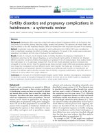
The selection criteria for UKA included medial unicom-
partmental disease (Figure 1A) with intact cruciate liga-
ments, as evaluated during the pre-operative clinical
consultation and confirmed intra-operatively. Patients
with anterior knee pain, either as a clinical complaint or
on pre-operative evaluation of knee extension against
resistance, were deemed not appropriate candidates. Full
institutional review board approval was granted for the
investigation of these patients, all of whom provided writ-
ten informed consent for participation in this study.
Clinical and Radiographic Evaluation
All patients were evaluated clinically and radiographically
pre-operatively, as well post-operatively at approximately
3 months, 6 months, 1 year, and annually thereafter. Clin-
ical evaluation was performed with use of the Knee Soci-
ety (KSS) rating system [6], encompassing both objective
and functional scores. Radiographic evaluation was per-
formed using antero-posterior, lateral, and Merchant view
radiographs of the knees (Figure 1B), with measurement
of femoral and tibial angles, alpha and beta angles, and
medial and lateral joint spaces as described by Villers and
Cartier [7]. Patients were additionally evaluated for the
presence of patellar osteophytes as an indicator of patel-
lofemoral arthritis that is easily identifiable on most
standard follow-up radiographs. Radiolucencies were
evaluated at post-operative follow-up visits using the zone
system described by Kennedy and White [8].
Surgical Technique and Postoperative Management
All surgeries were performed by a single surgeon (P.M.B.)
using a medial parapatellar approach. An M/G
®
(Zimmer
Inc., Warsaw Indiana) metal-backed unicompartmental
prosthesis was used in all cases. Unicompartmental pros-
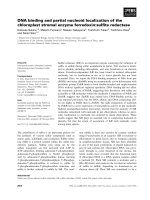
Pre-operative antero-posterior (A) and Merchant view (B) radiographs of a patient with medial compartment osteoar-thritis treated with a metal-backed unicompartmental knee arthroplastyFigure 1
Pre-operative antero-posterior (A) and Merchant
view (B) radiographs of a patient with medial com-
partment osteoarthritis treated with a metal-backed
unicompartmental knee arthroplasty.
Table 1: Patient Characteristics
Variables Sample (n = 80)
Mean age (years) 72 (44-91)
Men:Women (percent) 42:58
Mean body mass index (kg/m
2
) 27 (17-39)
Obesity (body mass index ≥ 30 kg/m
2
) (percent) 28
Pre-operative diagnosis (percent) -
Osteoarthritis 86
Osteonecrosis 14
Follow-up period (months) 60 (24-168)
Unicondylar knee implant failure (percent) 11
Journal of Orthopaedic Surgery and Research 2009, 4:39 />Page 3 of 7
(page number not for citation purposes)
theses represent approximately 5% of the total number of
knee arthroplasties performed by this surgeon in any
given year. The skin incisions ranged from 10 to 15 cen-
timeters. The patella was displaced laterally at the start of
the procedure to inspect the patellofemoral joint and the
lateral compartment, to evaluate the patella for the pres-
ence of osteophytes, and to confirm that the anterior and
posterior cruciate ligaments (ACL and PCL) were intact.
Inspection was done in both flexion and extension.
Varus releases were performed. Intramedullary instru-
mentation was used to make a distal femoral cut in 4
degrees of valgus orientation. The tibia was resected using
an extramedullary alignment jig, with a minimum of 2
millimeters of bone removed in the greatest depth of
deformity. A reciprocating saw was then used to make the
center cut just medial to the ACL footprint and this bone
fragment was removed. The leg was brought into exten-
sion to assess alignment. Next, femoral cuts were made
and sized relative to the tidemark to avoid patellofemoral
impingement. Finally, the chamfer, posterior, and peg
cuts were made.
The tibia was sized in both the anterior-posterior (AP) and
medial-lateral (ML) dimensions to optimize coverage
while avoiding implant overhang. A keel cut and two peg
cuts were made. Trial components were used to achieve 1
to 2 millimeters of laxity in full extension, with balanced
flexion. Occasionally, additional soft tissue releases were
required to achieve this aim.
Next, the metal implants were cemented into position,
starting with the tibial component followed by the femo-
ral prosthesis. A polyethylene trial was then placed on the
tibial tray, and the leg was brought into full extension to
allow the cement to harden. The trial was then removed,
and the residual cement was removed with an osteotome.
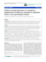
The final polyethylene spacer was then implanted, and
balancing and alignment of the knee was confirmed
throughout the full range of motion of the knee (Figure
2).
Data Analysis
Failure of UKA was defined as a revision to total knee
arthroplasty (TKA). In our center we do not treat sympto-
matic aseptic loosening with implantation of a new UKA
prosthesis; all these patients are revised to a TKA. The Kap-
lan-Meier method was used to estimate the survivorship
of the prosthesis used in the study cohort. The Wilcoxon
rank sum test was used to compare continuous variables
(such as age, BMI, Knee Society scores, and most of radio-
graphic measurements) between the failure and non-fail-
ure groups; a chi-squared or Fisher exact test was used to
compare categorical variables (such as male, obesity (BMI
≥ 30 kg/m
2
), diagnosis, and presence of preoperational
patella osteophytes) between the two groups. Similarly,
Wilcoxon matched-pair signed-rank tests and chi-squared
tests were used to compare continuous and categorical
variables, respectively, before and after the operations.
Seven factors were evaluated for association with implant
failure: patient age, gender, obesity (BMI ≥ 30 kg/m
2
), pre-
operative diagnosis (osteonecrosis or osteoarthritis), pre-
operative Knee Society objective and functional scores,
and the presence of patellar osteophytes prior to surgery.
A Cox proportional hazard model was used to examine
whether any of these factors were associated with the risk
of failure of UKA, with a hazard ratio over one indicating
that the factor was an independent predictor of a higher
risk of failure of UKA. Patients that died or were lost to fol-
low-up were excluded from this analysis.
Results
Clinical and radiographic outcomes
The mean preoperative Knee Society objective and func-
tional scores were 49 points (standard deviation, SD = 9)
and 48 points (SD = 10), respectively (Table 2). Both
scores had substantially improved at final follow-up, with
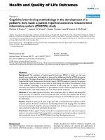
Post-operative antero-posterior radiograph of the same patient shown in Figure 1 at 6 week follow-up visitFigure 2
Post-operative antero-posterior radiograph of the
same patient shown in Figure 1 at 6 week follow-up
visit.
Journal of Orthopaedic Surgery and Research 2009, 4:39 />Page 4 of 7
(page number not for citation purposes)
a mean of 95 (SD = 4) and 92 (SD = 7) points, respec-
tively.
Radiographic analysis revealed that the femoral angle
increased by a mean of 0.9 degrees postoperatively (p <
0.01). However, there was no significant change in tibial
angle. Medial joint space also increased significantly from
a mean of 1.0 mm preoperatively to 3.0 mm postopera-
tively. At final follow-up, stable non-progressive lucent
lines less than 2 mm in size were present in five of the
unrevised patients (7%). One patient had a lucent line >2
mm in size but was asymptomatic and doing well at the
most recent follow-up, with Knee Society pain and func-
tion scores of 92 and 90 points, respectively. One patient
had progressive lucent lines in more than one zone and
was judged to have an impending component failure. A
revision was recommended to this patient but she refused
as she was asymptomatic at most recent follow-up with
Knee Society pain and function scores of 99 and 100
points respectively. A complete description of radio-
graphic characteristics can be found in Table 2.
Failure of UKA
Of the 80 knees that were treated with UKA, nine (11%)
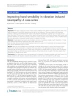
were revised to a TKA over the follow up period. Two cases
were due to component loosening, and three were attrib-
uted to patellofemoral/lateral pain (Figures 3A and 3B).
Other reasons for revision included polyethylene wear (n
= 2), progression of arthritis (n = 1), and a tibial plateau
fracture (n = 1). This tibial plateau fracture was non-trau-
matic in origin, and was likely due to implant subsidence
into visibly osteopenic bone. The mean time from the
date of UKA to revision to TKA was 48 months (range, 4
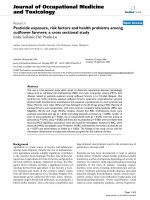
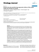
to 135 months). Kaplan-Meier survival analysis revealed
that the survival rate of UKA implant was 92% at 5 years
(95% CI: 83-96%), and 84% at 10 years (95% CI: 68-
93%), found in Figure 4.
Factors associated with failure of UKA
There were differences in some patient factors between the
failure and non-failure groups, but no independent pre-
dictors of failure were identified. There was a significant
difference in the mean age at index arthroplasty (73 versus
61 years; p < 0.01) between the non-failure and failure
groups, respectively. There was a higher proportion of
obese patients in the failure group compared to the non-
failure group (44% versus 20%) but this difference was
not significant (p = 0.11). Although the age difference was
significant between the failure and non-failure groups, the
hazard ratio of age was 0.94 (95% confidence interval, CI:
0.86-1.03), suggesting that age did not independently
affect the risk of failure of UKA. Consistent with the
descriptive analysis, obesity had a high hazard ratio of
Table 2: Clinical and radiographic characteristics before and after UKA
Variables Pre-operative [SD] Final follow-up [SD] P value
a
Knee Society Scoring System
Mean objective score (points) 49 [9] 95 [4] < 0.01
Mean functional score (points) 48 [10] 92 [7] < 0.01
Radiographic Characteristics
Mean femoral angle (degrees) 97 [2] 97 [3] < 0.01
Mean tibial angle (degrees) 84 [2] 84 [2] 0.81
Mean medial joint space (millimeters) 1.0 [1.0] 2.9 [1.6] < 0.01
Mean lateral joint space (millimeters) 6.0 [2.0] 5.7 [1.9] 0.19
Mean patellar medial joint space (millimeters) 2.6 [1.6] 2.7 [2.0] 0.98
Mean patellar central joint space (millimeters) 3.6 [2.1] 3.2 [2.2] 0.04
Mean patellar lateral joint space (millimeters) 2.7 [1.9] 2.3 [1.8] 0.07
Presence of patellar osteophytes (percent of patients) 48 38 0.31
Alpha angle (degrees) 87 [4]
Beta angle (degrees) 82 [6]
SD = standard deviation
a
p values were calculated based on Wilcoxon matched-pairs signed-ranks tests for continuous variables and chi-squared tests for categorical
variables
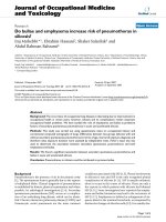
Antero-posterior (A) and Merchant view (B) radiographs of the same patient as Figures 1 and 2, taken at 41 month fol-low-upFigure 3
Antero-posterior (A) and Merchant view (B) radio-
graphs of the same patient as Figures 1 and 2, taken
at 41 month follow-up. The patient complained of increas-
ing patello-femoral pain, and was revised to a total knee
arthroplasty shortly thereafter.
Journal of Orthopaedic Surgery and Research 2009, 4:39 />Page 5 of 7
(page number not for citation purposes)
2.12 but the 95% CI included a hazard ratio of 1.0. A
more detailed comparison of the failure and non-failure
groups can be found in Table 3.
Discussion
Although patient selection is thought to influence the suc-
cess of UKA, controversy remains over which specific fac-
tors affect the outcome of this procedure. Patient age,
gender, and weight have been examined in previous stud-
ies without conclusive findings. Other factors, such as pre-
operative diagnosis Knee Society function scores and
patellar arthritis, have rarely been studied in relation to
failure of UKA implant. This study used prospectively col-
lected data to examine seven factors that may be associ-
ated with failure of UKA implants. We followed 80 knees
for an average of 60 months. The survivorship of the UKA
implants was 84% at 10 years follow up which is compa-
rable to those reported in the literature [9,10]. Overall, we
did not find any independent predictor of failure of UKA.
Traditionally, UKA was recommended for patients aged
60 years or over with a sedentary lifestyle [1]. However,
with a hazard ratio of 0.94, our results suggest that age is
not a predictor of failure of UKA. Gioe et al. examined the
survival of 1,047 knee arthroplasties in patients aged 55
years old or younger using a community registry and did
not find an association between age groups and survival
rate [11]. Although the mean age in the failure group of
the present study was 6 years younger than the non-failure
group, young age was not found to be an independent
predictor of failure. Several studies devote attention to
younger patients (less than 60 years of age) treated with
UKA, all of whom had excellent results. Schai et al. fol-
lowed 28 knees in 28 patients who had a mean age of 52
years; only two knees were revised over a maximum of six
years follow up [12]. Similarly, Pennington et al. reported
a survival rate of 92% at 10 years in a group of younger
patients [13]. Tabor and Tabor evaluated two patient
cohorts to compare the survivorship and functional out-
comes of UKA of patients aged 60 and over to those in a
younger age group, and did not find a significant differ-
ence [10]. However, there are also studies reporting a poor
survival rate in younger patients [14-16]. Additionally,
using a Cox proportional hazard model, two studies
found a hazard ratio of failure that favors superior out-
comes in older patients [14,16]. The difference in these
findings could be attributed to the age range of patients
and the skills of the surgeons.
To date, gender has not been used as an inclusion/exclu-
sion criterion for UKA, though some studies have found a
difference in outcomes between male and female patients
[10,17,18]. However, consistent with our findings, the
majority of the studies did not find gender as a significant
predictor of failure of UKA [11,14-16].
Plot of Kaplan Meier survivorship estimate based on the fail-ures of metal-backed unicompartmental knee arthroplasty components reported in the present studyFigure 4
Plot of Kaplan Meier survivorship estimate based on
the failures of metal-backed unicompartmental knee
arthroplasty components reported in the present
study.
Table 3: Comparison of patient characteristics between the failure and non-failure groups
Failure Non-failure Hazard Ratio
Variables (N = 9) (N = 71) P value
a
Ratio 95% CI
Mean age (years) 61 [8] 74 [9] < 0.01 0.94 0.86-1.03
Male gender (percent) 44 42 1.00 0.30 0.05-1.87
Mean body mass index (kg/m
2
) 28 [7] 27 [4] 0.71 - -
Obesity (body mass index ≥ 30 kg/m
2
) (percent) 44 20 0.11 2.13 0.34-13.3
Diagnosis (percent)
Osteoarthritis 100 85 0.35 - -
Osteonecrosis 0 15 0.00 0.00
Mean pre-op objective score (points) 53 [10] 49 [9] 0.21 1.01 0.92-1.11
Mean pre-op functional score (points) 51 [5] 47 [10] 0.30 1.07 0.96-1.19
Pre-op patellar osteophytes (percent of patients) 33 46 0.38 0.27 0.07-2.01
CI = confidence interval
a
p values were calculated based on Wilcoxon rank sum tests for continuous variables, and chi-squared or Fisher exact tests for categorical
variables.
Journal of Orthopaedic Surgery and Research 2009, 4:39 />Page 6 of 7
(page number not for citation purposes)
Weight and obesity are other factors to consider when
UKA is applied. A multi-center investigation by Heck et al.
reported mean BMIs in the failure and non-failure groups
of 33 kg/m
2
and 25 kg/m
2
, respectively [17]. However,
many other studies have not found an association
between weight and/or obesity and failure of UKA
[10,15]. One study even suggested that obese patients had
a better survival rate when compared to their non-obese
counterparts [18]. In addition, excellent survival rates
have been reported in studies that did not consider weight
when qualifying patients for UKA [19]. Despite some sur-
geons suggesting that patients over 80 kg or those who are
clinically obese should not be treated with UKA [5,20],
such criteria do not seem to be supported by the majority
of studies, including the findings in the present report.
Although most UKAs are performed to in patients with
osteoarthritis, it is not the only indication for UKA.
Osteonecrosis can be treated with UKA with good results.
Parratte et al. studied 31 osteonecrotic knees receiving
UKA with a minimum follow up of three years and
reported the survival rate of 96.7% at 12 years [21]. The
authors noted that the outcomes of UKA were similar to
those in primary osteoarthritis [12]. Similarly, Gioe et al.
reported that there is no difference in survival rate based
on diagnosis [11].
Preoperative Knee Society objective and functional scores,
and patellar osteophytes have rarely been studied as pre-
dictors for UKA failure. Although anterior knee pain is a
relative contradiction for UKA based on conventional sur-
gical criteria, a recent study found that it did not affect the
success of UKA using the Oxford phase 3 device [22]. Our
findings indicate that pain and function of the affected
knee are not related to failure of UKA. Patella osteophytes
were also not a risk factor for UKA failure.
UKA is an effective treatment for unicompartmental knee
disease. In addition to its clinical advantages, it may be
more cost-effective when compared to TKA [23]. Oppo-
nents of UKA cite the poor survival rate of UKA implant
relative to TKA. However, several studies have reported
excellent survival rates [19,24]. Patient selection is a criti-
cal issue to success with this treatment modality. Conven-
tional criteria suggest that patients should be over 60 years
of age, weigh no more than 82 kg, and not perform heavy
labor or be extremely physically active [20,25]. Although
careful selection of patients is a key to the success of UKA,
excessive restrictions will discount the benefits of the pro-
cedure and underplay its importance in treating unicom-
partmental knee disease. Better outcomes may be
achieved with expanded criteria as the surgical technique
and devices continue to be developed. Improvement in
our understanding of factors related to UKA failure will
shed light on patient selection criteria and help improve
surgical outcomes of UKA.
Several limitations are noted in this study. First, the sam-
ple size is relatively small. Certain patient factors, notably
obesity, trended towards significance in our analysis of
independent predictors of failure, and it is possible that a
larger study group would provide additional power to bet-
ter define the associations between the factors and risk of
failure of UKA. Additionally, because of the small and
diverse number of failures, we did not attempt to assess
hazard ratios for each individual cause for revision. It is
possible that such an analysis would reveal variability in
independent associations for some modes of failure.
Finally, the follow up time is relatively short compared to
some other studies on UKA. The average length of follow
up was five years, which affects the survival rate in this
study. In addition, long-term outcomes could not be
assessed.
Conclusion
Young age, gender, obesity, diagnosis, pre-operative
objective and functional scores and patella osteophytes
were not predictors of failure of a unicondylar knee
implant, although increased obsesity was association with
a high hazard ratio. The findings suggest that the standard
criteria for UKA may be expanded without compromising
the outcomes, although caution may be warranted in
patients with very high body mass index pending addi-
tional data to confirm our results.
Competing interests
No external financial support was received in support of
this study.
MAM is a consultant for Stryker Orthopaedics and Wright
Medical Technologies, and receives royalties from Stryker
Orthopaedics. PMB is a consultant for Stryker Orthopaed-
ics, and receives royalties from Stryker, Arthrocare,
Biomet, and Synthes.
None of the other authors have any financial or non-
financial competing interests to disclose.
Authors' contributions
TMS, MAM, LPL, JX, PMB designed the study. LPL, DRM,
PMB collected the data. TMS, LPL, JX, DRM, MGZ, ana-
lyzed the data. TMS, MAM, DRM, MGZ, prepared the
manuscript. MAM, JX, MGZ, PMB ensured the accuracy of
the data and analysis. All authors have read and approved
the final manuscript.
References
1. Griffin T, Rowden N, Morgan D, Atkinson R, Woodruff P, Maddern
G: Unicompartmental knee arthroplasty for the treatment
Publish with BioMed Central and every
scientist can read your work free of charge
"BioMed Central will be the most significant development for
disseminating the results of biomedical researc h in our lifetime."
Sir Paul Nurse, Cancer Research UK
Your research papers will be:
available free of charge to the entire biomedical community
peer reviewed and published immediately upon acceptance
cited in PubMed and archived on PubMed Central
yours — you keep the copyright
Submit your manuscript here:
/>BioMedcentral
Journal of Orthopaedic Surgery and Research 2009, 4:39 />Page 7 of 7
(page number not for citation purposes)
of unicompartmental osteoarthritis: a systematic study. ANZ
J Surg 2007, 77:214-221.
2. Bert JM: Unicompartmental knee replacement. Orthop Clin
North Am 2005, 36:513-522.
3. Suggs JF, Li G, Park SE, Steffensmeier S, Rubash HE, Freiberg AA:
Function of the anterior cruciate ligament after unicompart-
mental knee arthroplasty: an in vitro robotic study. J Arthro-
plasty 2004, 19:224-229.
4. Jeer PJ, Cossey AJ, Keene GC: Haemoglobin levels following uni-
compartmental knee arthroplasty: influence of transfusion
practice and surgical approach. Knee 2005, 12:358-361.
5. Deshmukh RV, Scott RD: Unicompartmental knee arthro-
plasty: long-term results. Clin Orthop Relat Res 2001:272-278.
6. Insall JN, Dorr LD, Scott RD, Scott WN: Rationale of the Knee
Society clinical rating system. Clin Orthop Relat Res 1989:13-14.
7. Villers P, Cartier P: [Method of radiological measurement for
adjusting partial prosthesis of the knee]. Ann Radiol (Paris) 1978,
21:545-546.
8. Kennedy WR, White RP: Unicompartmental arthroplasty of
the knee. Postoperative alignment and its influence on over-
all results. Clin Orthop Relat Res 1987:278-285.
9. Ackroyd CE, Whitehouse SL, Newman JH, Joslin CC: A compara-
tive study of the medial St Georg sled and kinematic total
knee arthroplasties. Ten-year survivorship. J Bone Joint Surg Br
2002, 84:667-672.
10. Tabor OB Jr, Tabor OB: Unicompartmental arthroplasty: a
long-term follow-up study. J Arthroplasty 1998, 13:373-379.
11. Gioe TJ, Novak C, Sinner P, Ma W, Mehle S: Knee arthroplasty in
the young patient: survival in a community registry. Clin
Orthop Relat Res 2007, 464:83-87.
12. Schai PA, Suh JT, Thornhill TS, Scott RD: Unicompartmental knee
arthroplasty in middle-aged patients: a 2- to 6-year follow-up
evaluation.
J Arthroplasty 1998, 13:365-372.
13. Pennington DW, Swienckowski JJ, Lutes WB, Drake GN: Unicom-
partmental knee arthroplasty in patients sixty years of age
or younger. J Bone Joint Surg Am 2003, 85-A:1968-1973.
14. Harrysson OL, Robertsson O, Nayfeh JF: Higher cumulative revi-
sion rate of knee arthroplasties in younger patients with
osteoarthritis. Clin Orthop Relat Res 2004:162-168.
15. Collier MB, Engh CA Jr, McAuley JP, Engh GA: Factors associated
with the loss of thickness of polyethylene tibial bearings after
knee arthroplasty. J Bone Joint Surg Am 2007, 89:1306-1314.
16. Eickmann TH, Collier MB, Sukezaki F, McAuley JP, Engh GA: Survival
of medial unicondylar arthroplasties placed by one surgeon
1984-1998. Clin Orthop Relat Res 2006, 452:143-149.
17. Heck DA, Marmor L, Gibson A, Rougraff BT: Unicompartmental
knee arthroplasty. A multicenter investigation with long-
term follow-up evaluation. Clin Orthop Relat Res 1993:154-159.
18. Tabor OB Jr, Tabor OB, Bernard M, Wan JY: Unicompartmental
knee arthroplasty: long-term success in middle-age and
obese patients. J Surg Orthop Adv 2005, 14:59-63.
19. Murray DW, Goodfellow JW, O'Connor JJ: The Oxford medial
unicompartmental arthroplasty: a ten-year survival study. J
Bone Joint Surg Br 1998, 80:983-989.
20. Kozinn SC, Scott R: Unicondylar knee arthroplasty. J Bone Joint
Surg Am 1989, 71:145-150.
21. Parratte S, Argenson JN, Dumas J, Aubaniac JM: Unicompartmen-
tal knee arthroplasty for avascular osteonecrosis. Clin Orthop
Relat Res 2007, 464:37-42.
22. Berend KR, Lombardi AV Jr, Adams JB: Obesity, young age, patel-
lofemoral disease, and anterior knee pain: identifying the
unicondylar arthroplasty patient in the United States. Ortho-
pedics 2007, 30:19-23.
23. Robertsson O, Borgquist L, Knutson K, Lewold S, Lidgren L: Use of
unicompartmental instead of tricompartmental prostheses
for unicompartmental arthrosis in the knee is a cost-effec-
tive alternative. 15,437 primary tricompartmental prosthe-
ses were compared with 10,624 primary medial or lateral
unicompartmental prostheses. Acta Orthop Scand 1999,
70:170-175.
24. Berger RA, Nedeff DD, Barden RM, Sheinkop MM, Jacobs JJ, Rosen-
berg AG, Galante JO: Unicompartmental knee arthroplasty.
Clinical experience at 6- to 10-year followup. Clin Orthop Relat
Res 1999:50-60.
25. Stern SH, Becker MW, Insall JN: Unicondylar knee arthroplasty.
An evaluation of selection criteria. Clin Orthop Relat Res
1993:143-148.