báo cáo hóa học:" Diagnostic challenge: bilateral infected lumbar facet cysts - a rare cause of acute lumbar spinal stenosis and back pain" pptx
Bạn đang xem bản rút gọn của tài liệu. Xem và tải ngay bản đầy đủ của tài liệu tại đây (467.02 KB, 5 trang )
CAS E REP O R T Open Access
Diagnostic challenge: bilateral infected lumbar
facet cysts - a rare cause of acute lumbar spinal
stenosis and back pain
Brett A Freedman, Tuan L Bui, S Timothy Yoon
*
Abstract
Symptomatic synovial lumbar facet cysts are a relatively ra re cause of radiculopathy and spinal stenosis. This case
and brief review of the literature, details a patient who presented with acutely symptomatic bilateral spontaneously
infected synovial facet (L4/5) cysts. This report highlights diagnostic clues for identifying infection of a facet cyst.
Introduction
Lumbar facet cysts are a less common but well docu-
mented cause of compressive radiculopathy and lumbar
spinal stenosis, with approximately 500 total cases
reported in the literature [1-5]. The lumbar facet is a
synovial-lined zygoaphophyseal joint, comprising the
articulation between the inferior and superior articulat-
ing processes of the spinal vertebrae. The facet joint,
like synovial lined joints of the appendicular skeleton,
are prone to cyst formation as a manifestation of
osteoarthritis. T o date, infection of a lumbar facet cyst
has not been report ed in the literature. This case illus-
trates the clinical findings and outcomes associated with
bilateral infected lumbar facet cysts.
Case Report
History
Our patient is a 63 year old overweight gentleman who
presented to the emergency room with a three day his-
tory of progressive low back pain and pain radiating
down the right worse than left leg in an L5 distribution.
He also noted an acute onset of drop foot. He rated his
pain as 10 out of 10. He reported that he has had a his-
tory of intermittent back pain, but no prior leg symp-
toms. He has diabetes, which was marginally controlled
(HgbA1C was 7.4), coronary artery disease and one
week prior to presentation he completed an 8 week
course of radiation therapy for prostate cancer.
Physical Examination
On examination, he was in significant pain. He had
bilateral lower extremity weakness. His motor strength
testing revealed 4/5 left and right iliopsoas and 4+/5 left
and right quadriceps, hamstrings and gastrocnemius
muscles, all of which appeared to be pain induced
reductions of strength. Additionally, he had 3/5 l eft and
right tibialis anterior (TA) and extensor hallucis longus
(EHL) function. Hi s peroneals were also weak (4-/5). He
had normal sensation to light touch and pin prick. His
deep tendon reflexes were 2+ bilaterally. He had a nor-
mal upper extremity neurological and digital rectal
exam.
Imaging and Labs
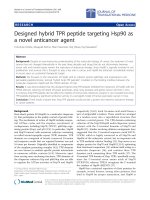
Plain radiographs and a CT scan demonstrated severe
arthrosis at the L4/5 facet joints. (Figure 1) MRI
revealed what appeared to be large degenerative bilateral
L4/5 facet cysts with extensions into the interspinous
and epidural space, causing severe compression of the
thecal sac. (Figure 1 and 2) There was paravertebral
muscle heterogenous hyperintensity on fat-suppressed
T2 images. He was afebrile and had normal white cell
count and blood sugars. Due t o the unusual acuity of
symptom presentation, potential for immune compro-
mise given his medical co-morbidities and subtle MRI
findings suggestive of local inflammatory response in
the paravertebral muscles, an ESR and CRP were
obtained. They were both markedly elevated. (ESR 103
mm/hr; CRP 33.2 mg/dL) His admission and subsequent
laboratory results are located in Table 1.
* Correspondence:
Department of Orthopaedic Surgery, Emory University School of Medicine,
Emory Spine Center, Altanta, GA 30329, USA
Freedman et al. Journal of Orthopaedic Surgery and Research 2010, 5:14
/>© 2010 Freedman et al; licensee BioMed Central Ltd. This is an Open Access article distributed under the t erms of the Creative
Commons Attribution License (http://cre ativecommons.org/licenses/by/ 2.0), which permits unrestricted use, distribution, and
reproduction in any medium, provided the original work is properly cited.
Operation and Pathological Findings
The clinical exam, imaging studies and laboratory find-
ings all sug gested this patient’s symptoms were due to a
L4/5 degenerative facet cyst causing sympt omatic lum-
bar stenosis; however the markedly elevated CRP and
ESR and inflammatory signal on MRI was worrisome for
an infectious etiology. He was admitted to the hospital
and taken to the OR the following day for a decompres-
sion and possible fusion of L4/5. Intraoperatively, expos-
ing the L4/5 facets revealed voluminous cysts that
expressed frank pus u pon incisio n. As a result, we
decided to stage this patient’s surgeries. At the first
stage we performed subtotal L4 and L5 laminectomies,
near-total facet capsulectomy, partial facetectomy an d
thorough lavage to widely eradicate the inf ected struc-
tures. The infection appeared to be completely con-
tained within the facet cysts. Tissue samples were sent
for culture and pathology, which grew out Methicillin-
Resistant Staphylococcus Aureus (MRSA). Pathology
showed chronic and acute inflammatory changes with-
out evidence of neoplasm. Infectious disease was con-
sulted and he was started on intravenous Vancomycin,
which was continued for a total of 6 weeks. Two days
following the initial procedure, he underwent a direct
lateral interbody fusion (X-LIF, NuVasive, Inc, San
Diego, CA) with BMP-2 (InFuse, Medtronic, Inc,
Minneapolis, MN) and posterior pedicle screw
instrumentation.
Figure 1 Advanced degenerative changes of the L4/5 facets are seen on these AP radiograph and CT scan images. (1A, B and D) Note
the subchondral sclerosis and cystic changes. The axial T2 MRI image shows a focal fluid-like collection in bilateral L4/5 facet joints with
contiguous extension into the midline dorsal epidural space (dotted line) (1C). Additionally, there is heterogeneous increased signal in the
paravertebral muscles. There is no evidence of spondylodiscitis or paravertebral muscle abcess.
Freedman et al. Journal of Orthopaedic Surgery and Research 2010, 5:14
/>Page 2 of 5
Postoperative Course
After his decompression surgery, the patient did well,
with cessation of radicular symptoms, and was dis-
charged on the 4
th
postoperati ve day following the sub-
sequent X-LIF. Upon dis charge from the ho spital, his
pain level improved to 5 /10, with pain solely in his
back. His motor strength normalized in all groups
except for EHL, TA and P, which only improved to 4/5.
However, this was sufficient to eliminate his drop foot
gait. At his 2 week clinic appointment, he was afebrile
and his wound was healing without complication.
Unfortunately, our patient was a visitor to this country
who p ermanentl y resided in the Virgin Islands. He had
been here for his cancer treatments, but upon resolution
of his back and leg symptoms, he returned to his home.
He was scheduled to return to our clinic 2 weeks, 6
weeks, and 3 and 6 months postoperatively, but only
camefora2and8weekvisit.Athis8weekvisit,he
had completed his IV antibiotic therapy two weeks prior
and denied any pain. He rated pain in his legs and back
as0/10.HestillhadsomeslightL5weakness(4+/5
EHL, TA) on examination; however, this did not affect
his gait. His laboratory values had normalized. (Table 1)
Due to his living situation, follow-up at 3 and 6 months
was obtained telephonically and demonstrated no evi-
dence of recurrent leg symptoms or infection.
Discussion
The clinical presentation, managemen t and o utcomes of
aseptic lumbar facet cysts have been reported [1-6]. In
2004, Epstein performed a comprehensive review of the
15 published case series, which provides defining char-
acteristics of this patho logy [2]. Facet cysts are detected
in 0.6 - 10% of MRI scans of the lumbar spine [1-7].
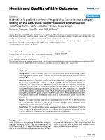
Figure 2 Right to left sagittal T2 MRI images with their associated CT sagittal reconstructions beneath, show the fluid-like (isodense
and isointense to CSF) signal in the right greater than left facet joints, as well as diffusely increased signal in the adjacent
paravertebral muscles. (2A and C) The midline sagittal MRI image shows the compressive epidural portion of the cyst, with a stalk that trails
into the interspinous space (dotted line), where it communicates with the facet cysts. (2B) The CT images show non-specific chronic destructive
changes to the L4/5 facet, typical of uncomplicated lumbar facet cysts. (2D, E and F)
Freedman et al. Journal of Orthopaedic Surgery and Research 2010, 5:14
/>Page 3 of 5
The L4/5 level is most commonly affected and cysts
most commonly occur in p atients 60-65 years of age
[1-3,5,8]. L5 ra diculopathy is the most common primary
complaint, although 95-100% of patients will have low
back pain, as well [1,2,9].
Facet sagittal orientation (> 45 degrees) and facet
arthrosis are present in over 77% of patients with symp-
tomatic lumbar facet cysts [9-11]. As in the appendicu-
lar skeleton, the primary response of synovial joints to
arthritis, is the over-produ ction of synovial fluid, which
in turn raises the intra-capsular pressure. In some
patients, weak or thin areas in the facet ca psule give
way, the result is a focal mushroom-like swelling [1,12].
With time cystic fluid dehydrates, and the cyst itself
undergoes myxoid degeneration. Classic cystic appear-
ance on MRI (isodense and isointense with CSF) occurs
in as few as 57% of facet cysts [9]. These space occupy-
ing lesions can compress nerve roots, causing radiculo-
pathy (57-75%) or neurogenic claudication (25%)
[2,9,13].
Although cases of successful nonoperative treatment
have been reported with steroid injections or image-
guided aspiration, for the most part, symptomatic syno-
vial cysts require excision [1,2,9,14]. While agreement
exists for surgical intervention of symptomatic facet
cysts, the extent of surgery needed is debatable. Sur-
geons tend to favor either decompression alone or
decompression and fusion, with decompression alone
being the more commonly reported approach [1,5].
Concurrent sp ondylo listhesis, especially in the presence
of significant low back pain, is the most common reason
for adding an arthrodesis [1,3,5,12]. The lack of prospec-
tive cohort studies require surgeons to base their treat-
ment plan on hypothesis and interpretation of case
series which report good-excellent results (in > 75% of
cases) for both approach es [1,5,12]. Those who advocate
arthrodesis tend to point to t wo primary issues. First,
the c yst is only an effect, the true cause is the underly-
ing facet arthrosis and possibly instability [3,12]. Simply
excising the cyst will not treat the cause. Conversely, the
rate of recurrence following laminectomy alone appears
to be quite low, averaging < 3% across published series
[1,13]. Second, patients with lumbar facet cysts
overwhelmingly have abnormal motion segments and
low back pain [2,3,9,12]. Excision and decompression
alone does not address these concomitant pathologies
and may worsen segmental instability [ 3,9]. However,
the rate of re-operation for symptomatic instability
appears to be low as well (2% in Lyons et al. series of
194 patients) [5].
Our pat ient had chronic low back pain, sagittally
oriented facets (> 45 degrees) with extensive cystic and
sclerotic changes. We performed significant facetec-
tomies and near-total capsulectomy to wide ly debride
the infection; thus we elected to fuse his spine. His
recent XRT exposure and his underlying marginally
controlled diabetes made him vulnerable to infection–
Class B host [15]. Hypertrophic synov ium in facet cysts,
devoid of a basement membrane, allowed MRSA to
localize and develop into a closed space infection. This
sequence of hematogenous seeding and subsequent
infection is common to other synovial joints [16]. This
case clearly demonstrates that this can occur in lumbar
facet joints, as well. Further, debridement of the infected
tissue, prolonged culture-specific antibiotic and stabiliza-
tion through instrumented spinal fusion can successfully
eradicate this rare form of infection and result in an
excellent clinical outcome (= complete symptom resolu-
tion, no recurrence) [1,12].
This case report highlights diagnostic clues that sug-
gest infection of an underlying facet cyst. The key find-
ings appear to be rapid progression of symptoms,
associated elevation in CRP and ESR and paravertebral
muscles edema. Symptomatic neurological compressio n
in uncomplicated facet cysts develops over time as
degeneration progresses. Only 7% of cases present
within 7 days of symptom onset, perhaps dues to intra-
cystic hemorrhage [1,9,17]. In patients presenting with
acutely progressive lumbar stenotic or radiculopathic
symptoms which are attributed to lumbar facet cysts,
the possibility of infection of the cysts should be consid-
ered and evaluated.
Consent
Written informed consent to pub lish could not be
obtained despite reasonable attempts. The patient can-
not be identified from the case report and there is no
reason to believe that they would object to its
publication.
Acknowledgements
We would like to acknowledge Bettie Cheek, RN for her assistance with this
project.
No funding was received in support of this project.
Authors’ contributions
All authors contributed in writing this case report, and have all read and
approved the final manuscript.
Table 1 Pertinent lab values
Emergency
Department
POD#1 At
Discharge
8 week
follow-up
WBC (/mcL) 9,800 7,700 7,200 7,600
CRP (mg/dL) 33.2 19.6 16.5 .27 (0.033
at 3mo)
HgA1C (%) 7.4
POD#1 = postoperative day one, WBC = white blood count (normal value
< 11,100/mcL), CRP = C-reactive Protein (normal value < 0.8 mg/dL), HgA1C
(Hemoglobin A1C, therapeutic goal < 6%).
Freedman et al. Journal of Orthopaedic Surgery and Research 2010, 5:14
/>Page 4 of 5
Competing interests
The authors declare that they have no competing interests.
Received: 18 July 2009 Accepted: 5 March 2010
Published: 5 March 2010
References
1. Boviatsis EJ, Staurinou LC, Kouyialis AT, Gavra MM, Stavrinou PC,
Themistokleous M, Selviaridis P, Sakas DE: Spinal synovial cysts:
pathogenesis, diagnosis and surgical treatment in a series of seven
cases and literature review. Eur Spine J 2008, 17(6):831-7.
2. Epstein NE: Lumbar Synovial Cysts. A Review of Diagnosis, Surgical
Management and Outcome Assessment. J Spinal Disord Tech 2004,
17:321-325.
3. Epstein NE: Lumbar laminectomy for the resection of synovial cysts and
coexisting lumbar spinal stenosis or degenerative spondylolisthesis: an
outcome study. Spine 2004, 29(9):1049-55.
4. Eyster EF, Scott WR: Lumbar synovial cysts: report of eleven cases.
Neurosurgery 1989, 24:112-115.
5. Lyons MK, Atkinson JL, Wharen RE, Deen HG, Zimmerman RS, Lemens SM:
Surgical evaluation and management of lumbar synovial cysts: the Mayo
Clinic experience. J Neurosurg 2000, 93(suppl 1):53-57.
6. Banning CS, Thorell WE, Leibrock LG: Patient outcome after resection of
lumbarjuxtafacet cysts. Spine 2001, 26:969-972.
7. Doyle AJ, Merrilees M: Synovial cysts of the lumbar facet joints in a
symptomatic population: prevalence on magnetic resonance imaging.
Spine 2004, 29:874-878.
8. Deinsberger R, Kinn E, Ungersböck K: Microsurgical treatment of juxta
facet cysts of the lumbar spine. J Spinal Disord Tech 2006, 19(3):155-60.
9. Metellus P, Fuentes S, Adetchessi T, Levrier O, Flores-Parra I, Talianu D,
Dufour H, Bouvier C, Manera L, Grisoli F: Retrospective study of 77
patients harbouring lumbar synovial cysts: functional and neurological
outcome. Acta Neurochir (Wien) 2006, 148:47-54.
10. Fujiwara A, Tamai K, An HS, Lim TH, Yoshida H, Kurihashi A, Saotome K:
Orientation and osteoarthritis of the lumbar facet joint. Clin Orthop Relat
Res 2001, , 385: 88-94.
11. Fujiwara A, Tamai K, An HS, Kurihashi T, Lim TH, Yoshida H, Saotome K: The
relationship between disc degeneration, facet joint osteoarthritis, and
stability of the degenerative lumbar spine. J Spinal Disord 2000,
13(5):444-50.
12. Khan AM, Synnot K, Cammisa FP, Girardi FP: Lumbar synovial cysts of the
spine: an evaluation of surgical outcome. J Spinal Disord Tech 2005,
18(2):127-31.
13. Howington JU, Connolly ES, Voorhies RM: Intraspinal synovial cysts: 10-
year experience at the Ochsner Clinic. J Neurosurg 1999,
91(2 Suppl):193-9.
14. Parlier-Cuau C, Wybier M, Nizard R, Champsaur P, Le Hir P, Laredo JD:
Symptomatic lumbar facet joint synovial cysts: clinical assessment of
facet joint steroid injection after 1 and 6 months and long term follow-
up in 30 patients. Radiology 1999, 210:509-513.
15. Cierny G, Mader JT:
Approach to adult osteomyelitis. Orthop Rev 1987,
16(4):259-70.
16. Zink BJ, Weber JE: Chapter 130 - Bone and Joint Infections. Rosen’s
Emergency Medicine: Concepts and Clinical Practice St Louis, MO: Mosby,
IncMarx JA , 5 2002, 1925-43.
17. Ramieri A, Domenicucci M, Seferi A, Paolini S, Petrozza V, Delfini R: Lumbar
hemorrhagic synovial cysts: diagnosis, pathogenesis, and treatment.
Report of 3 cases. Surg Neurol 2006, 65:385-390.
doi:10.1186/1749-799X-5-14
Cite this article as: Freedman et al.: Diagnostic challenge: bilateral
infected lumbar facet cysts - a rare cause of acute lumbar spinal
stenosis and back pain. Journal of Orthopaedic Surgery and Research 2010
5:14.
Submit your next manuscript to BioMed Central
and take full advantage of:
• Convenient online submission
• Thorough peer review
• No space constraints or color figure charges
• Immediate publication on acceptance
• Inclusion in PubMed, CAS, Scopus and Google Scholar
• Research which is freely available for redistribution
Submit your manuscript at
www.biomedcentral.com/submit
Freedman et al. Journal of Orthopaedic Surgery and Research 2010, 5:14
/>Page 5 of 5