báo cáo hóa học:" Metastatic breast carcinoma of the coracoid process: two case reports" pptx
Bạn đang xem bản rút gọn của tài liệu. Xem và tải ngay bản đầy đủ của tài liệu tại đây (1.26 MB, 4 trang )
CAS E REP O R T Open Access
Metastatic breast carcinoma of the coracoid
process: two case reports
Eric C Benson
1*†
, Darren S Drosdowech
2
Abstract
Background: The coracoid process of the scapula is a rare site of involvement for metastatic disease or for primary
tumors. We are unaware of any reports in the literature of pathologic coracoid process fractures and only one
report of metastatic disease to the coracoid.
Methods and Results: In this case report, we present two cases with metastatic breast carcinoma of the coracoid
process, one of which presented with a pathologic fracture of the coracoid.
Conclusions: An orthopaedic surgeon must be aware of the potential for metastatic disease to the coracoid as
they may be the first medical provider to encounter evidence of malignant disease.
Introduction
The coracoid process of the scapula is a rare site of
involvement for metastatic disease or for primary
tumors. Bone metastases are common in patients with
breast carcinoma, with an incidence as high as 73%
(range 47-85%) [1]. The exact mechanism of meta stases
to bone remains unknown.
We are unaware of any reports in the literature of
pathologic coracoid process fractures, and only one
report of metast atic disease to the coracoid [2]. We pre-
sent the cases of two patients with metastatic breast car-
cinoma of the coracoid process, one of w hich presented
with a pathologic fracture of the coracoid. We informed
the patients or their families that the data concerning
their cases wo uld be submitted for publication, and they
consented.
Case 1
A 40-year-old, right-hand dominant female who had a
known history of right breast carcinoma presented to our
clinic for evaluation for open biopsy of a lesion at the base
of the coracoid. Four months prior to clinic presentation,
she underwent right breast lumpectomy and lymph node
dissection. Surgical pathology revealed invasive mammary
carcinoma, SBR grade 2 with no involvement of the lymph
nodes. Resection margins were negat ive. She was Her-2-
neu negative, estrogen receptor negative, and progesterone
receptor positive. A bone scan revealed increased uptake
attheeighththoracicvertebraandintheregionofthe
coracoid in the right shoulder. Further CT imaging of
both regions indicated a fracture through the transverse
process of T8, though the patient was asymptomatic at
this level and had a prior history of a fall from a horse that
correlated with this finding. There was no history of any
shoulder pain resulting from or subsequent to that fall. CT
imaging of the scapula showed osteolytic change at the
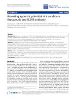
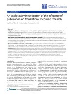
base of the coracoid. Radiographs and relevant CT scan
images are shown (Figures 1 and 2). She had received the
first cycle of adjuvant chemotherapy with FEC-100 but
further cycles were discontinued until further information
regarding the possible sites of metastases was collected.
Instead, she was placed on Tamoxifen and Clodronate.
She was otherwise healthy and took no other medications.
On physical exam there was no palpable mass in the
region of the right shoulder, no skin discoloration or
changes, and her range of motion and strength were
normal. She was nontender to palpation over the cora-
coid process. She had no tenderness to palpation over
T8 or elsewhe re throughout the spine. Upper and lower
extremity neurovascular exam showed no focal deficits.
The patient consented to open biopsy of the coracoid
and was taken to the operating room. Through a delto-
pectoral approach, the coracoid was identified and
* Correspondence:
† Contributed equally
1
Department of Orthopaedic Surgery and Rehabilitation, Division of Shoulder
and Elbow Surgery, MSC10 - 5600, 1 University of New Mexico, Albuquerque,
NM 87131, USA
Benson and Drosdowech Journal of Orthopaedic Surgery and Research 2010, 5:22
/>© 2010 Benson and Drosdowech; licensee BioMed Central Ltd. This is an Open Access article distributed under the terms of the
Creative Commons Attr ibution License ( which permits u nrestricted use, dis tribution, and
reproduction in any medium, provided the original work is properly cited.
biopsy specimens from the lesion at the base of the cor-
acoid were sent to pathology for frozen section and per-
manent sections. The intra-operative frozen section was
positive for adenocarcinoma.
The patient had no complications following the biopsy
and the surgical pathology report confirmed the lesion
was a metastatic breast adenocarcinoma. The immuno-
histochemical stains showed moderately to strongly
positive progesterone receptors in about 15% and mod-
erately positive estrogen receptors in about 2% of malig-
nant cells.
Approximately twenty months after her initial lum-
pectomy, the patient underwent right partial mastect-
omy for recurrent carcinoma. At most recen t follow-up,
two years after initial diagnosis, she is doing well with
no evidence of local recurrence or progression of meta-
static disease.
Case 2
A 23-year-old, right-hand dominant female sports coach
fell backwards onto outstretched arms while snowboarding
one week prior to presentation. She noted immediate left
shoulder pain, was seen at on outside Emergency Depart-
ment, and was referred to orthopedics for management of
her shoulder injury. She sustained no other injuries in the
fall. She noted no other previous complaints with regard
to her left shoulder. She took Naprosyn for pain relief.
Over the months leading up to the fall, she was treated
with NSAIDs at another center for chest wall pain pre-
sumed to be osteochondritis. Otherwise, she had no signif-
icant findings in review of her past medical history. Prior
surgeries included removal of a Bartholin’scyst.
Physical examination revealed isolated point tender-
ness over the tip of the coracoid. She had full neck,
shoulder, and elbow range of motion with some discom-
fort at the terminal range of internal and external rota-
tion of the shoulder. Her neurovascular exam showed
no focal deficits.
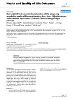
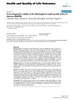
Radiographs showed a nondisplaced fracture of the
coracoid (Figures 3 and 4). These were compared to her
outside films taken immediately after her fall and
showed no interval change in position of the fragment.
We recommended non -op erati ve management of this
stable injury. Short-term immobilization using a sling
followed by initiation of physiotherapy was arranged.
Gentle strengthening was to start after approximately
four to six weeks as tolerated.
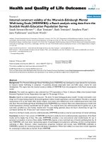
Figure 1 AP radiograph demonstrating the metastatic lesion of
the coracoid process.
Figure 2 CT scan showing the metastatic lesion at the base of
the coracoid.
Figure 3 AP radiograph of the nondisplaced pathologic
coracoid process fracture.
Benson and Drosdowech Journal of Orthopaedic Surgery and Research 2010, 5:22
/>Page 2 of 4
Tragically, this previously healthy, active, young
woman was admitted to an outside facility only two
weeks later with hypercalcemia, multiple sites of bone
metastases noted on skeletal survey, and an abnormal
liver scan. She was diagnosed with metastatic adenocar-
cinoma of the left breast. In addit ion to the cor acoid,
she had multiple metastatic lesions in her thoracic spine
and bilateral femurs as well as brain and liver metas-
tases. Over the course of the following four months she
suffered from e ncephalopathy, SIADH, leptomeningeal
carcinomatosis, and eventually passed away in her home
receiving palliative care.
Though the patient’s mech anism of injury was consis-
tent with an acute coracoid fracture, in retrospect her
injury was likely a pathologic fracture secondary to her
metastatic breast adenocarcinoma.
Discussion
Tumors of the coracoid process are rare. We could onl y
identify one report of a metastatic lesion to the coracoid
using a PubMed search of the literature [2]. Primary
bone tumors of the coracoid include osteoid osteoma,
osteosarcoma, giant cell tumor, chondrosarcoma, capil-
lary hemangioma, aneurysmal bone cyst, lymphoma, and
plasmacytoma [3]. In our PubMed literature search, we
found no reports of pathologic coracoid fractures.
Breast cancer’s propensity to metastasize to bone is
not clearly understood. Batson described the valveless
venous plexus co mmonly thought to contribut e to the
spread of breast and prostate carcinoma to sites in t he
axial and appendicular skeleton [4]. More recently, stu-
dies suggest some of the mechanisms for bone destruc-
tion once tumor cells have gained access to a distant
site. These include osteoclast activating factors such as
parathyroid hormone-related pr otein (PTH-rP), tumor
necrosis factor (TNF) a and b, epidermal growth factor
(EGF), and prostaglandins [5]. These changes to the
bone architecture lead to structural weakness, and typi-
cally, the radiographic appearance of breast metastases
to bone is one of mixed osteoblastic and osteolytic
appearance.
Often, the orthopaedic surgeon is the first medical pro-
vider to encounter evidence of malignant disease and as
such must be aware of potential sites of involvement.
When interpreting radiographs, especially in an area as
difficult as the coracoid, it is important to maintain an
index of suspicion for underlying pathologic processes,
especially since isolated fractures of the coraco id process
are rare [6-23]. When present, it may be difficult to iden-
tify the bony architecture at the fracture site secondary to
overlying structures. It may be prudent to obtain extra
imaging to clearly show the bony characteristics of the
injury. A 20 degree posterior oblique film with 20 degrees
of cephalad angulation can show coracoid fractures and
bone morphology more clearly if other views are incon-
clusive [24]. CT scans may also be useful.
The role of the orthopaedic surgeon may also include
recommendations for bisphosphonate use. In concert with
the consulting medical oncologist, administ ering bispho-
sphonates may reduce the risk of skeletal complications in
patients receiving systemic therapy who have lytic bone
metastatic lesions secondary to breast cancer [25,26].
The coracoid process of the scapula is a rare site of
acute isolated trauma, primary tumors, or of metastatic
disease. We present what we believe to be the first
repo rted case of a pathologic fracture of the coracoid in
one of two patients who presented with metastatic
breast carcinoma of the coracoid. Although rare, ortho-
paedic surgeons must be aware of the potential for a
pathologic process involving the coracoid.
Consent
Informed consent was obtained from the patient or
patient’s family for publication of this case report and
all accompanying radiographic images.
Author details
1
Department of Orthopaedic Surgery and Rehabilitation, Division of Shoulder
and Elbow Surgery, MSC10 - 5600, 1 University of New Mexico, Albuquerque,
NM 87131, USA.
2
University of Western Ontario, Division of Orthopedic
Surgery, Hand and Upper Limb Centre, St. Joseph’s Health Centre, 268
Grosvenor St, London, ON N6A 4V2, Canada.
Authors’ contributions
DD performed all clinical evaluations and interactions with the patients. EB
reviewed the case files, contacted the patients’ or patients’ families to obtain
informed consent, and prepared the manuscript and image files. Both EB
and DD read, revised, and approved the final manuscript.
Competing interests
The authors declare that they have no competing interests.
Received: 6 May 2009 Accepted: 26 March 2010
Published: 26 March 2010
Figure 4 Axillary radiograph showing the pathol ogic coracoid
fracture.
Benson and Drosdowech Journal of Orthopaedic Surgery and Research 2010, 5:22
/>Page 3 of 4
References
1. Galasko CSB: The anatomy and pathways of skeletal metastases. Bone
Metastasis Boston: GK HallWeiss L, Gilbert AH 1981, 49.
2. Kato Y, Numata A, Wada N, Iwata T, Saga Y, Hashimoto H, Kakizaki H: A
case of metastatic renal cell carcinoma to the ovary. Hinyokika Kiyo 2006,
52:923-7.
3. Ogose A, Sim FH, O’Connor MI, Unni KK: Bone Tumors of the coracoid
process of the scapula. Clin Orthop Relat Res 1999, 358:205-214.
4. Batson OV: The role of the vertebral veins in metastatic process. Ann
Intern Med 1942, 16:38.
5. Travers MT, Barrett-Lee PJ, Berger U, Luqmani YA, Gazet JC, Powles TJ,
Coombes RC: Growth factor expression in normal, benign and malignant
breast tissue. Br Med J 1988, 296:1621.
6. Asbury S, Tennent TD: Avulsion fracture of the coracoid process: a case
report. Injury 2005, 36:567-568.
7. Ogawa K, Yoshida A, Takahashi M, Ui M: Fractures of the coracoid process.
J Bone Joint Surg Br 1997, 79:17-19.
8. Guiral J, Real JL, Curto JM: Isolated fracture of the coracoid process of the
scapula. Acta Orthop Belg 1996, 62:60-61.
9. Eyres KS, Brooks A, Stanley D: Fractures of the coracoid process. J Bone
Joint Surg Br 1995, 77:425-428.
10. Bauer G, Fleischmann W, Dussler E: Displaced scapular fractures:
indication and long-term results of open reduction and internal fixation.
Arch Orthop Trauma Surg 1995, 114:215-219.
11. Gil JF, Hayday A: Isolated injury of the coracoid process: case report. J
Trauma 1991, 31:1696-1697.
12. Martin-Herrero T, Rodriguez-Merchan C, Munuera_Martinez L: Fractures of
the coracoid process: presentation of seven cases and review of the
literature. J Trauma 1990, 30:1597-1599.
13. Kopecky KK, Bies JR, Ellis JH: CT diagnosis of fracture of the coracoid
process of the scapula. Comput Radiol 1984, 8:325-327.
14. Goldberg RP, Vicks B: Oblique angled view for coracoid fractures. Skeletal
Radiol 1983, 9:195-197.
15. Heyse-Moore GH, Stoker DJ: Avulsion fractures of the scapula. Skeletal
Radiol
1982, 9:27-32.
16. Zilberman Z, Rejovitzky R: Fracture of the coracoid process of the scapula.
Injury 1981, 13:203-206.
17. Mariani PP: Isolated fracture of the coracoid process in an athlete. Am J
Sports Med 1980, 8:129-130.
18. Froimson AI: Fracture of the coracoid process of the scapula. J Bone Joint
Surg Am 1978, 60:710-711.
19. DeRosa GP, Kettelkamp DB: Fracture of the coracoid process of the
scapula: case report. J Bone Joint Surg Am 1978, 59:696-697.
20. Sandrock AR: Another sports fatigue fracture. Stress fracture of the
coracoid process of the scapula. Radiology 1975, 117:274.
21. Boyer DW Jr: Trapshooter’s shoulder: stress fracture of the coracoid
process of the scapula. J Bone joint Surg Am 1975, 57:862.
22. Rounds RC: Isolated Fracture of the Coracoid Process. J Bone Joint Surg
Am 1949, 31:662-663.
23. Petty OH: Fracture of the coracoid process of the scapula caused by
muscular action: with report of case. Ann Surg 1907, 45:427-430.
24. Goldberg RP, Vicks B: Oblique angled view for coracoid fractures. Skeletal
Radiol 1983, 9:195-197.
25. Hortobagyi GN, Theriault RL, Lipton A, Porter L, Blayney D, Sinoff C,
Wheeler H, Simeone JF, Seaman JJ, Knight RD, Heffernan M, Mellars K,
Reitsma DJ: Long-term prevention of skeletal complications of metastatic
breast cancer with pamidronate: Protocol 19 Aredia Breast Cancer Study
Group. J Clin Oncol 1998, 16:2038-2044.
26. Theriault RL, Lipton A, Hortobagyi GN, Leff R, Glück S, Stewart JF, Costello S,
Kennedy I, Simeone J, Seaman JJ, Knight RD, Mellars K, Heffernan M,
Reitsma DJ: Pamidronate reduces skeletal morbidity in women with
advanced breast cancer and lytic bone lesions: A randomized, placebo-
controlled trial. J Clin Oncol 1999, 17:846-854.
doi:10.1186/1749-799X-5-22
Cite this article as: Benson and Drosdowech: Metastatic breast
carcinoma of the coracoid process: two case reports. Journal of
Orthopaedic Surgery and Research 2010 5:22.
Submit your next manuscript to BioMed Central
and take full advantage of:
• Convenient online submission
• Thorough peer review
• No space constraints or color figure charges
• Immediate publication on acceptance
• Inclusion in PubMed, CAS, Scopus and Google Scholar
• Research which is freely available for redistribution
Submit your manuscript at
www.biomedcentral.com/submit
Benson and Drosdowech Journal of Orthopaedic Surgery and Research 2010, 5:22
/>Page 4 of 4