báo cáo hóa học:" Decrease in the expression of the type 1 PTH/PTHrP receptor (PTH1R) on chondrocytes in animals with osteoarthritis" potx
Bạn đang xem bản rút gọn của tài liệu. Xem và tải ngay bản đầy đủ của tài liệu tại đây (901.72 KB, 6 trang )
Becher et al. Journal of Orthopaedic Surgery and Research 2010, 5:28
/>Open Access
RESEARCH ARTICLE
BioMed Central
© 2010 Becher et al; licensee BioMed Central Ltd. This is an Open Access article distributed under the terms of the Creative Commons
Attribution License ( which permits unrestricted use, distribution, and reproduction in
any medium, provided the original work is properly cited.
Research article
Decrease in the expression of the type 1
PTH/PTHrP receptor (PTH1R) on chondrocytes in
animals with osteoarthritis
Christoph Becher
1
, Thomas Szuwart
2
, Philipp Ronstedt
2
, Sven Ostermeier
1
, Adrian Skwara
3
, Susanne Fuchs-
Winkelmann
3
and Carsten O Tibesku*
4
Abstract
Background: To evaluate the expression of the type 1 PTH/PTHrP receptor (PTH1R) on chondrocytes from hyaline
cartilage over the course of osteoarthritis (OA).
Methods: In 12 NZW rabbits, the anterior cruciate ligament (ACL) was resected to create anterior instability of the knee.
In 12 control rabbits, only a sham operation, without resection of the ACL, was performed. Four animals from each
group were killed at 3, 6, and 12 weeks. After opening the knee joint, OA was macroscopically graded and hyaline
cartilage of the load-bearing area was evaluated histologically according to the Mankin scale and by immunostaining
for PTH1R.
Results: There was a positive linear correlation between the time after surgery and the macroscopic and histologic OA
scores. The scores in the control group were constant over the time course. Immunostaining showed significantly less
expression of PTH1R in the experimental compared to the control group after 6 (P < 0.05) and 12 weeks (P < 0.01). In
the experimental group, a negative linear correlation between PTH1R expression and macroscopic and histologic
grades was found.
Conclusions: The results show an in vivo decrease in the expression of PTH1R on chondrocytes over the time course of
OA. Further studies are needed to evaluate whether new treatment approaches could evolve from this knowledge.
Introduction
The type 1 PTH/PTHrP receptor (PTH1R) belongs to a
family of G-protein-coupled receptors (GPCR) with
seven membrane-spanning helixes [1]. The PTH1R acts
as a common receptor for the Parathyroid hormone
(PTH) and the Parathyroid hormone-related peptide
(PTHrP) [1-3]. It is highly expressed in bone and kidney
and mediates in these tissues the PTH-dependent regula-
tion of mineral ion homeostasis [1]. The PTH1R is also
highly expressed in the prehypertrophic chondrocytes of
metaphyseal growth plates [4,5]. It was demonstrated
that PTH has a stimulatory effect on proliferation of
chondroprogenitor cells and inhibits collagen and matrix
synthesis and thus regulates cartilage growth and chon-
drocytic apoptosis [6].
PTHrP is known as an important local factor for chon-
drogenesis by regulating chondrogenesis in a manner that
attenuates chondrocyte hypertrophy [3,7]. By acting
through a complex signaling network, PTHrP allows the
bone to grow and elongate normally [1,7-9].
Osteoarthritis (OA) is one of the most common skeletal
disorders characterized by cartilage degradation, osteo-
phyte formation and thickening of the subchondral bone
in joints. Since little is known about the underlying
molecular mechanism, several experimental OA models
have been developed for investigation [10]. Whereas the
role of PTH1R in osteoarthritic chondrocytes remains
largely unanswered, it was shown that as disease severity
in OA progresses, PTH1R expression and protein levels
are reduced in human subchondral osteoblasts [11]. It
was suggested that OA osteoblasts may be responsible for
the initiation of this disease since it was indicated that
* Correspondence:
4
Sporthopaedicum Straubing, 94315 Straubing, Germany
Full list of author information is available at the end of the article
Becher et al. Journal of Orthopaedic Surgery and Research 2010, 5:28
/>Page 2 of 6
cocultures of OA osteoblasts with normal cartilage
explants may initiate cartilage degradation [12].
Furthermore, a clear reduction was shown in PTH1R
abundance in OA osteoblasts and that it was related to
disease severity [11]. The expression of PTHrP in carti-
lage and OA was addressed in several studies. It was
shown that PTHrP is released in high concentrations in
OA [13] and is more abundant in knees of patients with
OA than in normal human knee articular cartilage [14].
Furthermore, the expression of PTHrP in chondrocytes
obtained from cartilage tissues from patients with
osteoarthritis during hip replacement surgery was depen-
dent on the degree of cartilage degeneration. Cartilage
with moderate degeneration expressed more of this pep-
tide than mildly or severely degraded cartilage specimens
did [15]. However, all such studies dealt with late-stage
OA because human specimens were available only at the
time of surgery.
The present study was designed to evaluate the expres-
sion of the PTH1R on chondrocytes over the time course
of experimentally induced secondary OA in an animal
model as well as possible correlations between macro-
scopic and histologic findings in the degenerated hyaline
cartilage.
Materials and methods
Animal experiments were performed at the Department
of Orthopedics at the University of Muenster. All proce-
dures were performed with the permission of the local
government's animal rights protection authorities and in
accordance with the National Institute of Health guide-
lines for the use of laboratory animals (G49/2000).
Twenty-eight female NZW rabbits were used in the study.
All animals were fully grown, clinically healthy, and had a
mean ± SD weight of 4,193.75 ± 298.84 gm at the time of
surgery and a mean ± SD weight of 4,274.64 ± 462.49 gm
at the time of death.
Trial groups
The animals were divided into 3 groups. In 12 rabbits, the
anterior cruciate ligament (ACL) was resected to create
anterior instability of the knee (group 1). In 12 control
rabbits, only a sham operation was performed, without
resection of the ACL (group 2). The 4 animals of group 3
underwent no surgical treatment.
Surgical procedure
All operations were performed under general anesthesia
(intramuscular administration of ketamine and xylazine).
All animals received an intramuscular injection of broad-
spectrum antibiotics (Tardomyocel; Bayer, Leverkusen,
Germany) as prophylaxis against infection. For postoper-
ative pain medication, intramuscular metamizole (Noval-
gin; Aventis Pharmaceuticals, Bad Soden, Germany) was
administered. Operations on both knee joints were per-
formed under a single dose of anesthesia. The knee joints
were opened using a medial paramedian skin incision of 3
cm in length and a medial parapatellar incision of the
capsule. In animals of group 1, the ACL was resected
using a tapering scalpel. The patella was dislocated later-
ally. In animals of group 2 (control group), the joint and
skin were closed again with no further surgical treatment
(sham operation). An intraoperative Lachman test was
carried out to evaluate anterior instability. Before closure,
the joints were rinsed thoroughly. Postoperatively, all ani-
mals were allowed to move freely in their cages (which
measured 100 cm × 70 cm × 40 cm).
Macroscopic evaluation
Four animals from each group were killed 3, 6, and 12
weeks after the operations. The 4 animals from group 3
(no surgical treatment) were killed after they reached
maturity. The joints were evaluated macroscopically by
using a self-developed grading system consisting of 4 dif-
ferent criteria: fibrillations and ulcerations of the hyaline
cartilage, osteophyte formation, and joint effusion. Scores
for fibrillations ranged from 0 for intact hyaline cartilage
to 3 for marked fibrillations, scores for ulcerations ranged
from 0 for normal to 2 for a large area of ulceration,
scores for osteophyte formation ranged from 0 for no
osteophytes to 3 for marked osteophyte formation, and
scores for joint effusion ranged from 0 for no effusion to 3
for marked effusion. The total score ranged from 0 to 11,
with 0 being a macroscopically intact knee joint and 11
being late-stage OA. After macroscopic grading, both
distal femurs were immediately stored at ~80°C until fur-
ther examination.
Specimen preparation
Tissue samples (3-5-mm thick) were fixed in 4% buffered
paraformaldehyde for 2 days. After decalcification with
buffered EDTA (20% EDTA, pH 7.4), the samples were
dehydrated and embedded in paraffin. Sections were cut
at a thickness of 5 μm, mounted on poly-L-lysine-coated
glass slides, deparaffinized in xylene, and washed 3 times
with distilled water and then with Tris buffered saline
(TBS; pH 7.5) for 2 minutes each (washing procedure).
Some of the sections were stained with Safranin O or
with hematoxylin and eosin (H&E) to evaluate histologic
changes of the cartilage and bone tissue according to the
Mankin scale [16]. The Mankin score included assess-
ments of the structure, cellularity, Safranin stainability,
and integrity of the tidemark. The scores for structure
ranged from 0 for normal to 6 for complete disorganiza-
tion of the cartilage, scores for cellularity ranged from 0
for normal to 3 for hypocellularity, scores for Safranin O
stainability ranged from 0 for normal to 4 for no stainabil-
ity, and scores for tidemark integrity ranged from 0 for an
Becher et al. Journal of Orthopaedic Surgery and Research 2010, 5:28
/>Page 3 of 6
intact tidemark to 1 for blood vessels crossing the tide-
mark. The total score ranged from 0 to 14, where 0 =
intact hyaline cartilage and 14 = late-stage OA.
Immunohistochemical method for labeling PTH1R
Deparaffined sections were hydrated and incubated for 5
min in proteinase K (ready-to-use; Dako, Hamburg, Ger-
many). After washing in TBS, the slides were treated for
10 min with 3% H
2
O
2
and blocked for 30 min in 3%
bovine serum. A monoclonal mouse anti-rabbit PTH1R
was used as primary antibody (IgG1, clone VFF-18,
Bender MedSystems, Vienna, Austria). The sections were
incubated with the primary antibody in PBS + 1% bovine
serum albumin (diluted 1:50) over night at 4°C. After
washing once again sections were incubated for 30 min in
the secondary antibody anti-mouse IgG (K4001; Dako) at
room temperature. After washing in TBS, the reaction
was developed with aminoethylcarbazole chromogen
substrate (ready-to-use; Dako) for 30 minutes at room
temperature. To demonstrate specificity of binding of
mouse anti-rabbit PTH1R in control experiments, sec-
tions were incubated (a) without primary antibody or,
instead of primary antibody, (b) mouse IgG1 with irrele-
vant specificity (Aspergillus niger glucose oxidase; Dako)
was used at the same concentration as the primary anti-
body. In all controls tested the specificity no positive
staining of the tissue slices was visible.
Quantitative counting of cells
Pictures of histologic sections were taken with a Cool-
Snap-Pro Color camera (model A00J82025; Media Cyber-
netics, Silver Spring, MD). The cells were counted using
Image-Pro Plus software for Windows, version 4.1
(Media Cybernetics). First, the total number of chondro-
cytes in an immunohistochemically stained section was
determined. Afterward, the number of immunohis-
tochemically stained cells was counted in the same sec-
tion and measured in relation to the total number of
chondrocytes. Zonal attribution of PTH1R positive chon-
drocytes was done with respect to the typical shape of
chondrocytes at the different zones of articular cartilage
according to Buckwalter et al. [17].
Statistical analysis
Statistical analysis was performed using the Statistical
Package for Social Sciences, release 11.0 (SPSS, Munich,
Germany). Student's t-test was performed for compari-
son of scores in the control and experimental groups.
Spearman's coefficient was calculated to determine cor-
relations. P values less than 0.05 were considered signifi-
cant.
Results
Two animals developed postoperative hematomas,1 ani-
mal had a superficial wound infection and was treated
with a single injection of antibiotics, and 1 animal was
reoperated upon 2 days after surgery because of a wound
gap. All 4 wounds subsequently healed with no sequelae.
All other animals had no complications and, an average of
1 week after surgery, no joint effusion was found upon
clinical evaluation.
When the animals were killed, 11 of the 12 rabbits in
group 1 (ACL resection) did not have an ACL. One ani-
mal in group 1 had intact ACLs bilaterally and was taken
out of the experimental group. All animals in groups 2
and 3 had an intact ACL at the time of death.
Macroscopic grading of OA
A total of 54 joints were evaluated macroscopically. Signs
of degeneration according to our self-developed grading
system were found in all groups. The mean ± SD macro-
scopic grade of OA was 0.375 ± 0.518 in group 3 (control
group). In group 2 (sham operation), it was 0.375 ± 0.744
after 3 weeks, 0.75 ± 0.886 after 6 weeks, and 0.625 ±
0.518 after 12 weeks. In group 1 (ACL resection), the
mean ± SD macroscopic grade of OA was 6.625 ± 1.768
after 3 weeks, 6.5 ± 2.33 after 6 weeks, and 9.5 ± 1.049
after 12 weeks. Group 1 had statistically significantly
higher values than group 2 at every time point (P < 0.01).
The macroscopic grade of OA correlated positively with
the number of weeks after the operation (r
s
= 0.519, P <
0.05).
Histologic grading of OA
According to the Mankin score, group 1 (ACL resection)
showed a highly significant increase in the grade of OA
compared with the untreated control group and the
sham-operated group (group 2). In group 3 (no treat-
ment), the mean ± SD histologic grade of OA was 1.75 ±
1.581. In group 2 (sham operation), it was 3.25 ± 0.707
after 3 weeks, 1 ± 1.195 after 6 weeks, and 2.25 ± 1.165
after 12 weeks. In group 1 (ACL resection), the mean ±
SD histologic grade of OA was 7.25 ± 3.615 after 3 weeks,
8 ± 2.878 after 6 weeks, and 9.83 ± 3.061 after 12 weeks.
Group 1 had statistically significantly higher values than
group 2 at every time point (P < 0.01). The average histo-
logic grade of OA increased with the number of postop-
erative weeks. The macroscopic and histologic grades of
OA were strongly correlated (r
s
= 0.770, P < 0.01).
PTH1R staining by immunohistochemistry
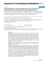
Immunohistochemical staining of PTH1R showed
marked staining of the cell membrane in all cases. PTH1R
was expressed mainly in the middle zones of cartilage. It
was rarely found in the superficial zones (Figure 1). In
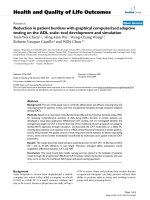
group 1 (ACL resection), the percentage of PTH1R+ cells
averaged 6.47% after 3 weeks, 15.14% after 6 weeks, and
9.15% after 12 weeks (Figure 2). In group 2 (sham opera-
tion), the percentage of PTH1R+ cells averaged 21.11%
after 3 weeks, 33.90% after 6 weeks, and 30.53% after 12
Becher et al. Journal of Orthopaedic Surgery and Research 2010, 5:28
/>Page 4 of 6
weeks (Figure 2). In group 3 (control group), the percent-
age of PTH1R+ cells averaged 30.85%.
The percentages of PTH1R+ cells differed significantly
between group 1 and 2 after 6 (P < 0.05) and 12 weeks (P
< 0.01), but not after 3 weeks (P = 0.059). The percentages
of PTH1R+ cells differed significantly between the ACL
resected group and the control group at 3, 6 and 12 weeks
respectively (P < 0.01).
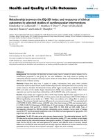
There was a statistically significant negative correlation
between the histologic grade of OA and the percentage of
PTH1R+ chondrocytes (r
s
= -0.601, P < 0.001) (Figure 3).
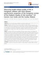
There was also a statistically significant negative correla-
tion between the macroscopic grade of OA and the per-
centage of PTH1R+ chondrocytes (r
s
= -0.541, P < 0.001)
(Figure 4).
Discussion
The use of experimental animal models in which joint
instability is induced through surgical intervention in an
effort to clarify the mechanisms whereby the mechanical
stress leads to OA development, have been widely used in
the literature [18-21]. Whereas the mechanisms of the
type 1 PTH/PTHrP receptor (PTH1R) in osteoblasts
were addressed in several studies [11,22-25], the expres-
sion of the PTH1R and its correlation with macroscopic
and histologic features over the time course in a model of
experimentally induced early OA are largely unknown.
Our results confirm the expression of PTH1R in nor-
mal articular cartilage chondrocytes as detected by
immunohistochemistry analysis [14]. We could demon-
strate a negative linear correlation between PTH1R
expression and macroscopic and histologic grades in the
early course of OA.
This is consistent with findings that as disease severity
in OA progresses, PTH1R expression and protein levels
are reduced in human subchondral osteoblasts. Cases of
severe, moderate, and mild OA patients who underwent
total knee replacement surgery indicated a progressive
Figure 1 Immunohistochemical staining of PTH1R. A, PTH1R ex-
pression 3 weeks after the sham operation. B, PTH1R expression 3
weeks after resection of the anterior cruciate ligament (ACL). C, PTH1R
expression 6 weeks after the sham operation. D, PTH1R expression 6
weeks after resection of the ACL. E, PTH1R expression 12 weeks after
the sham operation. F, PTH1R expression 12 weeks after resection of
the ACL. Magnification bar: 0,25 mm.
Figure 2 Time course of the development of PTH1R expression in
osteoarthritis. The percentage of PTH1R+ chondrocytes was signifi-
cantly increased in the anterior cruciate ligament (ACL) transection
group after 6 weeks and 12 weeks. Values are the mean and SD.* = P <
0.05.
Figure 3 Correlation between the histologic grade of osteoarthri-
tis (OA) according to the Mankin scale and expression of PTH1R.
A negative correlation was demonstrated, with a Spearman's coeffi-
cient of -0.601 (P < 0.001).
Becher et al. Journal of Orthopaedic Surgery and Research 2010, 5:28
/>Page 5 of 6
decrease in PTH1R levels from -10% (mild) to -60%
(severe) versus normal individuals [11]. Subchondral
bone sclerosis is a major pathophysiological manifesta-
tion of OA, and it still is unknown if it precedes cartilage
breakdown in OA. A study to determine the influence of
osteoarthritic phenotype of subchondral osteoblasts on
the phenotype of human chondrocytes demonstrated
that sclerotic osteoblasts, but not nonsclerotic osteo-
blasts induced a significant decrease of PTH1R gene
expression in chondrocytes [25]. In contrast, PTH1R
gene expression was depressed in sclerotic osteoblasts
[24]. Accordingly it was shown that proteinases like
matrix metalloproteinase 13 (MMP-13) who have been
proven to be the principal initiator of OA progression
were significantly up-regulated in sclerotic osteoblasts
compared with nonsclerotic osteoblasts [24]. MMP-13
are also highly expressed in the hypertrophic chondro-
cytes in response to joint instability [10]. Hypertrophic
differentiation and chondrocyte apoptosis are known to
be involved in OA development [26]. High levels of
PTHrP have been found in the synovial fluid of osteoar-
thritic joints [13] and showed to regulate chondrogenesis
in a manner that attenuates chondrocyte hypertrophy [7].
PTH participates in the regulation of cartilage growth
and chondrocytic apoptosis [6]. Thus, it can be assumed
that PTH1R participates in the underlying molecular
mechanisms between cartilage degradation and subchon-
dral bone remodelling and determines an important
appearance in osteoarthritic progression.
Whereas the diffuse distribution of the stained cells in
the middle zone in normal cartilage of our study was con-
sistent with findings by other researchers [14], the distri-
bution was not different in OA cartilage which is in
contrast to findings in OA human cartilage at the time of
joint replacement when receptor staining was relatively
restricted to areas near the cartilage surface [14]. How-
ever, we confirmed that PTH1R is less expressed in OA
cartilage with only a minority of cells expressing the
receptor than in normal cartilage. It was shown in young
adult bovine articular cartilage that chondrocytes of the
radial zone occupied twice the volume and surface area of
the chondrocytes of the superficial zone but were 10
times more synthetically active [27]. We hypothesize that
PTH1R is down-regulated in the early course of OA due
to the increased mechanical stimuli in the active radial
and transitional zone and may be found in the superficial
zone in late stages of OA when cartilage degradation is
advanced.
In summary, the present study shows a decrease in the
expression of the PTH1R receptor over the time course of
OA. Further studies are needed to determine 1) the phys-
iologic role of this receptor in normal articular cartilage,
2) whether PTH1R down-regulation is an underlying
cause or a repair response in OA, 3) whether this pattern
also applies to humans, and 4) whether new treatment
approaches could evolve from this knowledge.
Competing interests
In support of their research, none of the authors received grants or outside
funding. None of the authors received payments or other benefits or a com-
mitment or agreement to provide such benefits from a commercial entity.
Authors' contributions
All authors read and approved the final manuscript
CB drafted the manuscript and participated in data and statistical analysis.
TS participated in the conception of the study, participated in the specimen
preparation and was responsible for immunohistochemical staining.
PR participated in the surgical procedures, specimen preparation and macro-
scopic and histological grading of OA. He carried out the quantitative counting
of cells and participated in immunohistochemical staining.
SO participated in the statistical analysis and manuscript preparation.
AS participated in the surgical procedures, specimen preparation and macro-
scopic and histological grading of OA.
SFW participated in the conception of the study and supervised the protocol.
COT was responsible for the initial conception of the research question, super-
vising the protocol and manuscript preparation.
Author Details
1
Orthopaedic Department, Hannover Medical School, 30625 Hannover,
Germany,
2
Institute of Anatomy, Westfalian Wilhelms University, 48149
Muenster, Germany,
3
Department of Orthopaedics and Rheumatology,
Philipps University Marburg 35043 Marburg, Germany and
4
Sporthopaedicum
Straubing, 94315 Straubing, Germany
References
1. Mannstadt M, Juppner H, Gardella TJ: Receptors for PTH and PTHrP: their
biological importance and functional properties. Am J Physiol 1999,
277(5 Pt 2):F665-75.
2. Abou-Samra AB, Uneno S, Jueppner H, Keutmann H, Potts JT Jr, Segre GV,
et al.: Non-homologous sequences of parathyroid hormone and the
parathyroid hormone related peptide bind to a common receptor on
ROS 17/2.8 cells. Endocrinology 1989, 125(4):2215-7.
3. Amizuka N, Henderson JE, White JH, Karaplis AC, Goltzman D, Sasaki T, et
al.: Recent studies on the biological action of parathyroid hormone
(PTH)-related peptide (PTHrP) and PTH/PTHrP receptor in cartilage and
bone. Histol Histopathol 2000, 15(3):957-70.
4. Lee K, Deeds JD, Segre GV: Expression of parathyroid hormone-related
peptide and its receptor messenger ribonucleic acids during fetal
development of rats. Endocrinology 1995, 136(2):453-63.
Received: 6 October 2009 Accepted: 26 April 2010
Published: 26 April 2010
This article is available from: 2010 Becher et al; licensee BioMed Central Ltd. This is an Open Access article distributed under the terms of the Creative Commons Attribution License ( which permits unrestricted use, distribution, and reproduction in any medium, provided the original work is properly cited.Journal of Orthopaedic Surgery and Research 2010, 5:28
Figure 4 Correlation between the macroscopic grade of osteoar-
thritis (OA) and the expression of PTH1R. A negative correlation was
demonstrated, with a Spearman's coefficient of -0.541 (P < 0.001).
Becher et al. Journal of Orthopaedic Surgery and Research 2010, 5:28
/>Page 6 of 6
5. Lee K, Lanske B, Karaplis AC, Deeds JD, Kohno H, Nissenson RA, et al.:
Parathyroid hormone-related peptide delays terminal differentiation
of chondrocytes during endochondral bone development.
Endocrinology 1996, 137(11):5109-18.
6. Harrington EK, Lunsford LE, Svoboda KK: Chondrocyte terminal
differentiation, apoptosis, and type X collagen expression are
downregulated by parathyroid hormone. Anat Rec A Discov Mol Cell Evol
Biol 2004, 281(2):1286-95.
7. Vortkamp A, Lee K, Lanske B, Segre GV, Kronenberg HM, Tabin CJ:
Regulation of rate of cartilage differentiation by Indian hedgehog and
PTH-related protein. Science 1996, 273(5275):613-22.
8. Lanske B, Karaplis AC, Lee K, Luz A, Vortkamp A, Pirro A, et al.: PTH/PTHrP
receptor in early development and Indian hedgehog-regulated bone
growth. Science 1996, 273(5275):663-6.
9. Weir EC, Philbrick WM, Amling M, Neff LA, Baron R, Broadus AE: Targeted
overexpression of parathyroid hormone-related peptide in
chondrocytes causes chondrodysplasia and delayed endochondral
bone formation. Proc Natl Acad Sci USA 1996, 93(19):10240-5.
10. Kawaguchi H: Endochondral ossification signals in cartilage
degradation during osteoarthritis progression in experimental mouse
models. Mol Cells 2008, 25(1):1-6.
11. Hilal G, Massicotte F, Martel-Pelletier J, Fernandes JC, Pelletier JP,
Lajeunesse D: Endogenous prostaglandin E2 and insulin-like growth
factor 1 can modulate the levels of parathyroid hormone receptor in
human osteoarthritic osteoblasts. J Bone Miner Res 2001, 16(4):713-21.
12. Westacott CI, Webb GR, Warnock MG, Sims JV, Elson CJ: Alteration of
cartilage metabolism by cells from osteoarthritic bone. Arthritis Rheum
1997, 40(7):1282-91.
13. Kohno H, Shigeno C, Kasai R, Akiyama H, Iida H, Tsuboyama T, et al.:
Synovial fluids from patients with osteoarthritis and rheumatoid
arthritis contain high levels of parathyroid hormone-related peptide. J
Bone Miner Res 1997, 12(5):847-54.
14. Terkeltaub R, Lotz M, Johnson K, Deng D, Hashimoto S, Goldring MB, et al.:
Parathyroid hormone-related proteins is abundant in osteoarthritic
cartilage, and the parathyroid hormone-related protein 1-173 isoform
is selectively induced by transforming growth factor beta in articular
chondrocytes and suppresses generation of extracellular inorganic
pyrophosphate. Arthritis Rheum 1998, 41(12):2152-64.
15. Okano K, Tsukazaki T, Ohtsuru A, Osaki M, Yonekura A, Iwasaki K, et al.:
Expression of parathyroid hormone-related peptide in human
osteoarthritis. J Orthop Res 1997, 15(2):175-80.
16. Mankin HJ, Dorfman H, Lippiello L, Zarins A: Biochemical and metabolic
abnormalities in articular cartilage from osteo-arthritic human hips. II.
Correlation of morphology with biochemical and metabolic data. J
Bone Joint Surg Am 1971, 53(3):523-37.
17. Buckwalter JA, Rosenberg LC, Hunziker EB: Articular cartilage:
Composition, structure, response to injury, and methods of faciliating
repair. In Articular Cartilage and Knee Joint Function: Basic Science and
Arthroscopy Edited by: Ewing JW. New York: Raven Press Ltd; 1990:19-56.
18. Bluteau G, Gouttenoire J, Conrozier T, Mathieu P, Vignon E, Richard M, et
al.: Differential gene expression analysis in a rabbit model of
osteoarthritis induced by anterior cruciate ligament (ACL) section.
Biorheology 2002, 39(1-2):247-58.
19. Lorenz H, Wenz W, Ivancic M, Steck E, Richter W: Early and stable
upregulation of collagen type II, collagen type I and YKL40 expression
levels in cartilage during early experimental osteoarthritis occurs
independent of joint location and histological grading. Arthritis Res
Ther 2005, 7(1):R156-65.
20. Pond MJ, Nuki G: Experimentally-induced osteoarthritis in the dog. Ann
Rheum Dis 1973, 32(4):387-8.
21. Kamekura S, Hoshi K, Shimoaka T, Chung U, Chikuda H, Yamada T, et al.:
Osteoarthritis development in novel experimental mouse models
induced by knee joint instability. Osteoarthritis Cartilage 2005,
13(7):632-41.
22. Kawane T, Horiuchi N: Insulin-like growth factor I suppresses
parathyroid hormone (PTH)/PTH-related protein receptor expression
via a mitogen-activated protein kinase pathway in UMR-106
osteoblast-like cells. Endocrinology 1999, 140(2):871-9.
23. Kawane T, Mimura J, Yanagawa T, Fujii-Kuriyama Y, Horiuchi N:
Parathyroid hormone (PTH) down-regulates PTH/PTH-related protein
receptor gene expression in UMR-106 osteoblast-like cells via a 3',5'-
cyclic adenosine monophosphate-dependent, protein kinase A-
independent pathway. J Endocrinol 2003, 178(2):247-56.
24. Sanchez C, Deberg MA, Bellahcene A, Castronovo V, Msika P, Delcour JP, et
al.: Phenotypic characterization of osteoblasts from the sclerotic zones
of osteoarthritic subchondral bone. Arthritis Rheum 2008, 58(2):442-55.
25. Sanchez C, Deberg MA, Piccardi N, Msika P, Reginster JY, Henrotin YE:
Subchondral bone osteoblasts induce phenotypic changes in human
osteoarthritic chondrocytes. Osteoarthritis Cartilage 2005,
13(11):988-97.
26. Kuhn K, D'Lima DD, Hashimoto S, Lotz M: Cell death in cartilage.
Osteoarthritis Cartilage 2004, 12(1):1-16.
27. Wong M, Wuethrich P, Eggli P, Hunziker E: Zone-specific cell biosynthetic
activity in mature bovine articular cartilage: a new method using
confocal microscopic stereology and quantitative autoradiography. J
Orthop Res 1996, 14(3):424-32.
doi: 10.1186/1749-799X-5-28
Cite this article as: Becher et al., Decrease in the expression of the type 1
PTH/PTHrP receptor (PTH1R) on chondrocytes in animals with osteoarthritis
Journal of Orthopaedic Surgery and Research 2010, 5:28