báo cáo hóa học:" Paediatric biepicondylar elbow fracture dislocation - a case report" potx
Bạn đang xem bản rút gọn của tài liệu. Xem và tải ngay bản đầy đủ của tài liệu tại đây (678.44 KB, 3 trang )
CAS E REP O R T Open Access
Paediatric biepicondylar elbow fracture
dislocation - a case report
Mahendrakumar Meta
1*
, David Miller
2
Abstract
Paediatric elbow biepicondylar fracture dislocations are very rare injuries and have been only published in two
independent case reviews. We report a case of 13 years old boy, who sustaine d this unusual injury after a fall on
outstretched hand resulting in an unstable elbow fracture dislocation. Closed reduction was performed followed by
delayed ORIF (Open Reduction and Internal Fixation) with K wires. Final follow-up at 14 weeks revealed a stable
elbow and satisfactory function with full supinatio n-pronation, range of motion from 0°-120° of flexion and normal
muscle strength. This type of injury needs operative treatment and fixation to restore stability and return to normal
or near normal elbow function. The method of fixation (screws or K wires) may depend on size and number of
fracture fragments.
Background
Upper extremity injuries are more common in children
(65-75% of all fractures in children) as they tend to
protect themselves with their outstretched arms when
they fall [1]. Distal humerus fractures account for
approximately 86% of all fractures around elbow.
Whilst supracondylar fractures are the most common
elbow injuries, they are closely followed by fractures of
the lateral epicondyle and the medial epicondyle [1].
Medial epicondyle fractures are commonly associated
with elbow dislocations. Lateral epicondyle fracture s
are rare. Isolated injuries are reported sparsely and
mostly in textbooks like “Rockwood and Green’s Frac-
ture in Children” [1]. To our knowledge, biepicondylar
fractures with an associated elbow dislocation are only
reported twice in the literature [2,3].
Variations in appearance of different ossification cen-
ters around elbow add to the complex ity and difficult y
to diagnose and manage patients with this injury. The
medial epicondyle begins to ossify at approximately 5 to
6 yrs of age with fusion occurring at approximately
15 yrs of age. The lateral epicondyle appears at about
10 yrs of age and is not always visible [1]. Therefore
fractures may be easily overlooked due to its late and
unusual pattern of ossification [3-5].
The mechanism of injury is complex and still remains
to be resolved. Fifty percent of medial epicondyle frac-
tures are associated with elbow dislocations with the
ulnar collateral ligament causing an avulsion fracture.
When a child falls on outstretched hand with elbow in
full extension, the wrist and fingers are often hyperex-
tended, resulting in tension forces on the medial epicon-
dyle by the forearm flexors. In addition, normal valgus
carrying angle accentuate these avulsion forces. The
fracture fragment is incarcerated in the joint in 15-18%
of patients [1]. In contrast, lateral epicondyle fracture
can occur from a direct blow or avulsion forces from
the extensor muscles [1]. A plausible explanation for the
etiology of biepicondylar fractures could be the fact t hat
during fall on outstretched hand, valgus forces at the
elbow in combination with internal rotation of humerus
over planted f orearm and ha nd le ads to traction and
avulsion forces on both epicondyles [2].
Taylor et al [3] published the first case in a 9 yrs old
girl following a fall whilst horse riding in 1997. The
injury was treated with ORIF and K wires. The patient
recovered to a painless, stable elbow with full range of
motion at six months.
In 2008, Gani et al [2] reported a similar case of
13 yrs old girl with an unstable elbow joint following
closed reduction. The author proceeded to ORIF of
both epicondyles using screw fixatio n, which resulted in
satisfactory elbow function at 5 mo nths. Here the
* Correspondence:
1
Orthopaedic Registrar , Department of Orthopaedics, Royal Brisbane &
Women Hospital, Butterfield Street, Herston 4029, QLD Australia
Full list of author information is available at the end of the article
Meta and Miller Journal of Orthopaedic Surgery and Research 2010, 5:75
/>© 2010 Meta and Miller; lic ense e BioMed Central Ltd. This is an Open Access article distributed under the terms of the Creative
Commons Attribution License ( which permits unrestricted use, distribution, and
reproduction in any medium, provided the original work is properly cited.
mechanism was a direct injury to the elbow caused by
the fall of a heavy copper pot onto the involved elbow.
We report a case of biepicondy lar elbow fracture dis-
location in a 13- year-old boy, which was treated with
ORIF and K wire fixation.
Case Pre sentation
A 13 yrs old boy sustained a fall on his outstretched
hand. He presented with a grossly swollen and deformed
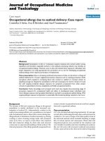
elbow. Radiographs demonstrated a posterolatera l elbow
dislocation with fractures of both the lateral and medial
epicondyles (Figures 1 and 2 - showing three different
views). The elbow dislocation was reduced and immobi-
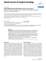
lized in the emergency department. Post-reduction
radiographs showed a reduced elbow with displaced
fractures of medial and lateral epicondyles (Figure 3-
Post reduction radiographs demonstrating AP and Lat-
eral views). However as the elbow remained clinically
highly unstable and the fractures were still markedly dis-
placed, operative intervention was deemed necessary.
ORIF of both the medial and lateral epicondyles was
performed using a separate medial and l ateral approach.
Due to the presence of fracture comminution and small
sized fragments of both epicondyles, screw f ixation was
deferred. K wire fixation using two 1.6 mm w ires for
each the lateral and medial epicondyle was preferred.
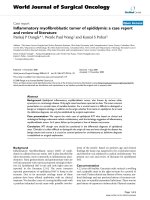
Post-operative radiographs showed satisfactory reduction
and fixat ion (Figu re 4- postopera tive radiographs show-
ing AP and lateral views after K wire fixation). Following
six weeks of immobilization in a plaster of Paris, active
Figure 1 Injury X-ray 1 (showing dislocat ed elbow with
biepicondylar fractures).
Figure 2 Injury X-ray 2.
Figure 3 Post reduction X-ray (showing reduced elbow with
displaced biepicondylar fractures).
Figure 4 Postoperative X-ray (showing fixation with K wires).
Meta and Miller Journal of Orthopaedic Surgery and Research 2010, 5:75
/>Page 2 of 3
elbow ROM (range of motion) was commenced by a
physiotherapi st. The patient receive d weekly phy-
siotherapist treatment until week 14. K wires were
removed a t postoperative week eight. At the final fol-
low-up 14 weeks postoperatively, satisfactory elbow
function (0°-120° flexion, full supination and pronation,
with normal strength an d stable elbow) was observed.
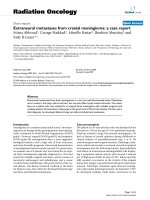
Radiographs demonstrated bony union and n o evidence
of myositis ossificans (Figure 5- Final follow up radio-
graphs showing AP and lateral views of elbow with
union of both epicondyles). Prophyl actic treatment for
myositis ossificans was not used.
Conclusion
Biepicondylar e lbow fracture dislocations are unstable
injuries. Open reduction and internal fixation of these
injuries is recommended to restore elbow stability and
function.
Consent
Written informed consent was obtained from the
patient’s pare nts for publication of this case report and
any accompanying images. A copy of the written con-
sent is available for review by the Editor-in-Ch ief of this
journal.
Author details
1
Orthopaedic Registrar , Department of Orthopaedics, Royal Brisbane &
Women Hospital, Butterfield Street, Herston 4029, QLD Australia.
2
Orthopaedic RMO, Department of Orthopaedics, Royal Brisbane & Women
Hospital, Butterfield Street, Herston 4029, QLD Australia.
Authors’ contributions
MM designed the study, collected data, wrote the manuscript and
performed literature review. DM assisted in writing manuscript, literature
review and obtained consent from parents. Both authors read and approved
the final manuscript.
Competing interests
The authors declare that they have no competing interests.
Received: 13 March 2010 Accepted: 15 October 2010
Published: 15 October 2010
References
1. Rockwood CA, Green DP, Bucholz RW, Heckman JD: Fractures in children.
Lippincott Williams & Wilkins, 7 2009, 475-477, 566-570, 577-578.
2. Gani NU, Rather AQ, Mir BA, Halwai MA, Wani MM: Humeral Biepicondylar
fracture dislocation in a child- a case report and review of literature.
Edited by: Cases J 2008, 1(1):163.
3. Taylor GR, Gent E, Clarke NM: Biepicondylar fracture dislocation of a
child’s elbow. Injury 1997, 28(1):71-2.
4. Silberstein MJ, Brodeur AE, Graviss ER: Some vagaries of the lateral
epicondyle. JBJS Am 1982, 64:444-448.
5. Joseph WCH, Lee FR, Harvey W, Mihvan OT: Injuries of the medial
epicondylar ossification center of the humerus. Am J Roentgenol 1977,
129:49-55.
doi:10.1186/1749-799X-5-75
Cite this article as: Meta and Miller: Paediatric biepicondylar elbow
fracture dislocation - a case report. Journal of Orthopaedic Surgery and
Research 2010 5:75.
Submit your next manuscript to BioMed Central
and take full advantage of:
• Convenient online submission
• Thorough peer review
• No space constraints or color figure charges
• Immediate publication on acceptance
• Inclusion in PubMed, CAS, Scopus and Google Scholar
• Research which is freely available for redistribution
Submit your manuscript at
www.biomedcentral.com/submit
Figure 5 Final follow-up X-ray (showing fully united medial
and lateral epicondyles).
Meta and Miller Journal of Orthopaedic Surgery and Research 2010, 5:75
/>Page 3 of 3