báo cáo hóa học:" Unicameral bone cyst of the lunate in an adult: case report" docx
Bạn đang xem bản rút gọn của tài liệu. Xem và tải ngay bản đầy đủ của tài liệu tại đây (1.42 MB, 3 trang )
CAS E REP O R T Open Access
Unicameral bone cyst of the lunate in an adult:
case report
Hakan Gündeş
*
, Mustafa Sahin, Tugrul Alici
Abstract
We report a case of a symptomatic unicameral (simple) bone cyst of the lunate in a 42-year- old woman. The
lesion was treated with curettage and cancellous autogenous iliac bone grafting. At five years of follow-up the
wrist was pain free, there were no limitations of motion, and the radiographs showed complete obliteration of the
cavity. To the best of our knowledge, no other unicameral bone cyst of the lunate has been reported in an adult.
Cysts with significant cavities at the carpal bones in an adult should be approached cautiously, as they may require
early curettage and bone grafting for healing, before collapse and degener ative changes occur.
Background
Unicameral bone cysts (UBCs), also known as simple
bone cysts are benign, fluid-filled lesions involving the
metaphysis of long bones [1-3]. On radiography they
demonstrate a centrally located lytic lesion with well-
defined margins [ 2]. The cyst wall is lined with a fibrous
membrane which contains serous yellow fluid [2]. 80%
of UBCs occur in the proximal humerus and proximal
femur [1,3]. Most UBCs occur in childhood where one
third of the cases will resolve spontaneously by skeletal
maturity [1-3]. Occurrence of a single symptomatic radi-
olucent lesion in the lunate is rare [4,5]. A differential
diagnosis of a painful radiolucent lesion in the lunate
would include intraosseou s ganglion, Kienböck’s disease,
osteoid osteoma, giant cell tumor, aneurysmal bone cyst
and enchondroma [4-9]. The incidence of UBCs invol-
ving the wrist bones and lunate has not been clearly
defined in the literature [10].
Case presentation
A 42-year-old woman was referred to the hand clinic
with dull pain and discomfort in her right dominant
wrist that had been present for six months. Pain was
steady and not aggravated by use. Examination revealed
very mild do rsal swelling o f the wrist, with tenderness
over lunate. The range of mot ion was slightly restricted
in all directions. A specific limitation on wrist flexion
and radial deviation was observed. A scaphoid shift test
was negative. Routine biochemical tests, blood count
and erythrocyte sedimentation rate (ESR) were within
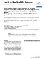
normal limits. AP and lateral radiograph of the wrist
revealed a radiolucent lesion measuring 11 mm in dia-
meter at the center of the lunate with round margins
(Figure 1). There was no scalloping, septae formation or
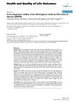
cortical thinning. Computed Tomography (CT) scans of
the wrist revealed a round hypodense cystic lesion of 10
mm in diameter without septae formation (Figure 2).
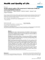
Magnetic Resonance (MR) imaging views on coronal
fat-suppressed and axial and sagittal T2 weighted
sequences have revealed a homogenous hyper-intense
cystic lesion in the lunate with smooth and round con-
tours (Figure 3).
A dorsal longitudinal incision of 6 cm was made on the
ulnar side of the Lister’s tubercle, extending proximally
and distally through the skin and subcutaneous tissue.
The extensor retinaculum was sectioned between the
third and fourth compartments, parallel to the incision.
The third and fourth compartments were connected.
Tendons were retracted and the capsule was exposed.
The capsule was cut open through an H-shaped incision ,
allowing the evaluation of the proximal part of the capi-
tate and the lunate fossa. Curettage was performed by
opening a dorsal 3 mm cortical window through the car-
tilage. After the fluid was aspirated, the fibrous mem-
brane-like tissue lining the cyst wall was curetted, and a
power burr was not used. The cyst was packed with can-
cell ous autogenou s iliac bone chips. We preferred to uti-
lize autogenous iliac bone over distal radius in order to
increase the chance of incorporation [1]. The wrist was
* Correspondence:
Maltepe University, School of Medicine, Department of orthopedics and
hand surgery. Istanbul, Turkey
Gündeş et al. Journal of Orthopaedic Surgery and Research 2010, 5:79
/>© 2010 Gündeşş et al; licensee BioMed Central Ltd. This is an Open Access article distributed under the terms of the Creative
Commons Attribution License (http://creat ivecommons.org/licenses/by/2.0), which permits unrestricted use, distribution, and
reproduction in any medium, provided the original work is properly cited.
protected with a well padded splint for two weeks to alle-
viate the pain and discomfort. After that, active and pas-
sive range of motion exercises and strengthening had
been instituted. The histopatholo gical diag nosis was uni-
cameral bone cyst. A radiograph that had been taken two
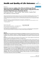
years after the operation showed solid incorporation o f
the graft. At five years of follow-up, the wrist was pain
free, there were no limitations of motion observed, and
the radiograph showed complete obliteration of the cav-
ity (Figure 4).
Conclusions
Most diagnosed UBCs occur in childhood [1-3]. UBC
etiology is unknown [ 1,3]. They account for 3% of all
bone tumors, and usually involve the metaphysis of long
bones, and have a predilection for the proximal humerus
and proximal femur [2,3]. A debate exis ts whether treat-
ment is necessary (because of spontaneous resolution)
and what treatment is most appropriate [1]. Patients
with UBCs usually present with a pathological fracture
or a complaint of mild pain in the affected reg ion [2,3].
Themainindicationforsurgeryistopreventortreat
pathological fracture [1]. Benign bone lesions are often
treated with intralesional curettage, and autogenously
bone grafts or various substitutes have been used to fill
the defect [11]. Curettage alone is often the standard
treatment for benign bone tumors giving the similar
recurrence and fracture rates [11]. Described treatment
options for a UBC include simple observation, curettag e
and grafting (autogenous or allogenous), steroids, demi-
neralized bone matrix, and bone marrow injection [1,3].
The incidence of UBCs involving the wrist bones and
Figure 1 PA radiograph of the rig ht wrist.Therewasa
radiolucent lesion measuring 11 mm in diameter at the center of
the lunate with round margins. There was no scalloping, septae
formation or cortical thinning.
Figure 2 Computed Tomography (CT) scans of the wrist
revealed a round hypodense cystic lesion of 10 mm in
diameter.
Figure 3 Magnetic Resonance (MR) imaging views on fat-
suppressed T2 weighted sequences have revealed a
homogenous hyper-intense cystic lesion in the lunate with
smooth and round contours.
Gündeş et al. Journal of Orthopaedic Surgery and Research 2010, 5:79
/>Page 2 of 3
lunate has not been clearly defined in the literature [10].
The differential diagnosis of a radiolucent lesion of the
lunate most co mmonly includes an intraosseous gang-
lion cyst or osteoid osteoma [7]. Kienböck’s disease,
osteoid osteoma, giant cell tumor, enchondroma, aneur-
ismal bone cyst (ABC), nonossifying fibroma and fibrous
dysplasia are less likely possibilities [4-9]. There are no
established guidelines for when and how to treat UBCs.
Injections of steroids, demine ralized bone matrix, and
bone marrow aspirate have been reported as methods of
treat ment with various success rates [ 1,3]. Standard sur-
gical treatment consists of curettage and cancellous
bone grafting [3]. The main indication for surgical inter-
vention is to prevent or treat a pathological fracture
[1,3].
In our case herein, in dications for surgery were clini-
cal history of pain and radiographic findings of a cystic
formation in the lunate.
UBCs of carpal bone in adulthood had been reported
before [10]. This was a case report of bilateral unicam-
eral bone cysts located in the hamate bones of a 22-
year-old man [10]. Our patient was unique in that she
had a UBC in her lunate bone. To the best of our
knowledge, no other unicameral bone cyst of lunate has
been reported in the literature. The etiology of this
symptomatic lesion remains unknown. Cysts with such
large cavities at the carpal bones in an adult should be
approached cautiously. They may require early curettage
and bone grafting for healing. Early treatment has its’
definitive benefits as it prevents collapse and degenera-
tive changes as in our case [8].
Authors’ contributions
HG carried out the operation, followed-up the patient and wrote the
manuscript. MS and TA participated in writing and design of the manuscript.
They also drafted the manuscript. All authors read and approved the final
manuscript.
Conflict of interest statement
Authors certifies that they have no commercial associations (e.g.,
consultancies, stock ownership, equity interest, patent/licensing
arrangements, etc.) that might pose a conflict of interest in connection with
the submitted article.
Consent
Written informed consent was obtained from the patient for publication of
this case report and accompanying images. A copy of the written consent is
available for review by the Editor-in-Chief of this journal.
Received: 21 May 2010 Accepted: 30 October 2010
Published: 30 October 2010
References
1. Sung AD, Anderson ME, Zurakowski D, Hornicek FJ, Gebhardt MC:
Unicameral Bone Cyst: A Retrospective Study of Three Surgical
Treatments. Clin Orthop Relat Res 2008, 466:2519-26.
2. Tey IK, Mahadev A, Lim KB, Lee EH, Nathan SS: Active unicameral bone
cysts in the upper limb are at greater risk of fracture. Journal of
Orthopaedic Surgery 2009, 17:157-60.
3. Yilmaz G, Aksoy MC, Alanay A, Yazici M, Alpaslan AM: Treatment of simple
bone cysts with methylprednisolone acetate in children Acta Orthop
Traumatol Turc. 2005, 39:411-15.
4. Bennet DC, Hauck RM: Intraosseous ganglion of the lunate. Ann Plast Surg
2002, 48:439-42.
5. Ikeda M, Oka Y: Cystic lesion in carpal bone. Hand Surg 2000, 5:25-32.
6. Athanasian EA: Aneurysmal bone cyst and giant cell tumor of bone of
the hand and distal radius. Hand Clin 2004, 20:269-81.
7. Baron J, Scharizer E: Tumors and tumor-like diseases of the carpal bones.
Handchir Mikrochir Plast Chir 1987, 19:195-205.
8. Oka Y, Umeda K, Ikeda M: Cyst-like lesions of the lunate resembling
Kienböck’s disease: a case report. J Hand Surg Am 2001, 26 :130-34.
9. Schmitt R, Christopoulos G, Kalb K, Coblenz G, Fröhner S, Brunner H,
Krimmer H, Lanz U: Differential diagnosis of the signal-compromised
lunate in MRI. Rofo 2005, 177:358-66.
10. Jasan M, House JH, Brand JC: Bilateral unicameral bone cysts in the
hamate bones. J Hand Surg Am 1990, 15(6):888-90.
11. Yanagawa T, Watanabe H, Shinozaki T, Takagishi K: Curettage of benign
bone tumors without grafts gives sufficient bone strength. A case-series
of 78 patients. Acta Orthopaedica 2009, 80:9-13.
doi:10.1186/1749-799X-5-79
Cite this article as: Gündeş et al.: Unicameral bone cyst of the lunate in
an adult: case report. Journal of Orthopaedic Surgery and Research 2010
5:79.
Figure 4 A radiograph taken five years after the operation
showed solid incorporation of the graft, and complete
obliteration of the cavity.
Gündeş et al. Journal of Orthopaedic Surgery and Research 2010, 5:79
/>Page 3 of 3