báo cáo hóa học:" Evaluation of unilateral cage-instrumented fixation for lumbar spine" doc
Bạn đang xem bản rút gọn của tài liệu. Xem và tải ngay bản đầy đủ của tài liệu tại đây (736.11 KB, 7 trang )
RESEARC H ARTIC L E Open Access
Evaluation of unilateral cage-instrumented
fixation for lumbar spine
Ti-Sheng Chang
1,2
, Jia-Hao Chang
3
, Chien-Shiung Wang
1
, Hung-Yi Chen
1
, Ching-Wei Cheng
1*
Abstract
Background: To investigate how unilateral cage-instrumented posterior lumbar interbody fusion (PLIF) affects the
three-dimensional flexibility in degenerative disc disease by comparing the biomechanical characteristics of
unilateral and bilateral cage-instrumented PLIF.
Methods: Twelve motion segments in sheep lumbar spine specimens were tested for flexion, extension, axial
rotation, and lateral bending by nondestructive flexibility test method using a nonconstrained testing apparatus.
The specimens were divided into two equal groups. Group 1 received unilateral procedures while group 2 received
bilateral procedures. Laminectomy, facectomy, discectomy, cage insertion and transpedicle screw insertion were
performed sequentially after testing the intact status. Changes in range of motion (ROM) and neutral zone (NZ)
were compared between unilateral and bilateral cage-instrumented PLIF.
Results: Both ROM and NZ, unilateral cage-instrumented PLIF and bilate ral cage-instrumented PLIF, transpedicle
screw insertion procedure did not revealed a significant difference between flexion-extension, lateral bending and
axial rotation direction except the ROM in the axial rotation. The bilateral group’s ROM (-1.7 ± 0. 8) of axial rotation
was decreased significantly after transpedicle screw insertion procedure in comparison with the un ilateral group
(-0.2 ± 0.1). In the unilateral cage-instrumented PLIF group, the transpedicle screw insertion procedure did not
demonstrate a significant difference between right and left side in the lateral bending and axial rotation direction.
Conclusions: Based on the results of this study, unilateral cage-instrumented PLIF and bilateral cage-instrumented
PLIF have similar stability after transpedicle screw fixation in the sheep spine model. The unilateral approach can
substantially reduce exposure requirements. It also offers the biomechanics advantage of construction using
anterior column support combined with pedicle screws just as the bilateral cage-instrumented group. The
unpleasant effect of couple motion resulting from inherent asymmetry was absent in the unilateral group.
Introduction
Chronic discogenic back pain caused by degenerative
disc disease is a common ailment in the general popula-
tion [1]. The degenerative often results from arthritic
changes in the intervertebral discs, facet joints, and liga-
ments surrounding the vertebral canal [2]. In clinical
practice, lateral recess stenosis and foramina l stenosis
may induce nerve root compression which can cause
unilateral symptoms. Unilateral PLIF may be satisfactory
in patients with unilateral symptom. This study evalu-
ated unilateral fusion in patients with unilateral
symptoms.
Posterior lumbar interbody fusion (PLIF) has proven
successful for relieving motion-induced discogenic pain
and was once considered standard treatment for degen-
erative disc disease [3-5]. A successful PLIF can restore
disc height, decompress the dura l sac and nerve roots,
immobilize the unstable intervertebral disc, and main-
tain load-bearing to anterior structure [6]. Bilateral PLIF
is associated with increased complication rates [7-9].
Elias and coworkers [7] reported a 15% incidence of
dural tear and postoperative radiculopathy. It typically
caused by excessive epidural bleeding and prolong or
excessive retraction. In 1982, Harm et al. [10] popular-
ized the surgical technique of transforaminal lumbar
interbody fusion (TLIF). Its advantages are posterior epi-
dural approach with interbody support and bilateral pos-
terior segmental pedicle screw fixation. Biomechanically,
* Correspondence:
1
Department of Bio-industrial Mechatronics Engineering, National Chung
Hsing University, Taichung, Taiwan
Full list of author information is available at the end of the article
Chang et al . Journal of Orthopaedic Surgery and Research 2010, 5:86
/>© 2010 Chang et al; licensee BioMed Centr al Ltd. This is an Open Access a rticle distributed under t he terms of th e Creative Commons
Attribution License (http://cre ativecommons.org/licenses/by/2.0), which permits unrestricted use, distribution, and reproduction in
any medium, provided the original work is properly cited.
preserving longitudinal ligaments is though to preserve
stability, to prevent implant dislocation, and to place a
compressive force on adequately sized intervetebral
implants [11-13]. This technique also avoids r etraction
of the ligamentum flavum as well as scarring of the
spinal canal. Nevertheless, its long learning curve, pro-
long radiation exposure and high cost make it unsuita-
ble for health insurance coverage in Taiwan.
Performing unilateral PLIF using a single interbody
cage has several advantages. Inserting a single interbody
cage through a unilateral approach compromises fewer
anatomic structures than two cages inserted through a
bilateral approach. However, it is u nknown that unilat-
eral PLIF can achieve biomechanical stability as the
bilateral PLIF. Suke et al. [14] found that unilateral
pedicle screw fixation was as effective as bilateral pedicle
fixation in lumbar spinal fusion independent of one or
two levels. Their conclusions cannot extend to the cage-
instrumented PLIF due to the different boundary of
decompression (especially facetectomy). Tencer et al.
[15] demonstrated that two PLIF structural devices pro-
duced a greater reduction in torsional stiffness than sin-
gle PLIF device. Chen et al. [16] demonstrated that
unilateral fixation with two c age insertion is a feasible
alternative in s pinal surgery. Wang et al. [17] showed
that an oblique insertion of a single BAK cage in instru -
men ted PLIF might reduce exposure and enable precise
implantation. These articles coul d not pro vide enough
evidence to support that unilateral fusion can supply
similar stability as the bilateral fusion. That is why uni-
lateral PLIF did not become routine procedures in the
treatment of degenerative lumbar spine disease. Goel
[18] demonstrated that the unilateral plate system
causes coupled motions due to its inherent asymmetry
and was unlikely to provide sufficient rigidity in fresh
cadaveric human spines. Rotational deformity of lumbar
spine might develop if inherent asymmetry persisted.
They considered complete excision of the disc was
required. Unilateral cag e-instrumented PLIF might over-
come inherent asymmetry. From the previous review,
this study was conducted with two aims. One was to
know the b iomechanical stability between unilateral and
bilateral cage-instrumented PLIF. The other was to
determine the unpleasant effect of couple motion result-
ing from inherent asymmetry was present or not in the
unilateral group.
Methods
Specimen Preparation
Twelve motion segments of L4/5 from twelve sheep
lumbar spines were studied for this in-vitro investiga-
tion. At the time of salvage, the animals were 12-18
months old and weighted 60 kg (53 to 65 kg). Following
preparation, the specimens were stored frozen at -20°C
then thawed at room temperature for 24 hours prior to
testing. Care was taken to completely preserve the bony
and ligamentou s structures of the locomotor segment of
each specimen, and only muscular and fatty tissue was
removed. The cranial and caudal vertebrae of each func-
tional spinal unit was anchored with stainless-steel
screws and embedded with custom-designed metal fix-
tures using polyester resin. The intervening segments
were left unconstrained.
Instrumentations
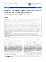
Flexion-extension, left-right lateral bending and left-
right axial rotation of each specimen were analyzed by
the spinal tester (Figure 1) [19]. The reliability test of
this tester was published in the literature [19]. The
servo motor (SMART MOTOR 2315D, ANIMATICS,
USA) and planetary reduction gearbox (AD042.S2.P1,
APEX DYNAMICS, TW) combined to form the drive
apparatus. A six-axis load cell (SI-660-60, ATI Indus-
trial automation, USA) measured the moments and the
forces during testing procedures. Below the load cell,
an X-Y table was used to achieve pure moment on
each specimen to prevent shear force. The signal from
the load cell was conditioned and connected to a
Figure 1 The testing machine and mounting system with a
specimen during performance of a flexibility test. Various
component of this tester is illustrated.
Chang et al . Journal of Orthopaedic Surgery and Research 2010, 5:86
/>Page 2 of 7
computer to provide a feedback signal for displacement
control testing.
Surgical procedures
The specimens were divided into two equal groups.
Group1 included specimens that were tested intact. The
following surgical procedures were then performed:
1. left hemilaminectomy,
2. left medial facectomy,
3. left discectomy,
4. left cage insertion (one, 8*8*12 mm), The interver-
tebral disc and approximately 2-3 mm of endplate
and subchronal bone of both vertebral bodies was
removed with curret and disc forcep. Care was taken
not to place the intervertebral disc under distraction
and compression. Custom-made titanium cage
(8*8*12 mm, Synthes) was impacted and gently
pushed into the intervertebral lumen. The cage was
within the intervertebral space about 2 mm below
the end plate. No scoliotic deformity was found after
this procedure.
5. left transpedicular screw fixation (two screws-
4.75*25 cm and one rod- ISOLA system),
The group 2 specimens underwent the same sequence
of procedures as the Group 1 except the bilateral sides,
which included:
1. total laminectomy,
2. bilateral medial facetectomy,
3. bilateral discectomy,
4. two cage insertion (8*8*12 mm), The interverteb-
ral disc and approximately 2-3 mm of endplate and
subchronal bone of both vert ebral bodies was
removed with curret and disc forcep. Care was taken
not to place the intervertebral disc under distraction
and compressio n. Cust om-made titanium cage
(8*8*12 mm, Synthes) was impacted and gently
pushed into the intervertebral lumen. The cage was
within the intervertebral space about 2 mm below
the end plate. No scoliotic deformity was found after
this procedure.
5. bilateral transpedicle screws fixation (four screws-
4.75*25 cm and two rods- ISOLA system)
All procedures were performed by a single experi-
enced spinal surgeon and co-author (TSC).
Testing procedures
The lower vertebrae were centered over the load cell
and maintained in neutral position with respect to the
set coordinate system described previously [19]. After
the specimen was mounted on the spinal tester, left-
right lateral bending, flexion-extension and left-right
axial rotation of the specimen were conducted at a con-
stant speed of 1°/sec in sequence before and after differ-
ent surgical procedures. A compressive preload of 100
N was applied. It represented that the spine supports
standing posture under external load. The direction was
reversed until the moment detected by the load cell
reached ±2 Nm. Depending on the specific purpose of
the study, the typical load used for biomechanical test-
ing is 6-10 Nm [20-22]. The load applied in the current
study was 2 Nm since this lo ad produced the wide
range of destruction including facetectomy and discect-
omy (which are similar to effects observed in clinical
practice). In the preliminary investigation, specimen fail-
ure occurred at loads exceeding 2 Nm. Krijnen et al.
[23] used 1 Nm in a goat model segmental stability with
stand-alone cage.
The load cell provided a feedback signal to the com-
puter through RS-232 interface with 40 Hz sampling
rate. The load and displacement data were collected and
recorded during testing. A real-time graphical display of
servo motor angle a nd applied moment was available
during the test.
Data and Statistical Analysis
The curvilinear regression analysis of force (Y-axis) and
displacement (X-axis) with fourth-order polynomials
were used to eliminate small variation in force. The
ROM of flexion-extension, lateral bending and axial
rotation were interpolated at 2 Nm from the moment-
angle relation. The NZ is the displacement at t he zero-
load point measured f rom the neutral position. The EZ
is the displacement from the zero-load point to the
maximum load point (2Nm). ROM at t he operative L4/
5 level was defi ned as the summation of NZ and EZ at
the fifth loading cycle.
To determine the effects of unilateral and bilateral
cage-instrumented PLIF differences on outcome mea-
sures (ROM, NZ) over the fifth repetition procedures
were computed and used for statistical analyses. We
used a simple contrast in repeated measurement analysis
of variance (rmANO VA) to compare the difference
between intact and the rest five procedures. Indepen-
dent sample T test was performed to co mpare the dif-
ference in ROM and NZ, wa s calculated by subtracting
the previous status from the data after surgical proce-
dure, between unilateral and bilateral cage-instrumented
PLIF of flexion-extension, lateral bending and axial rota-
tion. We c ompared individual procedure through this
way. Paired sample t test was used to compare the dif-
ference in ROM, was calculated by subtracting the intact
status from that data after the surgical procedure,
between right and left side of lateral bending and axial
rotation in the unilateral cage-instrumented PLIF. The
Chang et al . Journal of Orthopaedic Surgery and Research 2010, 5:86
/>Page 3 of 7
statistical data analyses were performed using SPSS for
Windows version 13.0 (SPSS. Inc. Chicago, Illinois) soft-
ware package. A p level less than 0.05 w as considered
statistically significant.
Results
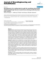
1. Effects of destructive and stabilizing procedures
on the ROM and NZ of unilateral and bilateral
group (Figure 2)
Both unilateral and bilateral group, the ROM and
NZ of flexion-extension and lateral bending were
decreased significantly a fter transpedicle screw in
comparison with intact status. In the axial rota-
tion direction, the ROM and NZ did not
decreased after transpedicle screw fixation on
both groups.
2. Comparison between unilateral and b ilateral
group in ROM and NZ (Table 1)
Figure 2 Effects of destructive and stabilizing procedures on the ROM and NZ of unilateral and bilateral group.(L:Laminectomy,F:
Facetectomy, D: Discetomy, C: Interbody Cage, P: Pedicle screw ).
Chang et al . Journal of Orthopaedic Surgery and Research 2010, 5:86
/>Page 4 of 7
The bilateral group’s ROM and NZ of flexion-exten-
sion and lateral bending did not significantly differ
from transpedicle screw procedure in comparison
with the unilateral group. The bilateral group’s ROM
of axial rotation was decreased significantly after
transpedicle screw insertion (-1.7 ± 0.8 vs -0.7 ± 0.4,
p < 0.05) procedure in comparison with the unilat-
eral group. The bilateral group’s NZ of axial rotation
did not significantly differ from transpedicle screw
procedure in comparison with the unilateral group.
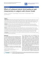
3. Symmetry of ROM in the unilateral group:
(Table 2, Figure 3)
The difference in ROM of right lateral bending (6.45
± 2.40 vs 6.29 ± 2.17) and axial rotation (0.23 ± 0.21
vs 0.28 ± 0.31) did not significantly differ after trans-
pedicle screw as compared with the left side.
Discussion
The availability of human vertebral column seg ments is
often limited due to the ethical, religious reasons as well
as increasing legal restrictions [24]. Therefore, readily
available animal vertebral columns were used for testing.
Calf, pig and sheep are the three popular species used
models to t est spinal implants [25,26]. The calf is the
species mainly used in vitro test for the pedicle screw
system. Similar to the calf, the pig is also often used in
vitro test to measure intradiscal pressure. The sheep and
pig is mainly used in vivo test for biomechanical experi-
ments [26]. Sheep spines have also been used in vitro to
study the initial stabilizing effect of spinal implants in
the lumbar [27]. The cross-section of six to seven lum-
bar vertebrae in sheep resembles that in humans
[28,29]. The sheep lumbar spine and the human spine
also have similar intervertebral disc morphology and
spi nal musculature anato my [28]. Sheep pedicles have a
smaller diameter, pedicle screw for example need to be
shortened to fit the sheep vertebral dimension [25].
Considerable progress in research is apparently needed
to achieve satisfactory understanding of the biomecha-
nics of the human lumbar spine before clinical applica-
tion under the result of animal experiment [25].
In vitro testing is an essential for studying spinal
mechanics [30,31]. Several parameters may be obtained
through biomechanical tests of flexibility to quantify
Table 1 The difference of ROM and NZ between unilateral and bilateral cage-instrumented PLIF over flexion-
extension, lateral bending and axial rotation
Procedures L F D C P
unilateral bilateral unilateral bilateral unilateral bilateral unilateral bilateral unilateral bilateral
Flexion Extension
(°)
ROM
(°)
1.9 ± 0.9 2.1 ± 0.8 0.5 ± 0.6 1.7 ±
0.7*
4.2 ± 1.1 7.1 ± 7.3 -10.4 ±
4.4
-13.6 ±
3.3
-3.7 ± 3.8 -5.8 ± 4.8
NZ (°) 1.2 ± 0.8 1.8 ± 1.2 0.4 ± 0.6 0.3 ± 0.6 1.9 ± 2.4 2.3 ± 5.3 -5.8 ± 1.6 -5.8 ± 3.9 -0.2 ± 1.5 -1.0 ± 2.8
Lateral Bending (°) ROM
(°)
0.4 ± 0.4 0.2 ± 0.5 0.7 ± 0.7 0.1 ± 0.5 1.8 ± 1.6 3.7 ± 1.0* -11.5 ±
2.6
-13.2 ±
2.2
-4.1 ± 1.7 -4.2 ± 1.7
NZ (°) 0.4 ± 0.8 0.9 ±0.9 0.0 ± 0.8 0.0 ± 0.7 -0.6 ± 2.7 -1.6 ± 1.5 -3.1 ± 2.1 -1.5 ± 1.3 -0.1 ± 0.2 -0.2 ± 0.1
Rotation (°) ROM
(°)
0.4 ± 0.3 0.2 ± 0.3 0.4 ± 0.3 0.3 ± 0.4 0.7 ± 0.4 0.2 ± 0.3* -0.6 ± 0.6 1.1 ± 0.9* -0.7 ± 0.4 -1.7 ±
0.8*
NZ (°) 0.1 ± 0.0 0.0 ±
0.1*
0.0 ± 0.1 0.0 ± 0.1 0.1 ± 0.1 -0.1 ±
0.1*
0.0 ± 0.2 0.3 ± 0.1* -0.1 ± 0.1 -0.2 ± 0.1
L: Laminectomy, F: Facetectomy, D: Discetomy, C: Interbody Cage, P: Pedicle screw
Note:
L = (L-I): ROM and NZ of the specimen after laminectomy minus intact status
F = [(I +L+F)-(I+L) ]: ROM and NZ of the specimen after facectomy minus laminectomy status
D = [(I+L+F+D)-(I +L+F)]: ROM and NZ of the specimen after discectomy minus facetectomy status
C = [(I+L+F+D+C)-(I+L+F+D)]: ROM and NZ of the specimen after interbody cage insertion minus discectomy status
P = [(I+L+F+D+C+P)-(I+L+F+D+C)]: ROM and NZ of the specimen after tranpedicle screw fixation minus interbody cage insertion status
*: significant: p <.05 compared with unilateral group
Table 2 The difference of ROM between right and left
side over lateral bending and axial rotation in the
unilateral group
Procedures ROM of lateral bending ROM of axial rotation
Lt cage-PLIF Right (°) Left (°) Right (°) Left (°)
L-I 0.35 ± 0.22 0.26 ± 0.26 0.31 ± 0.13 0.23 ± 0.2
L+F-I 0.82 ± 0.57 0.36 ± 0.15 0.60 ± 0.29 0.28 ± 0.2
L+F+D-I 1.68 ± 1.31 1.38 ± 1.49 0.85 ± 0.62 0.71 ± 0.45
L+F+D+C-I -4.65 ± 3.4 -4.47 ± 2.45 0.68 ± 0.41 0.43 ± 0.27
L+F+D+C+P-I -6.45 ± 2.4 -6.29 ± 2.17 0.23 ± 0.21 0.28 ± 0.31
L: Laminectomy, F: Facetectomy, D: Discetomy, C: Interbody Cage, P: Pedicle
screw
Note:
L-I: ROM of the specimen after laminectomy minus intact status
(L+F)-I: ROM of the specimen after facectomy minus intact status
(L+F+D)-I: ROM of the specimen after discectomy minus intact status
(L+F+D+C)-I: ROM of the specimen after interbody cage minus intact status
(L+F+D+C+P)-I: ROM of the specimen after tranpedicle screw minus intact
status
Chang et al . Journal of Orthopaedic Surgery and Research 2010, 5:86
/>Page 5 of 7
mechanical properties [30]. Such parameters include
range of motion (ROM) and neutral zone (NZ). In bio-
mechanical study, the ROM represents the stability of
the specimen before and after additional procedures
(including destructive and stabilizing procedures). The
NZ indicates the laxity around the neutral position of
a motion segment as well as residual deformation after
removing a defined pure moment load from a motion
segment. Mimura et al. [31] revealed that, in flexion-
extension and lateral bending, ROM decreases and NZ
increases during disc degeneration. In the early stage
of disc degeneration, ROM increases while in the late
stage of disc degeneration, ROM decreases. If only
ROM is used as the measurement parameter, misinter-
pretations are likely. Previous in vitro studies indicated
that NZ typically increases after experimentally
induced injuries [32,33], and that it decreases with the
addition of muscle forces and spinal instrumentation
[34]. In the ROM and NE, bilateral cage-instrumented
group’ s transpedicle s crew insertion procedure did not
demonstrate significantly difference in comparison
with the unilateral group in flexion-extension, lateral
bending and axial rotation direction except the ROM
of the rotation. Fortunately, rotation movement occu-
pied little portion of the daily activity. The major por-
tion of daily activity is flexion-extension and lateral
bending. Unilateral cage-instrumented PLIF have simi-
lar biomechanical stability to bilateral cage-instrumen-
ted PLIF in this sheep spine model. Studies have
demonstrated increased complications associated with
bilateral PLIF [7-9]. Bilateral instrumental fusion tech-
nique requires wide dissection, which jeopardizes the
function of the paraspinal muscle. The unilateral
approach can substantially reduce exposure require-
ments. This technique is easier than routine bilateral
cage-instrumented PLIF. When treating unilateral scia-
tica patients, the cage can be placed from the sympto-
matic side so as to avoid retraction of the nerve root
and dura sac of t he asymptomatic side.
Goel [18] demonstrated the difference in rigidity
between unilateral and bilateral instrumentat ion in fresh
cadaveric human spines. They reported that the unilat-
eral plate system causes coupled motions due to its
inherent asymmetry and was unlikely to provide suffi-
cient rigidity for decompressive procedures and would
therefore require complete excision of the disc. Unilat-
eral instrumentation is unadvised due to the effect of
inherent asymmetry . In the curren t study, no significant
differences were shown between right and left side in
the lateral bendi ng and axial rotation in the fusion pro-
cedures. This method may achieve sufficient stability.
The reason is that the inserted cage can be held by the
screw-rod system, and the ca ge can maintain adequate
disc space, which is particularly effective for correcting
instability associated with degenerative disc disorder.
There are some limitations in this study. First, the
paraspinal muscles were remved; however, these muscles
had an in vivo stabilizing effect therefore, our results
probably overestimated the destabilizing effect of the
segmental bone and other soft tissue alternation. Sec-
ond, in this in vitro test, the results only reflect acute
postoperative stability with relatively few loading cycles
and are not necessarily indicative of repetitive loading
cycles in the human spine. The biological effects of the
potential healing process are unpredictable. Third, subsi-
dence is a complication of cage sinking with loss of nor-
mal intervertebral height. The mean subsidence was
greater in the one cage model because of the less con-
tact area than the two cage model. An in vivo study
may be necessary to show whether unilateral group had
higher incidence of subsidence or not.
In conclusion, based on the results of this study, uni-
lateral cage-instrumented PLIF and bilateral cage-instru-
mented PLIF have similar stability in the sheep spine
model. The unilateral approach can substanti ally reduce
exposure requirements. It also offers the biomech anics
advantage of c onstruction using anterior column sup-
port combined with pedicle screws just as the bilateral
Figure 3 ThedifferenceofROMbetweenrightandleftsideoverlateral bending and axial rotation in the unilateral group.(L:
Laminectomy, F: Facetectomy, D: Discetomy, C: Interbody Cage, P: Pedicle screw ).
Chang et al . Journal of Orthopaedic Surgery and Research 2010, 5:86
/>Page 6 of 7
cage-instrumented PLIF. The unpleasant effect of couple
motion resulting from inherent asymmetry was absent
in the unilateral group.
Acknowledgements
The authors would like to thank the Armed Force Taichung general hospital
research and developmental center (plan number 9607) for financially
supports.
Author details
1
Department of Bio-industrial Mechatronics Engineering, National Chung
Hsing University, Taichung, Taiwan.
2
Department of Neurosurgery, Armed
Forces Taichung General Hospital, Taichung, Taiwan.
3
Department of Physical
Education, National Taiwan Normal University, Taipei, Taiwan.
Authors’ contributions
Author contributions to the study and manuscript preparation include the
following. Conception and design: TSC, JHC, CWC; Acquisition of data: CSW;
Analysis and interpretation of data: TSC, JHC, CWC; Critically revising the
article: TSC, JHC; Reviewed final version of the manuscript and approved it
for submission: TSC, JHC; Statistical analysis: HYC; Administrative/technical/
material support: TSC, JHC, CWC; Study supervision: CWC. All authors read
and approved the final manuscript.
Competing interests
The authors declare that they have no competing interests.
Received: 27 September 2009 Accepted: 11 November 2010
Published: 11 November 2010
References
1. Shekelle PG, Markovich M, Louie R: An epidemiologic study of episodes of
back pain care. Spine 1995, 20:1668-1673.
2. Shier D, Butler JL, Lewis R: Hole’s Human Anatomy and Physiology.
McGraw-Hill;, 10 2004.
3. Yi CH, Wang CT, Chen PQ: Instrumented posterior lumbar interbody
fusion in adult spondylolisthesis. Clin Orthop Relat Res 2008,
466:3034-3043.
4. Leufven C, Nordwall A: Management of chronic disabling low back pain
with 360 degrees fusion. Results from pain provocation test and
concurrent posterior lumbar interbody fusion, posterolateral fusion, and
pedicle screw instrumentation in patients with chronic disabling low
back pain. Spine 1999, 19:2042-2045.
5. Vamvanij V, Fredrickson BE, Thorpe JM, Stadnick ME, Yuan HA: Surgical
treatment of internal disc disruption: an outcome study of four fusion
techniques. J Spinal Disord 1998, 11:375-382.
6. Krijnen MR, Mensch D, Dieen JHV, Wuisman IP, Smit TH: Primary spinal
segment stability with a stand-alone cage- in vitro evaluation of a
successful goat model. Acta Orthopaedica 2006, 77:454-461.
7. Elias WJ, Simmons NK, Kaptain GJ, Chadduck JB, Whitehill JB, Whitehill R:
Complications of posterior lumbar interbody fusion when using a
titanium threaded cage. J Neurosurg 2000, 93:338-340.
8. Okuyama K, Suzuki T, Tamma Y, Chiha M, Sato K: Posterior interbody
fusion: a retrospective study of complications after facet joint excision
and pedicale screw fixation in 148 cases. Acta Ortho Scand 1999,
70:329-334.
9. Molinari RW, Sloboda J, Johnstone FL: Are 2 cages needed with
instrumented PLIF ? A compsrison of 1 versus 2 interbody cages in a
military population. Am J Orthop 2003, 32:337-343.
10. Harms T, Rolinger H: A one-stage procedure in operative treatment of
spondylolietheses: dorsal traction-reposition and anterior fusion. Z
Orthop Ihre Grenzgeb 1982, 120:343-347, (Germ).
11. Harms T, Jeszensky D: The unilateral transforaminal approach for
posterior lumbar interbody fusion. Orthop Traumatol 1998, 6:88-99.
12. Lowe TG, Tahernia , O’Brien MF, Smith DA: Unilateral transforaminal
posterior lumbar interbody fusion (TLIF): indicattons, technique, and 2-
year results. J Spinal Disord Tech 2002, 15:31-38.
13. Salehi SA, Tawk R, Ganju A: Transforaminal lumbar interbody fusion:
surgical technique and results in 24 patients. Neurosurgery 2004,
54:368-374.
14. Suke KS, Lee HM, Kim NH, Ha JW: Unilateral versus bilateral pedicle screw
fixation in lumbar spinal fusion. Spine 2000, 25
:1843-1847.
15. Tencer AF, Hampton D, Eddy S: Biomechanical properties of threaded
inserts for human interbody spinal fusion. Spine 1995, 20:2408-2414.
16. Chen HH, Cheung BS, Wang WK, Li A, Li KC: Biomechanical analysis of
unilateral fixation with interbody cages. Spine 2005, 30:E92-96.
17. Wang ST, Goel VK, Fu CY, Kubo S, Choi W, Liu CL, Chen TH: Posterior
instrumentation reduces differences in spine stability as a result of
different cage orientations: an in vitro study. Spine 2005, 30:62-67.
18. Goel VK, T-H Lim, Gwon J, Chen JY, Winterbottom JM, Park JB,
Weinstein JN, Ahn JY: Effects of rigidity of an internal fixation device. A
comprehensive biomechanical investigation. Spine 1991, 16:S155-161.
19. Chang TS, Cheng CW, Wang CS, Chen HY, Chang JH: A new multi-
directional tester for the evaluation of spinal biomechanics. JMBE 2009,
29:7-13.
20. Oxland TR, Lund T: Biomechanics of the stand-alone cages and cages in
combination with posterior fixation: a literature. Eur Spine J 2000,
9:95-101.
21. Vadapalli S, Robon M, Biyani A, Sairyo K, Khandha A, Goel VK: Effect of
lumbar interbody cage geometry on construct stability: A cadaveric
study. Spine 2006, 31:2189-2194.
22. Cheng BC, Gorden J, Cheng J, Welch WC: Immediate biomechanical
effects of lumbar posterior dynamic stabilization above a circumferential
fusion. Spine 2007, 32:2551-2557.
23. Krijnen MR, Mensch D, Dieen JHV, Wuisman PI, Smit TH: Primary spinal
segment stability with a stand-alone cage- in vitro evaluation of a
successful goat model. Acta Orthopaedica 2006, 77:454-461.
24. Wilke HJ, Krischak ST, Wenger KH, Claes LE: Load-displacement properties
of the thoracolumbar calf spine: experiment results and comparison to
know human data. Eur Spine J 1997, 6:130-137.
25. Kettler A, Liakos L, Haegele B, Wilke HJ: Are the spines of calf, pig and
sheep suitable models for pre-clinical implant tests? Eur Spine J 2007,
16:2186-2192.
26. Alini M, Eisenstein SM, Ito K, Little C, Kettler AA, Masuda K, Ralphs J,
Stokes I, Wilke HJ: Are animal models useful for studying human disc
disorders/degeneration? Eur Spine J 2008, 17:2-19.
27. Nagel RA, Kramers PC, Rahn BA, Cordey J, Perren SM: A paradigm of
delayed union and nonunion in the lumbosacral joint- S study of
motion and bone grafing of he lumbosarcral spine in sheep. Spine 1991,
16:553-559.
28. Wilke HJ, Kettler A, Wenger KH, Claes LE: Anatomy of the sheep spine and
its comparison to the human spine. Anat Rec 1997, 247
:542-555.
29. Wilke HJ, Kettler A, Claes LE: Are sheep spines a valid biomechanical
model for human spines ? Spine 1997, 22:2365-2374.
30. Panjabi MM: The stabilizing system of the spine. Part II. Neutral zone and
instability hypothesis. J. Spinal Disord 1992, 5:390-397.
31. Mimura M, Panjabi MM, Oxland TR, Criso TJ, Yamamoto I, Vasavada A: Disc
degeneration affects the multidirectional flexibility of the lumbar spine.
Spine 1994, 19:1371-1380.
32. Panjabi MM, Kifune M, Liu W, Arand M, Vasavada A, Oxland TR: Graded
thoracolumbar spine injuries: development of multidirectional instability.
Eur Spine J 1998, 7:332-339.
33. Oxland TR, Panjabi MM: The onset and progression of spinal injury: a
determination of neutral zone sensitivity. J Biomech 1992, 25:1165-1172.
34. Wilke HJ, Wolf S, Claes LE, Arand M, Wiesuel A: Stability increase of the
lumbar spine with different muscle groups. A biomechanical in vitro
study. Spine 1995, 20:192-198.
doi:10.1186/1749-799X-5-86
Cite this article as: Chang et al.: Evaluation of unilateral cage-
instrumented fixation for lumbar spine. Journal of Orthopaedic Surgery
and Research 2010 5:86.
Chang et al . Journal of Orthopaedic Surgery and Research 2010, 5:86
/>Page 7 of 7