báo cáo hóa học:" Osteomyoplastic transtibial amputation: technique and tips" doc
Bạn đang xem bản rút gọn của tài liệu. Xem và tải ngay bản đầy đủ của tài liệu tại đây (1.87 MB, 4 trang )
TEC H N I C AL NOT E Open Access
Osteomyoplastic transtibial amputation:
technique and tips
Benjamin C Taylor
*
, Attila Poka
Abstract
Treatment of severe lower extremity trauma, diabetic complications, infections, dysvascular limbs, neoplasia,
developmental pathology, or other conditions often involves amputation of the involved extremity. However,
techniques of lower extremity amputation have largely remained stagnant over decades.
This article reports a reproducible technique for transtibial osteomyoplastic amputation.
Background
Amputation osteomyoplasty, or bone bridging, is a tech-
nique developed in 1920 to better correct the residual
limb to a normal physiological status [1]. Proponents of
this technique state t hat the bone bridging between the
tibia and fibula creates a larger and more stable end-
bearing construct as well as preventing the fibular
instability that occurs secondary to loss of the ankle
mortise [2-7]. Vascularity of the residual limb is
improved by sealing the intramedullary canal, which has
been shown in angiographic studies to reestablish intra-
medullary pressure, improve medullary blood flow com-
parable to healthy volunteers and increase the blood
flow to the residual limb [3,8-10]. The myoplasty or
myodesis component of the procedure recreates the
normal length-tension of the muscles [2,4,7], incre ases
and stabilizes the surface area available for prosthetic fit-
ting[11], normalizes muscle function as viewed with
EMG testing [12], and improves both the arterial and
venous circulation of the residual stump [8,13,14].
Results
The patient is placed in the supine position and a gen-
eral anesthetic administered. A pneumatic tourniquet is
placed on the proximal thigh and a bump under the
ipsilateral buttock is helpful to control rotation of the
limb.
Incision site and flap creation will depen d on location
of scars, deformities, wounds,orpreviousamputations.
Approximately twelve to fifteen centimeters of r esidual
tibia should be the goal in an average patient; distal
third amputations should be avoided due to poor soft
tissue coverage. Seventeen to tw enty-two centimeters
between the end of limb and the ground is required for
the use of most modern integrated high-impact foot and
pylon shock-absorbing systems. Preoperative discussion
with the patient’s prosthetist is recommended to inte-
grate the fitting needs into the surgical plans.
Although vascular-based skew flaps, fish mouth flaps,
long medial flaps or sagittal flaps may be used, we prefer
a long posterior flap. For creation of a long posterior
flap, the anterior incision is made at the approximate
level of resection, whereas the posterior incision is made
at a level one to two centimeters distal than the dia-
meter of the leg at the level of bone division (Figure 1).
The anterior flap is carried down anteromedially to just
above the periosteum as a single layer and the anterolat-
eral muscles are divided down to the intramuscul ar sep-
tum. The anterior tibial vessels and deep peroneal
neurovascular structures are individually ligated and
divided as they are encountered.
A periosteal flap is created from the anteriomedial and
anterolateral surfaces of the tibia from distal to proxi-
mal; this is elevated to a level just proximal to the
desired tibial cut. If no substantial perio steum is seen,
an osteoperiosteal flap can be created with use of an
osteotome to lift 1-2 mm of cortical bone on its limited
attachment. Proximal attachment of this periosteal flap
is desired to ensure maintenance of vascular supply. The
tibia is then sectioned with the fibular cut being made
approximately three centimeters distal to the level of the
tibial cut. The d istal tibial piece is then levered ante-
riorly as the posterior tibia and fibula are released to the
* Correspondence:
Department of Orthopaedic Surgery, Grant Medical Center, 285 East State
Street, Suite 500, Columbus, OH, 43215, USA
Taylor and Poka Journal of Orthopaedic Surgery and Research 2011, 6:13
/>© 2011 Taylor and Poka; licensee BioMed Central Ltd. This is an Ope n Access article distributed under the terms of the Creative
Commons Attribution License (http:/ /creativecommons.org/licenses/by/2.0), which permits unrestricted use, distribution, and
reproduction in any medium, provided the original work is properly cited.
level o f the posterior flap incision. The nerves and
vessels are again individually ligated and divided, and
the posterior incision is then carried through in a full-
thickness manner.
Aprovisionalnotchtoreceivethefibulaismadein
the distal tibia with a high-speed burr (Figure 2). A peri-
osteal flap is then ele vated from the remaining fibula
and reflected proximally to a level just above the tibial
cut. The resting distance between the tibia and fibula at
the tibial cut level is then measured (usually between
1-1.5 cm). A second fibular osteotomy is then made; the
lateral cortex i s osteotomized at the level of the tibial
cut with the medial cortex being osteotomized in a
step-cut fashion more proxi mally, to allow an improved
fit of the fibular strut. The free fibular piece is then
shortened to fit appropriately when laid in a transverse
fashion and the tibial groove modified with the high-
speed burr as necessary to create a tight fit (Figure 3).
The fibular strut is then attached to the fibula and tibia
with heavy non-absorbable suture via 2 mm drill holes.
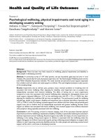
A high-speed burr is then used on the distal tibia, fibula
and bridge to round and bevel any edges (Figure 4). All
periosteal flaps are then carried distally around the bone
bridge as a sleeve, and sutured in position.
The tourniquet is released at this time and all bleeding
points are clamped and ligated or electrocoagulated
appropriately. The peroneal muscles are cut at an
appropriate length and brought medially, where they are
sutured to the deep fascia and periosteum overlying the
anteromedial tibia (Figure 5). A djunct osteobiological
agents may be used i n the bony bridge area at this time;
the authors have used rhBMP-2, platelet rich plasma,
allograft bone, autologous cancellous bone, and c ombi-
nations thereof in various scenarios. Autograft may
also be obtained from the distal stump at this time
(Figure 6). A closed suction drain is then placed superficial
to the peroneal musculature and carried out of the skin on
the anterolateral aspect of the distal stump. The posterior
myocutaneous flap is brought anteriorly, evaluated for
length and trimmed appropriately. The gastrosoleus mus-
cle complex is then beveled posteriorly as n eeded, and
Figure 1 Skin incision marked to create long posterior flap.
Figure 2 Provisional notch created in the distal tibia to receive
the fibular strut.
Figure 3 Fibular strut fitting into the tibial and fibular notches
created by the high-speed burr.
Figure 4 Fibular strut securely sutured in place via bone
tunnels through the fibular strut, distal tibia and fibula.
Taylor and Poka Journal of Orthopaedic Surgery and Research 2011, 6:13
/>Page 2 of 4
rotated anteriorly, where it is sutured into the anterior
muscle compartment, deep anterior fascia, and perios-
teum. Skin flaps are fashioned as necessary for a smooth
closure without tension and s utured together with inter-
rupted nonabsorbable sutures (F igure 7). Any dog-ears
should be trimmed sparingly as to minimize vascular
insults to the remaining skin.
Discussion
The efforts of creating a distal bone bridge and the osteo-
myoplasty does add time and potential morbidity to the
transtibial amputation procedure, but is directed at creat-
ing a more functional and physiological residual extre-
mity. Patient reported outcomes from this procedure are
encouraging and generally higher than that for traditional
transtibial amput ees, with improved rate of return to
work as well as patient-reported outcomes [1,2,7,15].
Indica tions for this procedure include acute trauma as
well as sequelae from tumor, trauma, previous surgery,
and congenital deformities. Although traditional thought
is that diabetic or dysvascular patients should not
undergo this procedure, several reports of these patients
included in larger groups reveal that they can undergo
this procedure successfully but may not perform as well
on functional testing [1,2,4,6,7].
Conclusions
The foot is a very unique end-bearing organ, and the
removal of the distal limb creates several difficulties.
Traditional transtibial amputation creates a smaller and
possible less stable area for weightbearing with sur-
rounding soft tissues that are not designed to resist the
compressive and shearing forces of weightbearing. This
procedure was developed to help create a more
enhanced and physiological weightbearing platform.
Consent
Written informed consent was obtained from the patient
for publication of this report and accompanying images.
A copy of the written consent is available for review by
the Editor-in-Chief of this journal.
Acknowledgements
We would like to thank John Hays, the prosthetist for many of these
patients, for contributing to their care and providing photography for the
technique described above.
Authors’ contributions
BCT was the primary author of the manuscript. AP contributed to the
manuscript and described his technique of amputation. All authors have
read and approved the manuscript.
Competing interests
The authors declare that they have no competing interests.
Received: 28 October 2010 Accepted: 7 March 2011
Published: 7 March 2011
References
1. Condie DN: Electromyography of the lower limb amputee. Medicine and
Sport 1973, 8, Biomechanics 3; 482-488, Karger, Basle.
Figure 5 The peroneal myoplasty is seen in its completed
state, with the optimal resting length and tension of the
muscles restored.
Figure 6 Harvesting cancellous autograft from the removed
aspect of the limb should be considered if the bone is free of
infection and graft is needed.
Figure 7 Final closure wit hout significant tension on wound
edges; suction drain also shown in place.
Taylor and Poka Journal of Orthopaedic Surgery and Research 2011, 6:13
/>Page 3 of 4
2. DeCoster T, Homedan S: Amputation Osteoplasty. Iowa Orthop J 2006,
26:54-9.
3. Deffer PA: Ertl osteoplasty at Valley Forge General Hospital. Amputee
Clinics Newsletter 1969, 1(1):1.
4. Ertl J: Prosthetic Primer: Pain and the Inactive Residual Extremity
Syndrome. [ />5. Ertl J: Über amputationsstümpfe. Chirug 1949, 20:218-224.
6. Ertl JW, Ertl JP, Ertl WJ, Stokosa J: The Ertl Osteomyoplastic Transtibial
Amputation Reconstruction: Description of Technique and Long Term
Results. [ />7. Hansen-Leth C: Muscle blood flow after amputation with special
reference to the influence of osseous plugging of the medullary cavity.
Acta Orthop Scand 1976, 47:613-8.
8. Hansen-Leth C: The vascularization in the amputation stumps of rabbits.
A microangiographic study Acta Orthop Scand 1979, 50:399-406.
9. Hansen-Leth C, Reiman I: Amputations with and without myoplasty on
rabbits with special reference to the vascularization. Acta Orthop Scand
1972, 43:68-77.
10. Langhagel J: Angiographische Untersuchung der Stumpfdurchblutung
bei Beinamputierten. Arbeit und Gesundheit, Georg Thieme Verlag, Stuttgart
1968.
11. Loon HE: Below knee amputation surgery. Artificial Limbs National
Academy of Sciences-National Research Council 1963, 6(2):86.
12. Pinto M, Harris WW: Fibular segment bone bridging in trans-tibial
amputation. Prosthet Orthot Int 2004, 28:220-4.
13. Pinzur MS, Guedes S, Saltzman M, Batista F, Gottschalk F, et al: Health
related quality of life in patients with transtibial amputation and
reconstruction with bone bridging of the distal tibia and fibula. Foot
Ankle Int 2006, 27(11):907-11.
14. Pinzur MS, Smith DG, Guedes S, Smith DG: Controversies in amputation
surgery. Instr Course Lect 2003, 52:445-454.
15. Taylor BC, French B, Poka A, Blint A, Mehta S: Osteomyoplastic and
Traditional Transtibial Amputations in the Trauma Patient: Perioperative
Comparisons and Outcomes. Orthopedics 2010, 33(6):390.
doi:10.1186/1749-799X-6-13
Cite this article as: Taylor and Poka: Osteomyoplastic transtibial
amputation: technique and tips. Journal of Orthopaedic Surgery and
Research 2011 6:13.
Submit your next manuscript to BioMed Central
and take full advantage of:
• Convenient online submission
• Thorough peer review
• No space constraints or color figure charges
• Immediate publication on acceptance
• Inclusion in PubMed, CAS, Scopus and Google Scholar
• Research which is freely available for redistribution
Submit your manuscript at
www.biomedcentral.com/submit
Taylor and Poka Journal of Orthopaedic Surgery and Research 2011, 6:13
/>Page 4 of 4