báo cáo hóa học:" The impact of tensioning device mal-positioning on strand tension during Anterior Cruciate Ligament reconstruction" ppt
Bạn đang xem bản rút gọn của tài liệu. Xem và tải ngay bản đầy đủ của tài liệu tại đây (621 KB, 7 trang )
RESEARC H ARTIC LE Open Access
The impact of tensioning device mal-positioning
on strand tension during Anterior Cruciate
Ligament reconstruction
Rajesh Maharjan
1†
, John J Costi
2†
, Richard M Stanley
2†
, David Martin
3†
, Trevor C Hearn
1†
and John R Field
1*†
Abstract
Background: In order to confer optimal strength and stiffness to the graft in Anterior Cruciate Ligament (ACL)
reconstruction, the maintenance of equal strand tension prior to fixation, is desired; positioning of the tensioning
device can significantly affect strand tension This study aimed to determine the effect of tensioning device mal-
positioning on individual strand tension in simulated cadaveric ACL reconstructions.
Methods: Twenty cadaveric specimens, comprising bovine tibia and tendon harvested from sheep, were used to
simulate ACL reconstruction with a looped four-strand tendon graft. A proprietary tensioning device was used to
tension the graft during tibial component fixation with graft tension recorded using load cells. The effects of the
tensioning device at extreme angles, and in various locking states, was evaluated.
Results: Strand tension varied significantly when the tensioning device was held at extreme angles (p < 0.001) or
in ‘locked’ configurations of the tensioning device (p < 0.046). Tendon position also produced significant effects (p
< 0.016) on the resultant strand tension.
Conclusion: An even distribution of tension among individual graft strands is obtained by maintaining the
tensioning device in an unlocked state, aligned with the longitudinal axis of the tibial tunnel. If the maintenance of
equal strand tension during tibial fixation of grafts is important, close attention must be paid to positioning of the
tensioning device in order to optimize the resultant graft tension and, by implication, the strength and stiffness of
the graft and ultimately, surgical outcome.
Background
Surgeons increasingly favour reconstruction of the ante-
rior cruciate ligament with the multi-strand tendon auto-
graft in preference to bone-patella tendon-bone grafts
(BPTB) because of the relatively low complication rate
[1] and availability of improved fixation methods; equally
tensioned quadrupled hamstring tendon (QHT) grafts
have been shown stronger and stiffer than BPTB grafts
[2-4]; Initial graft tension plays a vital role in maintaining
joint kinematics and in situ forces in the graft during
knee motion [5,6]. The application of excessive int ra-
operative tension can precipitate joint stiffness, the devel-
opment of abnormal stresses on the articular cartilage
and menisci, and which may also interfere with graft
revascularization [7-9]. Conversely, inadequate graft ten-
sion will lead to excessive joint laxity [3]. To maintain
optimum biomechanical properties it appears important
to generate, and maintain, similar tension in all four
strands of the QHT graft at the time of graft tensioning
and tibial fixation [10-12].
Currently, there is no consensus regarding the amount
oftensiontoapplytoagraftwhenitissecured[1].An
initial tension of 44N is considered optimum by some,
but there is no empirical evidence for this argument
[13,14]. Restoration of anterior translation to within 3
mm of the native ACL condition, after cyclic loading,
required approximately 68 N initial tension to be applied
[15]. Graft tensioning has been evaluated in numerous
cadaveric studies [7,15-19], with considerable variation in
graft tension observed between surgeons, prompting the
suggestion that graft tension should be more accurately
* Correspondence:
† Contributed equally
1
Comparative Orthopaedic Research Surgical Facility, School of Medicine,
Flinders University, Bedford Park, 5042, South Australia, Australia
Full list of author information is availabl e at the end of the article
Maharjan et al. Journal of Orthopaedic Surgery and Research 2011, 6:33
/>© 2011 Maharjan et al; licensee BioMed Central Ltd. This is an Open Access article distributed under the terms o f the Creative
Commons Attribution License (http://creativecomm ons.org/lic enses/by/2.0), which permits unrestricted us e, distribution, and
reproduction in any medium, provided the original work is pro perly cited.
measured and contr olled intra-o peratively [17]; Gertel et
al [20] demonstrat ed that the direction of tension ing and
the flexion angle of the knee at which the tension was
applied also plays a significant role in the initial graft
tension.
Various techniques have been used to maintain uniform
tension in all strands of a QHT graft. Bellemans et al [21]
demonstrated the use of one spiked staple to fix the ham-
string tendon to the tibia in order to maintain the appro-
priate tension prior to introduction of an interference
screw. Hamner et al [10] produced equal tension in the
strands by applying weights. Commercially available ten-
sioning devices can reportedly produce and maintain
equal tension in the strands of QHT. In principle, when
the tensioning device is pulled it exerts equal tension in all
of the strands. However, when the tensioning device is
deviated from tha t axis, which may occur while inserting
an interference screw, strand tension may alter. This may
have an adverse impact on the biomechanical properties
of the graft, which in turn may affect the surgical outcome.
This study aimed to quantify the effects, on individual
strand tension and stress, on tensioning device mal-
positioning. The null hypotheses were as follows:
1. Individual strand tensions, during looped four-
strand tendon graft ACL reconstruction, are equal
when using a tensioning device in line with the long-
itudinal axis of the tibial tunnel.
2. Angulation of the tensioning d evice, with respect
to the long axis of the tibial tunnel, will result in
equal strand tension.
3. Locking the tensioning device at extreme angles
will result in equal strand tension.
Methods
Simulation of ACL reconstruction with a looped four-
strand tendon graft was performed using cadaveric
bovine tibiae and sheep superficial digital flexor (SDF)
tendons harvested from skeletally mature individuals.
The utilization of animal-derived tissues was approved
by the Institutional Animal Welfare Committee.
To obtain a study power of 0.8 with an alpha of 0.05,
the required sample size was determined to be n = 20.
To this end 20 cadaveric reconstructions were performed
and tested.
The ACL Tie Tensioner (Mitek, Johnson and Johnson,
USA) was evaluated for its ability to apply reproducible
individual strand tension when positioned as might occur
in clinical practice (Figure 1).
Retrieved tendon strands were whipstitched using No.
1 braided polyester suture (Ethibond, Ethicon, Inc.,
USA); Suture loops were attached to hooks connected
to each load cell. The diameter of the graft composite
was measured by passing it through an incremental
sizing block to achieve a b undled strand diameter of
8.00 mm. The femoral aspect of the graft was stabilized
at the level of the tibial plateau using a circular rod
passed through the centre of the tendon loops and
which rested on the tibial plateau.
Biomechanical tests were performed with an Instron
materials testing system (Instron Pty Ltd, High Wycombe,
UK). Once placed in the testing system, with the tibial
tunnel at zero degrees (vertical), each tendon suture loop
was attached to a 25 kg (223 N) load cell (AL Design Inc,
Buffalo, New York, USA model ALD.75 DIA UTC MINI-
50 lb). All four load cells were then attached to the ten-
sioning device such that each arm supported two tendons
and their accompanying load cells (Figure 2). Load cells
were then balanced before applying tension to the tendon
strands. These were loaded to 150 N in tension for 10
sinusoidal cycles at 0.1 Hz., allowing the tendons to reach
a steady state of hysteresis and reduce the effects of creep
and stress relaxation found in viscoelastic tissue. Once
completed the Instron was kept in load control to main-
tain a tension of 150 N on the tendons.
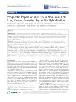
Figure 1 Schematic showing approximate position of the
strand bundle when undergoing tensioning in the various
planes. This figure does not reflect tensioning device ‘locking state’.
Maharjan et al. Journal of Orthopaedic Surgery and Research 2011, 6:33
/>Page 2 of 7
The tibia was then moved to place the tunnel at the
positions described below. At each position, the Instron
load was allowed to return to 150 N. The po sition was
maintained for five seconds before moving to the next
position. This allowed time for the tendons to undergo
creep recovery from their prior location and which also
served as a reference point for the next sequence of
tibial tunnel angulations.
The tensioning device was first evaluated in the
unlocked position (longitudinal alignment with axis of
tibial tunnel) then locked clock wise (CW) followed by
counter-clockwise (C-CW) locking (Figure 3). The tests
were repeated at each of the seven predetermined posi-
tions (tensioning device angle) for each of the locking
states.
The load in each tendon strand and actuator displace-
ment, was recorded for subsequent data analysis. Statis-
tical analysis was performed wit h SPSS (SPSS Inc.,
Illinois, USA). Repeated measures analysis of variance
(ANOVA) was used to evaluate the da ta. The indepe n-
dent variables, tensioning device state (unlocked, locked
clockwise, and locked counterclockwise), ten sioning
device position (7 positions, 01, D30, P30, 02, M30, L30,
03) and tendon position (4 positions; bottom lateral
[BL], top medial [ TM], top lateral [TL] and bottom
medial [BM]) were considered as within-subject factors.
The dependant variable was the tension in each strand.
For all statistical comparisons, a probability level of p <
0.05 was considered significant.
Results
Mean strand tensions for each test are displayed in
Table 1 and presented graphically in Figure 4. These
provide a synopsis of the strand bundle response to
each of the positions adopted and also reflect the ‘ lock-
ing state’ of the tensioning device.
When the tensioning device is utilized in the unlocked
position (aligned with the longitudinal axis of the tun-
nel), the angle at which the tensioning device is held
produces a significant effect (p < 0.0001) on the out-
come measures. Conversely, tendon position does not
produce a significant effect (p = 0.051). The interaction
between tensioning device angle and tendon position is
significant (p < 0.001) with BL significantly greater than
TM at all angles (p < 0.025).
Figure 2 The component arrangement for testing of
reconstructions: The tensioning device is positioned in series with the
reconstruction, four load cells and the load-train of the Instron as
depicted.
Figure 3 Tensioning device locking state:Thearmsofthe
tensioning device are shown in the locked clockwise position with
the central ring firmly pressed against the tensioning tube.
Maharjan et al. Journal of Orthopaedic Surgery and Research 2011, 6:33
/>Page 3 of 7
Table 1 The data displayed represents the mean strand tension (Newtons) ± standard deviation compiled from testing
each reconstruction (n = 20)
Unlocked 01 D30 P30 02 M30 L30 03
Top-medial 36.3 ± 1.4 36.7 ± 2.2 35.6 ± 1.6 36.1 ± 1.9 34.2 ± 1.8 37.7 ± 2.0 35.4 ± 1.7
Top-lateral 36.9 ± 1.6 36.9 ± 2.6 36.1 ± 2.5 37.1 ± 1.8 38.5 ± 2.7 34.6 ± 2.3 37.6 ± 1.7
Bottom-medial 36.8 ± 3.1 36.8 ± 2.7 35.6 ± 1.6 36.8 ± 2.8 38.5 ± 2.9 35.6 ± 2.4 37.8 ± 2.3
Bottom-lateral 38.1 ± 3.0 38.3 ± 2.8 38.8 ± 2.9 37.8 ± 2.9 36.7 ± 2.9 40.0 ± 2.3 37.2 ± 2.6
Locked-clockwise 01 D30 P30 02 M30 L30 03
Top-medial 39.6 ± 1.4 35.6 ± 3.2 38.4 ± 2.1 39.6 ± 1.6 35.8 ± 2.4 39.5 ± 2.2 39.0 ± 1.5
Top-lateral 40.6 ± 2.2 35.9 ± 3.3 38.6 ± 3.7 40.9 ± 1.3 40.7 ± 2.9 36.1 ± 3.3 41.3 ± 1.3
Bottom-medial 33.7 ± 3.0 38.7 ± 5.2 34.8 ± 3.2 33.6 ± 2.5 37.7 ± 3.1 33.1 ± 2.2 34.4 ± 2.3
Bottom-lateral 34.3 ± 3.1 39.2 ± 3.0 35.8 ± 3.5 33.8 ± 2.7 33.4 ± 3.0 39.5 ± 3.4 33.5 ± 2.8
Locked-counterclockwise 01 D30 P30 02 M30 L30 03
Top-medial 33.9 ± 1.9 33.4 ± 2.2 33.4 ± 2.2 33.7 ± 1.8 32.1 ± 2.5 34.9 ± 2.1 33.3 ± 1.6
Top-lateral 34.7 ± 1.9 33.3 ± 2.4 33.5 ± 3.6 35.0 ± 1.4 35.6 ± 2.6 32.1 ± 2.8 35.1 ± 1.5
Bottom-medial 39.5 ± 2.9 40.2 ± 3.3 39.6 ± 2.3 39.6 ± 2.7 41.9 ± 2.8 38.2 ± 2.3 40.2 ± 2.3
Bottom-lateral 40.4 ± 2.6 42.2 ± 2.9 41.5 ± 3.7 40.0 ± 2.1 38.8 ± 3.6 43.5 ± 2.1 39.8 ± 2.5
Testing was performed with the tensioning device held in three locking states (unlocked, locked-clockwise and locked-counterclockwise. Individual strand
response to loading (top-medial, top-lateral, bottom medial and bottom lateral) are recorded at each position (01, 02, 03: neutral; D30, P30: distal or proximal
excursion; M30, L30: lateral or medial excursion).
Figure 4 Strand tension: Graphical representation of individual strand tension (Newtons - N) in response to tensioning device and tendon
position. Standard deviations are not assigned to the figure to reduce its complexity.
Maharjan et al. Journal of Orthopaedic Surgery and Research 2011, 6:33
/>Page 4 of 7
The results of tensioning device locking produced sig-
nificant main effects with tensioning dev ice angle (p <
0.001), locking state ((p < 0.046) and tendon position
(p < 0.016) al l producing significant effects on the resul-
tant strand tension. The interaction between tensioning
device angle and tendon position was significant (p <
0.001) as was locking state and tendon p osition ((p <
0.001) with the interaction between locking state, ten-
sioning device angle and tendon position also produ cing
significant effects (p < 0.001)
Discussion
ACL reconstruction with the looped four-strand tendon
graft has gained popularity. Although clinical outcomes
[4,15,17,18] are similar to BPTB grafting there appear to
be fewer complications with optimal fixation techniques
now available [13,14]. The distribution of tension in all
strands of the graft, an integral factor in its success, is
gaining widesp read attention [15,18]. Due to the compo-
site nature of the graft, it appears essential to apply equal
tension to all the strands during tibial fixation [8,15]. This,
it is suggested, will provide optimal strength and stiffness
to the graft leading to a better surgical outcome [5,6,22]. It
is further proposed that any disparity in the tension
between strands may lead to disproportionate tensile load-
ing and which, may ultimately lead to early rupture of the
strands, weakening the entire reconstruction.
Brown et al [23] evaluated the manual application of
tension to grafts followed by fixation with 4.5 mm corti-
cal screws in combination with plastic, spiked washers.
In order to produce equal tension in all four strands,
suture loops were created from the graft ends; no data
was presented to confirm equality in strand tension.
Hammer et al [10 ], produced equal tension in strands
by applying known weight s. This study showed that
when strands were clamped, they exhibited better tensile
properties. The mean maximum load obtained for four
strand grafts was 2831 ± 538 N when the tension had
been applied manually and 4590 ± 674 N when it had
been applied with a weight. However, tension in indivi-
dual strands was not documented.
The objective in performing this cadaveric study was to
quantify the level of tension applied to all strands of a
looped four-strand tendon graft before tibial fixation.
This was undertaken to investigate the impact of mal-
positioning of the tensioning device on the resultant
strand tension. The analysis was conducted at three neu-
tral positions (01, 02, and 03) and with the tensioning
device helf in various positions (medial and lateral excur-
sion - 30
0
; proximal and distal excursion - 30
0
)and
locking states (Figures 1, 2, and 3).
Our first null hypothesis was shown, in part, to be
correct; strand tensions were not significantly different
when the tensioning device was at the 01 and 02 neutral
positions. However, strand tension differed significantly
at the third neutral position, 03 (Figure 4). One possible
explanation was that this position (03) follow ed medio-
lateral excursion of the tensioning device which may
have indiced residual tendon deformation, altering their
biomechanical behavior.
Our second null hypothesis evaluated the effect of
extreme angulation of the tensioning device, when
deviated to 30° from the neutral position in all four
planes, in the unlocked state (Figure 4). Strand tensions
were recorded at four positions; distal 30 (D30) , proxi-
mal 30 (P30), medial 30 (M30) and lateral 30 (L30).
Minimal variations in strand tension were observed
when data was recorded with e ither proximal or distal
excursion of the strands. A possible interpretation is
that at D30 and P30 the tendons are deviated proximally
and distally from their longitudinal axis, which may have
reduced impact on changes to their biomechanical prop-
erties. The plane of proximal-distal rotation lies closer
to the longitudinal axis of the tunnel and hence the
tendons.
Conversely, strand tension showed a significant differ-
ences when the tensioning device was deviated to M30
and L30 allowing rejection of the second null hypothesis
in this specific situation. A possible explanation is that
at M30 and L30 there is medio-lateral excursion of the
tendons away from their longitudinal axis. Such a varia-
tion, in the direction of load application, may result in
sig nificant structural deformation of the tendons, which
in turn will have a tangible impact on their biomechani-
cal behavior.
We rejected our third null hypothesis in that locking
state of the tensioning device produced a significant
impact on strand tension (Figure 4). The locked coun-
ter-clockwise state showed a greater significant diff er-
ence to its other locked counterpart. A possible reason
could be the shifting of the body of the tensioning
device in relation to its a rms, as occurs during locking;
this may impact on the direction of tension transmission
during tensioning. In the unlocked state, the body of the
device is positioned centrally between the arms. This
arrangement may contribute to a more uniform distri-
bution of strand tension. However, when the device is
locked, the body of the device moves in proximity to
either the proximal or distal end of the arms, depending
on the locking state. Hence, in the clockwise direction,
with the tendons situated proximally, TM and TL may
experience greater tensile force (39.6 and 40.6 N) as the
armsofthedevicemoveawayfromthem.Indistally
positioned tendons, BM and BL, may be subject to les-
ser tension (33.7 and 34.2 N) as the arms move towards
them. Their alig nment with the tunnel axis was alter ed,
and they were displaced distally by the moving arms.
Thus, locking the device causes significant variation in
Maharjan et al. Journal of Orthopaedic Surgery and Research 2011, 6:33
/>Page 5 of 7
strand tension, which may influence the biomechanical
behavior of the graft strands.
Although the strand tensions in the unlocked state were
uniformly distributed, stresses were not equal because
strand cro ss-sectional areas were different. Strand cross-
sectional area had a significant impact on the resultant
stress generated within the strand. Inequality of stresses
may lead to early rupture of the smaller strands as they
bear the greater tension per unit area. A possible solution
may be the harvest of tendons having similar cross-
sectional area. This would allow distribution of stresses
more uniformly across all strands, ultimately providing a
more optimal mechanical environment for the composite
graft.
Their remains no agreement regarding the quanti fica-
tion of tissue viscoelasticity nor reliable modelling [24];
difficulty arises in the delineation of viscoelastic and
pre-conditioning effect s, as both are manifest by similar
response features. The efficacy of anterior cruciate liga-
ment reconstruction, using eithe r QHT or BPTB grafts
is thought to depend on the relative amounts of graft
elongation or creep; hysteresis and creep eff ects appear
highest during the first few loading cycles with more
than 160 cycles required to reach a stead y state, beyond
which there was no further creep and hysteresis almost
constant [25,26]. It appears that the effect of cycli c pre-
conditioning is the progressive recruitment of fibres
[23,26].
Inthecurrentstudywehavearbitrarilychosento
allow a 5 second period of relaxation between tests; this
may lead to conjecture regarding our experimental
methodology and possible impact of creep on the resul-
tant data. It has been shown [27] that contr action dura-
tion significantly affects tendon strain at all levels of
applied force. In response to these findings it is appro-
priate, in order to compare tendon mechanical proper-
ties, that the duration of loading be standardized as it
has been in the current study.
A recent study [28], further complicates the situation
with the suggestion that equal-stress tensioning may
provide an alternative to equal-tension tensioning as
performed in the current study; data derived suggested
that equal-stress tensioning of tendon grafts resisted
graft creep significantly better, raising the issue of the
utilization of graft material having equal cross sectional
areas.
Conclusion
The findings of this study provide useful information for
ACL reconstructive surgery, in which a looped four-
strand tendon graft is utilized. It appears, that the opti-
mal position to induce and maintain uniform strand
tensi on, with a tensioni ng device, is along the longitudi-
nal axis of the tibial tunnel. Any deviation from this
axis, more so in t he medial and lateral planes, appears
to result in a significant variation in strand tension.
Similarly, superior strand tension was obtained by main-
taining the tensioning device in an unlocked state.
This study is a simulation of the human surgical pro-
cedure for graft tensioning. The reconstructions per-
formed in this study, using animal-tissue, do not
therefore provide a completely analogous system for
comparison. However, it does appear that surgeons
should consider closer attention to optimal alignment of
tensioning devices in use; if this is done, a more uniform
distribution of forces may b e generated in the four loop
components of the QHT reconstruction providing aug-
mented mechanical characteristics of t he reconstruction
and, by implication, possibly improve graft longevity and
effectiveness.
Acknowledgements
The authors wish to acknowledge the contribution of David Carney,
Johnson and Johnson, Adelaide, Australia for his, and his company’s support
of this study
Author details
1
Comparative Orthopaedic Research Surgical Facility, School of Medicine,
Flinders University, Bedford Park, 5042, South Australia, Australia.
2
Flinders
Medical Devices and Technologies - Biomechanics and Implants Group,
School of Computer Science, Engineering and Mathematics, Flinders
University, South Australia, Australia.
3
Sportsmed, Stepney, South Australia,
Australia.
Authors’ contributions
Authors contributed variably to the concept, design and performance of this
study. All authors have read and approved the final manuscript.
Competing interests
The authors declare that they have no competing interests.
Received: 10 January 2010 Accepted: 28 June 2011
Published: 28 June 2011
References
1. Amis AA, Jacob RP: Anterior cruciate ligament graft positioning,
tensioning and twisting. Knee Surg sports Traumatol Arthros 1998, 6:s2-s12.
2. Corry IS, Webb JM, Clingeleffer AJ, et al: Arthroscopic reconstruction of
the anterior cruciate ligament. A comparison of patellar tendon
autograft and four-strand hamstring tendon autograft. Am J Sports Med
1999, 27:444-454.
3. Yasuda K, Ichiyama H, Kondo E, et al: An in vivo biomechanical study on
the tension-versus-knee flexion angle curves of two grafts in anatomic
double-bundle anterior cruciate ligament reconstruction. Arthroscopy
2007, 23(8):869-876.
4. Wagner M, Kaab MJ, Schallock J, et al: Hamstring tendon versus patella
tendon anterior cruicate ligament reconstruction using biodegradable
interference fit fixation: a prospective matched-group analysis. Am J
Sports Med 2005, 33(9):1327-1336.
5. Samuelsson K, Andersson D, Jarlsson J: Treatment of anterior cruciate
ligament injuries with special reference to graft type and surgical
technique: an assessment of randomized controlled trials. Arthroscopy
2009, 25(10):1139-1174.
6. Arneja S, McConkey MO, Mulpuri K, et al: Graft tensioning in anterior
cruciate ligament reconstruction: a systematic review of randomized
controlled trials. Arthroscopy 2009, 25(2):200-207.
7. Mae T, Shino K, Nakata K, et al: Optimization of graft fixation at the time
of anterior cruciate ligament reconstruction. Part i: effect of initial
tension. Am J Sports Med 2008, 36(6):1087-10093.
Maharjan et al. Journal of Orthopaedic Surgery and Research 2011, 6:33
/>Page 6 of 7
8. Figueroa D, Calvo R, Vaisman A, et al: Effect of tendon tensioning: an in
vitro study in porcine extensor tendons. Knee 2010, 17(3):245-248.
9. Scheffler SU, Schmidt T, Gangey I, et al: Fresh-frozen free-tendon allografts
versus autografts in anterior ligament reconstruction: delayed
remodelling and inferior mechanical function during long-term healing
in sheep. Arthroscopy 2008, 24(4):448-458.
10. Hammer DL, Brown CH, Steiner ME, et al: Hamstring tendon grafts for
reconstruction of the anterior cruciate ligament: Biomechanical
evaluation of the use of multiple strands and tensioning techniques.
J Bone Joint Surg Am 1999, 81-A(4):549-557.
11. Grover DM, Howell SM, Hull ML: Early tension loss in an anterior cruciate
ligament graft. A cadaver study of four tibial fixation devices. J bone
Joint Surg Am 2005, 87-A(2):381-390.
12. Nurmi JT, Kannus P, Sievanen H, et al: Interference screw fixation of soft
tissue grafts in anterior cruciate ligament reconstruction: Part 2; effect of
preconditioning on graft tension during and after screw insertion. Am J
Sports Med 2004, 32(2):418-424.
13. Fu FH, Bennett CH, Lattermann C: Current trends in Anterior cruciate
ligament reconstruction-Part 1. Am J Sports Med 1999, 27(6):821-830.
14. Fu FH, Bennett CH, Ma CB, Menetrey J, Lattermann C: Current trends in
Anterior cruciate ligament reconstruction-Part l1. Am J Sports Med 2000,
28(1):124-136.
15. Boylan D, Greis PE, West JR, et al: Effects of initial graft tension on knee
stability after ACL reconstruction using hamstring tension: A cadaveric
study. Arthroscopy 2003, 19(7):700-705.
16. Chungfu C, Noorani S, Vercillo F, Woo SL: Tension patterns of the antero-
medial and postero-lateral grafts in double-bundle anterior cruciate
ligament reconstruction. J Orthop Res 2009, 27(7):879-884.
17. Cunningham R, West JR, Greis PE, Burks RT: A survey of the tension
applied to a doubled hamstring tendon graft for reconstruction of the
anterior cruicate ligament. Arthroscopy 2002, 18(9):983-988.
18. Fleming BC, Brady MF, Bradley MP, Banerjee R, Hulstyn MJ, Fadale PD:
Tibiofemoral compression force differences using laxity and force based
initial graft tensioning techniques in the anterior cruciate ligament-
reconstructed cadaveric knee. Arthroscopy 2008, 24(9):1052-1060.
19. Hoshino Y, Kuroda R, Nagamune K, Nishimoto K, Yagi M, Mizuno K,
Yoshiya S, Kurosaka M: The effect of graft tensioning in anatomic 2-
bundle ACL reconstruction on knee joint kinematics. Knee Surg Sports
Traumatol Arthrosc 2007, 15(5):508-514.
20. Gertel TH, Lew WD, Lewis JL, et al
: Effect of anterior cruciate ligament
graft tensioning, direction, magnitude, and flexion angel on knee
biomechanics. Am J Sports Med 1993, 21(4):572-580.
21. Bellemans J, Eid T, Fabry G: A modified technique for tibial interference
screw fixation of hamstring anterior cruciate ligament grafts. Arthroscopy
1999, 15(6):669-671.
22. Labs K, Perka C, Schneider F: The biological and biomechanical effect of
different graft tensioning in anterior cruciate ligament reconstruction:
An experimental study. Arch Orthop Trauma Surg 2002, 122(4):193-199.
23. Brown CH, Hammer D, Hecker AT, et al: Biomechanics of semitendinosus
and gracilis tendon grafts. Sports Medicine, Stockholm 1995, 31-40.
24. Einat R, Yoram L: Recruitment viscoelasticity of the tendon. J Biomech Eng
2009, 131(11):111-118.
25. Simonian PT, Levine RE, Wright TM, et al: Response of hamstring and
patellar tendon grafts for anterior cruciate ligament reconstruction
during cyclic tensile loading. Am J Knee Surg 2000, 13(1):8-12.
26. Schatzmann L, Brunner P, Staubli HU: Effect of cyclic preconsitioning on
the tensile properties of human quadriceps tendons and patellar
ligaments. Knee Surg Sports Traumatol Arthrosc 1998, 6(Suppl 1):S56-61.
27. Pearson SJ, Burgess K, Onambele GN: Creep and the in vivo assessment of
human patellar tendon mechanical properties. Clin Biomech 2007,
22(6):712-717.
28. Conner CS, Morris RP, Vallurupalli S, et al: Tensioning of anterior cruciate
ligament hamstring grafts: comparing equal tension versus equal stress.
Arthroscopy 2008, 24(12):1323-1329.
doi:10.1186/1749-799X-6-33
Cite this article as: Maharjan et al.: The impact of tensioning device
mal-positioning on strand tension during Anterior Cruciate Ligament
reconstruction. Journal of Orthopaedic Surgery and Research 2011 6:33.
Submit your next manuscript to BioMed Central
and take full advantage of:
• Convenient online submission
• Thorough peer review
• No space constraints or color figure charges
• Immediate publication on acceptance
• Inclusion in PubMed, CAS, Scopus and Google Scholar
• Research which is freely available for redistribution
Submit your manuscript at
www.biomedcentral.com/submit
Maharjan et al. Journal of Orthopaedic Surgery and Research 2011, 6:33
/>Page 7 of 7