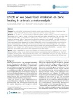
Kwon et al. Journal of Orthopaedic Surgery and Research 2010, 5:4 pdf
Bạn đang xem bản rút gọn của tài liệu. Xem và tải ngay bản đầy đủ của tài liệu tại đây (1014.11 KB, 6 trang )
RESEA R C H ARTIC L E Open Access
Lateral femoral traction pin entry: risk to the
femoral artery and other medial neurovascular
structures
John Y Kwon
1*
, Catherine E Johnson
1
, Paul Appleton
2
, Edward K Rodriguez
2
Abstract
Background: Femoral skeletal traction assists in the reduction and transient stabilization of pelvic, acetabular, hip,
and femoral fractures when splinting is ineffective. Traditional teaching has recommended a medial entry site for
insertion of the traction pin in order to minimize injury to the femoral artery as it passes through Hunter’s canal.
The present anatomical study evaluates the risk to the femoral artery and other medial neurovascular structures
using a lateral entry approach.
Methods: Six embalmed cadavers (twelve femurs) were obtained for dissection. Steinman pins were drilled from
lateral to medial at the level of the superior pole of the patella, at 2 cm, and at 4 cm proximal to this point. Medial
superficial dissection was then performed to identify the saphenous nerve, the superior medial geniculate artery,
the adductor hiatus, the tendinous insertion of the adductor magnus and the femoral artery. Measurements
localizing these anatomic structures relative to the pins were obtained.
Results: The femoral artery was relatively safe and was no closer than 29.6 mm (mean) from any of the three
Steinman pins. The superior medial geniculate artery was the medial structure at most risk.
Conclusions: Lateral femoral traction pin entry is a safe procedure with minimal risk to the saphenous nerve and
femoral artery. Of the structures examined, only the superior medial geniculate artery is at a risk of iatrogenic injury
due to its position. The incidence of such injury in clinical practice and its clinical significance is not known. Lateral
insertion facilitates traction pin placement since it minimizes the need to move the contralateral extremity out of
the way of the drilling equipment or the need to elevate or externally rotate the injured extremity relative to the
contralateral extremity.
Background
Skeletal traction via a femoral or tibial traction pin
assists in the reduction and transient stabilization of
acetabular fractures with or without concomitant hip
dislocation, pelvic vertical shear injuries, foreshortened
femoral shaft fractures, and other pelvic, hip or femur
injuries where splinting is not effective. Placement of a
femoral o r a tibial traction pin involves the risk of liga-
mentous knee injury, intramedullary canal contamina-
tion, vascular and/or nerve injury, intra-articular
contamination, and generation of a stress riser [1,2].
Traditional teaching has recommended a medial entry
site with blunt dissection for insertion of the traction
pin to minimize risk of injury to the femoral artery as it
passes through Hunter’s canal [3]. However, a review of
the literature reveals no anatomic justification for this
practice. In addition, medial entry for traction pin place-
ment can be technically more demanding as the contral-
ateral extremity often blocks drill positioning. This often
requires manipulation of the injured extremity to either
elevate it relative to the contralateral extremity or to
externally rotate it. Alternatively the contralateral extre-
mity needs to be moved out of the way. The objectives
of this anatomical study were to evaluate the risk to the
femoral artery and other medial neurovascular struc-
tures using a lateral pin entry approach , and to evaluate
the optimal position for lateral entry traction pin
placement.
* Correspondence:
1
Harvard Combined Orthopaedic Residency Program, Department of
Orthopaedic Surgery, Massachusetts General Hospital, 55 Fruit Street, Boston,
MA 02114, USA
Kwon et al. Journal of Orthopaedic Surgery and Research 2010, 5:4
/>© 2010 Kwon et al; licensee BioMed Centra l Ltd. This is an Open Access article distributed under the terms of the Creative Commons
Attribution License (http: //creativecommons.org/licenses/by/2.0), which perm its unrestricted use, distri bution, and reproduction in
any medium, provided the original work is properly cited.
Methods
Six embalmed cadavers (twelve femurs) were obtained
for dissection. For each leg, the superior pole of the
patella (SPP) was palpated and the skin marked with a
transverse line. Similar marks were made at 2 cm and 4
cm proximal to the SPP. The knee was held in full
extension with neutral extremity rotation. The distal
femur was palpated laterally for the midline position of
the femur in the anterior/posterior plane. A small skin
incision was made at the midline and a 4 mm Steinman
pin was drilled from lateral to medial exiting the medial
skin. This was repeated at the 2 cm and 4 cm marks
with additional pins (Figure 1).
Medial superficial dissection was then performed and
the saphenous nerve was identified (Figure 2). Measure-
ments of the direct ant erior to posterior distance from
each of the 3 Steinman pins to the saphenous nerve
were obtained. Further dissection was then performed
and the superior medial geniculate artery wa s identified
(Figure 3). Similar measurements were obtained from
each of the 3 pins. Additional dissection was performed
to verify that the pins exited from the mid femur in the
anterior posterior plane and did not skive anterior or
posteriorly.
The adductor hiatus, the tendinous insertion of the
adductor magnus and the femoral artery were then
identified. The area at which the femoral artery crossed
the adductor hiatus (FAAH) was visualized in each case.
Measurements characterizing this anatomic landmark
relativetothepinswereobtained.Theseincludethe
distance (dB) from a line drawn from the SPP Steinman
pin to a n anterior to posterior line extending from the
FAAH, the diagonal distance (dC) from each Steinman
pin to the FAAH, and the anterior-posterior distance
(dA) from each Steinman pin to the femoral artery
(either proximal or distal to the point w here the artery
crosses the adductor hiatus) (Figure 4).
The small amount of cadavers precludes a statistical
analysis or anatomical differences.
Results
Six cadavers w ere dissected for a total of 1 2 femurs.
There were 5 male cadavers and 1 female cadaver.
Cadaver #4 was found to have a right total knee
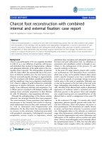
Figure 1 Photograph showing 4 mm Steinman pins inserted at the distal f emur from lateral to medial at the level of the superior
pole of the patella (SPP), at 2 cm, and at 4 cm proximal to SPP.
Kwon et al. Journal of Orthopaedic Surgery and Research 2010, 5:4
/>Page 2 of 6
arthroplasty and the superior medial geniculate artery
was unidentifiable. All other neurovascular structures
were identified in the remaining cadavers.
The mean distance (dB) from the superior pole of the
patella SPP to the anterior to posterior line extending
from the FAAH was 55.5 mm.
The mean anterior to posterior distances from the
SPP pin, the 2 cm pin, and the 4 cm pin t o the saphe-
nous nerve were 36.8 mm, 35.2 mm and 33.8 mm
respectively.
The mean anterior to posterior distances from the
SPP pin, th e 2 cm pin, and the 4 cm pin to the superior
medial geniculate artery were 9.4 mm, 11.5 mm and
12.9 mm respectively.
The mean diagonal distances (dC) from the SPP pin,
the 2 cm pin, and the 4 cm pin to the FAAH diagonally
were 59.8 mm, 44.5 mm and 33.9 mm respectively.
The mean anterior to posterior distance (dA) from the
SPPpin,the2cmpin,andthe4cmpintothefemoral
artery were 35.8 mm, 31.3 mm and 29.6 mm respectively.
Discussion
Femoral skeletal t raction has been used for over a cen-
tury with advent and wide spread use during the World
Wars. The initial use of tongs was improved by Fritz
Steinmann in 1907 who advocated the use of two pins
driven into the femoral condyles [4].
Tradi tional teaching has recommended a medial entry
site with blunt dissection for insertion of the traction
pin due to concerns of iatrogenic injury to the femoral
artery as it passes through Hunter’s canal. Although a
widely accepted technique , there has been no prior ana-
tomic study to justify this practice. This study, to our
knowledge, is the first and only study reported in the lit-
erature addressing this.
The femoral artery is relatively safe when pin place-
ment is performed from a lateral entry point. Instead,
the superior medial genicu late artery is the medial neu-
rovascular structure at most risk with lateral pin entry.
Theaveragedistancesfromapinplacedatthesuperior
patella pole to the superior medial geniculate artery
Figure 2 Cadaveric dissection of the medial knee showing the anatomic location of the saphenous nerve (asterisk) in relation to the 3
Steinman pins.
Kwon et al. Journal of Orthopaedic Surgery and Research 2010, 5:4
/>Page 3 of 6
were 9.4 mm, 11.5 mm from 2 cm proximal, and 12.9
mm from 4 cm proximal. In contrast, the average dis-
tances from a pin placed at the superior patella pole to
the saphenous nerve were 36.8 mm, 35.2 mm from 2
cm proximal, and 33.8 mm from 4 cm proximal. Simi-
larly, the femoral artery had a relatively wide safe zone
with the average distances from a pin placed at the
superior patella pole to the femoral artery being 35.8
mm, 31.3 mm from 2 cm proximal, and 29.6 mm from
4 cm proximal.
We propos e that lateral pin entry for femora l traction
pins is safe. The superior medial geniculate artery is the
structure at most risk of injury, particularly when pin
placement is done at the level of the superior patella
pole. The safe distance from the superior medial genicu-
late artery was increased as the pin was placed more
proximally at 2 cm and 4 cm from the superior patella
pole. While a more proximal pin position could be
advocated to protect the superior medial geniculate
artery, with the safest location for the entry point being
4 cm proximal to the superior patella pole, this may
result in a stress riser through the meta-diaphyseal area
of the femur once the pin is removed. Also, the safe
zone for the femoral artery decreases with more proxi-
mal pin placement. We therefore advocate that pin pla-
cement be made 2 cm proximal to the superior patellar
pole when performing a lateral approach. This will
increase the margin of safety for the superior medial
geniculate artery while preserving a generous safe zone
for the saphenous nerve and the femoral artery.
The incidence of injury to the superior medial genicu-
late artery and its clinical significance during trad itional
femoral traction pin placement is unknown. Ashok
Reddy, et al. demonstrated in a cadave ric study that the
medial femoral condyle is supplied primary by the
superior medial geniculate artery and other lesser
branches from the popliteal artery [5]. While the lateral
femoral condyle enjoys a rich intraosseous supply, the
intraosseo us supply to the medial femoral cond yle
appeared to consist of a single nutrient vessel supplying
Figure 3 Cadaveric dissection of the medial knee showing the anatomic location of the superior medial geniculate artery (asterisk)
and femoral artery passing through the adductor hiatus (arrow head) in relation to the 3 Steinman pins.
Kwon et al. Journal of Orthopaedic Surgery and Research 2010, 5:4
/>Page 4 of 6
the subchondral bone with an apparent watershed a rea
of limited supply. Theoretically i atrogenic injury d uring
traction pin placement could result in avascular necro-
sis. Whether lateral versus medial pin placement offers a
decreased risk to the geniculate artery and the clinical
sequela is unknown.
The authors recognize potential weaknesses in this
study. Our sample size was relatively small, 5 out of 6
cadavers were male, and race was unknown. Inherent to
any cadaveric anatomic study is the applicability of the
data collected across age, sex and race. However, the
anatomic relationships of the various neurovascular
structures studied were consistent enough in the 12
knees dissected that the findings are still useful and may
apply to a larger study population.
Another arguable weakness is the lack of lateral dis-
section. However, this study does not purport to identify
all structures at risk during transcutaneous o sseous
insertion of distal femoral traction pins nor to be an
anatomic study of the distal femur. Injury to the femoral
artery and the potential for devastating vascular injury is
the true rational for traditional medial entry and chal-
lenging this long-held belief was the purpose of our
work. Current practice of medial entry offers protection
to the femoral artery by percutaneous blunt dissection
down to bone medially before inserting the pin disre-
garding the safety of the lateral neurovascular structures
as pins come out bl indly through the lateral side. In our
study we offer the same protection to any lateral struc-
tures that may be at risk when implementing our lateral
insertion technique by using the same technique.
Conclusions
Although our results suggest that lateral pin entry is
safe, one must still use caution. Proper sterile technique
is essential as is careful blunt dissection of soft tissues
prior to pin entry. The femoral midpoint in the ante-
rior-posterior plane should be identified atraumatically
as vigorous pin movement can endanger the lateral vas-
cular structures. The pins should be drilled level with
the extremity in neutral alignm ent as pins directed in a
medial inferior direction may in fact endanger the
femoral artery.
Acknowledgements
None.
Author details
1
Harvard Combined Orthopaedic Residency Program, Department of
Orthopaedic Surgery, Massachusetts General Hospital, 55 Fruit Street, Boston,
MA 02114, USA.
2
Department of Orthopaedic Surgery, Orthopaedic Trauma,
Beth Israel-Deaconess Medical Center, 330 Brookline Avenue, Boston, MA
02215-5491, USA.
Authors’ contributions
JK: performed dissections and data collection and analysis, writing of
manuscript.
CJ: performed dissections and data collection and analysis.
EKR: intellectual contribution, edited manuscript.
PA: intellectual contribution, edited manuscript.
Competing interests
The authors declare that they have no competing interests.
Received: 14 May 2009
Accepted: 22 January 2010 Published: 22 January 2010
Figure 4 Schematic drawing of distances characterizing the position of the femoral artery relative to the pins.Distance(dB)isfrom
superior pole of patella (SPP) to femoral artery crossing the adductor hiatus (FAAH). Distance (dC) is diagonally from each Steinman pin to the
FAAH. Distance (dA) is posteriorly from each Steinman pin to the femoral artery.
Kwon et al. Journal of Orthopaedic Surgery and Research 2010, 5:4
/>Page 5 of 6
References
1. Althausen PL, Hak DK: Lower extremity Traction Pins: Indications,
Technique, and Complications. American Journal of Orthopaedics 2002,
31(1):43-47.
2. Mustard W, Simmons E: Experimental arterial spasm in the lower
extremities produced by traction. J Bone Joint Surg 1953, 35B:437-441.
3. Beaty H, Kasser J: Rockwood and Wilkins’: Fractures in Children. Lippincott
Williams & Wilkinschapter 22:952-953.
4. Peltier L: A Brief History of Traction. J Bone Joint Surg Am 1968,
50:1603-1617.
5. Reddy A, Frederick R: Evaluation of the Intraosseous and Extraosseous
Blood Supply to the Distal Femoral Condyles. American Journal of Sports
Medicine 1998, 26(3):415-419.
doi:10.1186/1749-799X-5-4
Cite this article as: Kwon et al.: Lateral femoral traction pin entry: risk to
the femoral artery and other medial neurovascular structures. Journal of
Orthopaedic Surgery and Research 2010 5:4.
Submit your next manuscript to BioMed Central
and take full advantage of:
• Convenient online submission
• Thorough peer review
• No space constraints or color figure charges
• Immediate publication on acceptance
• Inclusion in PubMed, CAS, Scopus and Google Scholar
• Research which is freely available for redistribution
Submit your manuscript at
www.biomedcentral.com/submit
Kwon et al. Journal of Orthopaedic Surgery and Research 2010, 5:4
/>Page 6 of 6