Lam et al. Journal of Orthopaedic Surgery and Research 2010, 5:18 potx
Bạn đang xem bản rút gọn của tài liệu. Xem và tải ngay bản đầy đủ của tài liệu tại đây (453.7 KB, 9 trang )
RESEARC H ARTIC L E Open Access
Skeletal nutrient vascular adaptation induced by
external oscillatory intramedullary fluid pressure
intervention
Hoyan Lam
1
, Peter Brink
2
, Yi-Xian Qin
1,2*
Abstract
Background: Interstitial fluid flow induced by loading has demonstrated to be an important mediator for
regulating bone mass and morphology. It is shown that the fluid movement generated by the intramedullary
pressure (ImP) provides a source for pressure gradient in bone. Such dynamic ImP may alter the blood flow within
nutrient vessel adjacent to bone and directly connected to the marrow cavity, further initiating nutrient vessel
adaptation. It is hypothesized that oscillatory ImP can mediate the blood flow in the skeletal nutrient vessels and
trigger vasculature remodeling. The objective of this study was then to evaluate the vasculature remodeling
induced by dynamic ImP stimulation as a function of ImP frequency.
Methods: Using an avian model, dynamics physiological fluid ImP (70 mmHg, peak-peak) was applied in the
marrow cavity of the left ulna at either 3 Hz or 30 Hz, 10 minutes/day, 5 days/week for 3 or 4 weeks. The
histomorphometric measurements of the principal nutrient arteries were done to quantify the arterial wall area,
lumen area, wall thickness, and smooth muscle cell layer numbers for comparison.
Results: The preliminary results indicated that the acute cyclic ImP stimuli can significantly enlarge the nutrient
arterial wall area up to 50%, wall thickness up to 20%, and smooth muscle cell layer numbers up to 37%. In
addition, 3-week of acute stimulation was sufficient to alter the arterial structural properties, i.e., increase of arterial
wall area, whereas 4-week of loading showed only minimal changes regardless of the loading frequ ency.
Conclusions: These da ta indicate a potential mechanism in the interrelationship between vasculature adaptation
and applied ImP alteration. Acute ImP could possibly initiate the remodeling in the bone nutrient vasculature,
which may ultimately alter blood supply to bone.
Introduction
Bone mass and morphology accommodates changes in
mechanical demands by regulating the site-specific
remodeling processes which consist of resorption of
bone, typically followed by bone formation. The active
processes of bone remodeling are responsible for bone
turnover, repair, and regeneration [1,2]. Yet, the uncou-
pling of bone formation and resorption can have serious
consequences, as demonstrated by stress fractures in
military recruits and athletes [3-6]. The mechanical
influence on bone adaptation remains a key issue in
determining the etiology of stress injury to bone. From
mechanotransduction point of view, bone remodeling is
regulated by various parameters within the mechanical
milieu, i.e., strain magnitude, frequency, duration, rate,
and cycle number [7,8] Stress injuries were initially
thought to emerge from repetitive vigorous activity,
inducing an accumulation of fatigue microfractures and
resulting in material failure [9]. However, this hypothesis
of repetitive loading related fatigue microdamage as the
sole causative factors for stress injuries has been shown
to be inconsistent based on two key findings: a) the
number of loading cyc les associated with stress fracture
in recruits and athletes are well below the fatigue frac-
ture threshold, and that there is not enough duration
for an accumulation of microdamage to contribute to
material failure within the early onset of stress fractures
[9-11], and b) the stress fracture site tends to occur
* Correspondence:
1
Department of Biomedical Engineering, Stony Brook University,
Bioengineering Building Stony Brook, NY 11794, USA
Lam et al. Journal of Orthopaedic Surgery and Research 2010, 5:18
/>© 2010 Lam et al; licensee B ioMed Central Ltd. This is an Open Access article distributed under the terms of the Creative Commons
Attribution License ( which permits unrestricted use, distribution, and reproduction in
any medium, provided the original work is properly cited .
close to the neutral axis of bending at the mid-diaphysis
rather than the site with maximum strain magnitude
[12,13].
The skeletal vascular system supplies nutrients to and
remove wastes from bone tissue, marrow cavity and
periosteum, in which blood flow is directly coupled with
general status of bone health. The vasculature also regu-
lates intramedulla ry pressure(ImP)viacirculation.The
principal nutrient artery pierces the diaphysis at the
nutrient foramen, penetrates through the cortex and
branch proximally and distally within the medullary cav-
ity to the metaphyseal regions, supplying the inner two-
thirds of the cortex [14,15]. During fracture he aling, the
amount of bone remodeling was significantly reduce d if
intracortical fluid flow, along with blood flow, was pre-
vented [12,13]. There exist a close correlation between
systemic blood pressure and ImP under the normal con-
ditions. In various animal models, the ImP is approxi-
mately ranged 20-30 mmHg, while nearby systemic
blood pressure is about 100-140 mmHg, which is
approximately 4 folds higher than associated ImP
(Table 1). Under the external loading condition, ImP is
increased and/or alternated [13,16-20]. In a rat hindlimb
disuse model, increased ImP by 68% via femoral vein
ligation could significant increase the femoral bone
mineral content and trabecular density [21]. Others
have shown that increasing pressure gradient within the
vasculature can induce new bone formation at the peri-
osteal, endosteal and trabecular surfaces [14]. External
skeletal muscle contraction can substantially increase
ImP and subsequently enhance bone adaptation, even in
adisusemodel[18,22-24].Mechanicalintervention
through vibratory knee joint l oading can trigger bone
formation. These experiments have evidently verified the
critical role of the change in fluid pressure within the
marrow cavity and the s keletal vasculature on bone
adaptation [14,25] (Table 2).
Skeletal vasculat ure remodeling is critical for main-
taining adequate tissue perfusion and is responsible for
regulating interstitial fluid pressure. Arterial adaptation
is often associated with hypertrophy of th e vessel, redis-
tribution of t he extracellular mat rix and smooth muscle
cells (SMCs) [26,27]. The tunica media is the thickest
layer in nutrient artery, which comprises of layers of
SMCs embedded in a network of connect ive tissue. This
layer provides tensile strength, elasticity and contractility
to the vessel [28]. Its structure and morphology also
play a critical role in maintaining blood pressure [28,29].
In human hypertension, histological analyses showed
that there is a greater media/lumen ratio in untreated
hypertensi ve subjects [30]. Th e greater media/lume n
ratio is a result of either higher vessel wall area and/or
smaller lumen area, or both.
It is hypothesized that bone fluid flow induced by ImP
can regulate the nutrient arterial adaptation. Thus, the
objective of this study is to evaluate nutrient vessel
remodeling under dynamic stimulation by evaluating the
morphologic changes on the nutrient arterial wall with
increased mechanical-induced ImP, and to discuss their
potential role in regulating fluid flow through nutrient
vessels,
Methods
Animals and Experimental Preparations
All surgical and experimental protocols were approved
by the University’s Lab Animal Use Committee. The
surg ical protocol was previously described and modified
slightly for this study [13,19]. In brief, under isoflurane
anesthesia, surgical procedures were perfor med on bot h
left and right ulnae of twenty-nine adult skeletally
mature male turkeys. For the left ulna, a 3-mm diameter
hole was drilled and tapped through the cortex of the
dorsal side, appro ximately 2 cm from the proximal end .
A specially designed fluid loading device, with an inter-
nal fluid chamber approximately 0.6 cm
3
, was inserted
into the bone with an O-ring seal to prevent leakage.
The fluid loading device was attached to a surgical plas-
tic tube with an inner diameter approximately 2 mm
wide and 12 cm in length, filled with saline as external
oscillatory loading fluid. A diaphragm was placed in the
center of the fluid chamber, separating the internal mar-
row from external oscillatory loading fluid. The bone
marrow and external flow media were completely
Table 1 Blood pressure and nearby ImP
Animal Blood Pressure (mmHg) ImP (mmHg)
Dog [23,33,50] 110-140 Femoral, Carotid arteries 17-40 Femoral diaphysis and metaphysic (mean)
Rabbit [23] ~120 Carotid artery 20-20 Femoral diaphysis (mean)
Rat [16,18] 20-30 Femoral arteries 5 Femoral marrow (peak-peak)
Turkey [19] 40-80 Ulna and femoral arteries 15-25 Ulna and femoral marrow (peak-peak)
Table 2 ImP induced by mechanical stimulation
Animal Location Type of loading ImP (mmHg)
(peak-peak)
Turkey [7,13,19] ulna ~600 με axial 90-150
Rat [20] femur Venous ligation 60
Rat [16,18] femur Muscle stimulation 40
Rat [2] femur Knee loading 22
Lam et al. Journal of Orthopaedic Surgery and Research 2010, 5:18
/>Page 2 of 9
isolated from one another to avoid co ntamination and
infection. The plastic tube extended through the skin
and coupled the fluid loading device to the oscillatory
loadingunit.Theexternalportionofthedevicewas
flushed and cleaned each day, while antibiotic cream
was applied to the surrounding tissue to furthe r prevent
infection. The contra-lateral right ulnae served as sham
control. With similar surgical procedures as the left, a
3-mm diameter hole was drilled and tapped at the prox-
imal end o f the right cortex. A titanium screw with an
O-ring was used in replacement of the fluid loading
device.
Additional four turkeys were sacrificed at the end of
the experiment without undergo any surgical operation.
These age-matched controls were needed to examine
the handedness, if any, between the left and right ulnae.
Dynamic Fluid Flow Stimulation
The loading system was calibrated based on previous
study 13. In brief , with the same surgical procedure as
above, an additional tube was connected at the distal
end of the ulnae, where a 50-psi pressure transducer
(Entran EPX-101W) was placed into the marrow cav-
ity. The ImP was measured within the physiological
magnitude of 10-180 mmHg and at a range of fre-
quency, 1-40 Hz. A standard graph of marrow pressure
at different frequencies was generated and was used to
calibrate the loading system.
After surgery, the animals were monitored closely dur-
ing normal activities. Fluid pressure stimulation began
on the second day subsequent to the surgery. A sinusoi-
dal fluid pressure was applied to the marrow cavity of
the left ulna through an external fluid oscillatory loading
unit. The loading unit was controlled to generate
changes in the fluid pressure within the intramedullary
canal, by varying magnitudes and frequencies. Based on
the calibration data, the pressure magnitudes applied
were between 50 mmHg to 90 mmHg, which have
shown to be under physiological range [7,13,19]. The
sinusoidal ImP was applie d for 10 minutes per day,
5 days per week, at 3 Hz for 3-weeks (n = 7), 3 Hz for
4-weeks (n = 5), 30 Hz for 3-weeks (n = 6), and 30 Hz
for 4-weeks (n = 11).
Histomorphometry Analyses
Immediately after the animals were sacrificed, the prin-
ciple nutrient arteries from both left and right ulnae
were loca ted. Under the mi croscope, the nutrie nt
arteries were carefully dissected starting from the lumen
with approximately 8 mm in length, and fixed in 10%
formalin solution immediately. The adaptive responses
of the arteries were analyzed through a standard soft tis-
sue histology procedure. The fixed arteries were
embedded in paraffin wax. In order to obtain arterial
cross sections, each vessel was oriented so that it was
straight and perpendicular to the cutting surface. The
paraffin blocks were then sectioned transversely to pro-
duce 8 μm thin slices (RM2165 Microtome, Leica, IL).
Each section was stained with hematoxin and eosin
(H&E, Polyscien ce, PA), dehydrated with a series of
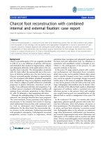
ethanol and cleared with xylene. A representative H&E
stained cross sectional nutrient artery i mage is shown in
Figure 1.
The arterial wall area, lumen area, and wall thickness
of each section were measured using OsteoMeasure
(Version 2.2, SciMeasure, GA) by manually contouring
the inner and o uter boundaries of the tunica media
layer of the nutrient artery. Six cross-sections were ana-
lyzed for each nutrient artery. The full cross section of
the nutrient artery was viewed under a digitized micro-
scope at 40× magnification.
The SMC layer numbers were assessed via sector ana-
lysis, in which the arterial cross-section was divided into
six equal sect ors. An image was captured at each sector
using an inverted microscope (Zeiss, Ax ioVision 4, Ger-
many) under 200× magnification. The numbers of
SMCs were quantified by drawing a line across the
arterial wall, perpendicular to SMC stretch, and count-
ing the nucleus along the line. All the analyses were per-
formedbyasingleoperatortoensureconsistencyof
measurements.
Statistical Analyses
Statistical Analysis System (SAS, Cary, NC) software was
used for data analyses. Experimental data is expressed as
means ± standard error (SE) of each group. The non-
parametric Wilcoxon test was performed and
Figure 1 A representative cross-sectional image of a nutrient
artery from turkey ulna. TM, tunica media; L, lumen; E, endothelial
cells; SMC, smooth muscle cells. The scale bar at the bottom left
corner is 100 μm.
Lam et al. Journal of Orthopaedic Surgery and Research 2010, 5:18
/>Page 3 of 9
sig nificance lev el was considered at p < 0.05. Data from
each histomorphometric parameter was compared in
two ways: (1) each stimulated group was compared to
the average of all controls (age-matched controls and
sham) to show the effect of ImP on the nutrient arterial
morphology, and (2) the ImP stimulated groups were
compared between the various loading regimes to
demonstrate the importance of the loading parameters.
Results
Nutrient Arterial Wall Area
The tunica media of the nutrient arteries demonstrated
up to a 50% increase in area when subjected to ImP sti-
mulation (Figure 2). It is important to point out that
nutrient arteries from the age-matched animals showed
an average of 4% natural differences in arterial wall area
between the left and the right ulnae (0.121 ± 0.01 mm
2
vs. 0.126 ± 0.015 mm
2
), demonstrating minimal left and
right handedness in turkeys. There was no significant
difference between the age-matched controls and sham
controls. Thus, the average o f the age-matched and
sham arterial wall area was calculated to serve as an
overall control and compared that to each experimental
wall area. The cross sectional arterial wall area for load-
ing conditions at 3 Hz and 30 Hz for 3-weeks were
0.199 ± 0.2 mm
2
and 0.207 ± 0.3 mm
2
,whichweresig-
nificantly increased by 46% (p < 0.01) and 51% (p <
0.05), respectively, comparing to the control (0.136 ±
0.05 mm
2
). Furthermore, comparisons between experi-
men tal groups showed significant changes between ImP
loadings applied at 30 Hz for 3-weeks versus 4-weeks
and 3 Hz, 3-weeks vs. 30 Hz, 4-weeks (p < 0.05)
(Figure 2). There was no significant change between
experimental and control in 4-weeks loading for both
3 Hz and 30 Hz.
Nutrient Arterial Lumen Area
Similar to the arterial wall area, the difference in lumen
area between the left and right ulnae in the age-matched
animals was approximately 4%. The age-matched and
sham data were pooled and compa red to experimental
groups. Fluid loadings for 3-weeks showed 14% and 3%
increase in cross sectional lumen area at 3 Hz and 30
Hz stimulations when compared to controls, yielding
lumen area of 0.045 ± 0.01 mm
2
and 0.039 ± 0.01 mm
2
,
respectively (Figure 3). Yet, loadings for 4-weeks showed
decrease in lumen area at both frequencies, yielding area
of 0.027 ± 0.007 mm
2
for 3 Hz (-28%) and 0.022 ± 0.007
mm
2
for 30 Hz (-42%). Though the trends in lumen
area changes were seen between 3-weeks and 4-weeks
stimulations, no statistical significance was found within
and between groups due to the large variabilit y within
the samples.
Nutrient Arterial Wall Thickness
The thickness of the tunica media increased for all load-
ing conditions, ranging from 4-28% (Figure 4). The
arterial wall thickness after 3-weeks stimul ation at 3 Hz
was 146 ± 11 μm significantly increased by 28% from
the control, 113 ± 4 μm (p < 0.05). Although the per-
centage changes in thickness wa s not as great as those
of area, it suggested that augment in thickness might
0
0.05
0.1
0.15
0.2
0.25
0.3
Controls 3Hz, 3w k s 3Hz, 4w k s 30Hz, 3w k s 30Hz, 4w k s
Loading Conditions
Arterial Wall Area (mm^2)
* *
***
Figure 2 Arterial wall area histomorphological analysis of the nutrient arteries, subjected to 3 Hz or 30 Hz ImP stimulation, 10
minutes/day, 5 days/week for 3-week or 4-week. Comparison between the experimental arteries and the pooled average of age-matched
and sham controls. Values are mean ± SE. Significant difference between the ImP stimulated nutrient artery and pooled controls (* p < 0.05 & **
p < 0.01).
Lam et al. Journal of Orthopaedic Surgery and Research 2010, 5:18
/>Page 4 of 9
partially contribute to the arterial wall area changes.
Despite there was no significant difference between the
sham and age-matched controls, the sham thickness
value for the animals subjected to 3-weeks ImP loading
at 30 Hz (100 ± 7 μm) was smaller than other controls
(116 ± 7 μm) (P > 0.5). Thus, when compared to its
sham operated arteries, the 30 Hz stimulation induced
an 18% augmentation at the wall thickness. Lastly, ImP
stimulation at 3 Hz and 30 Hz for 4-weeks showed a
slight increase (9% and 6%, respectively) (P > 0.5), which
may be due to the reduction of the lumen area seen in
Figure 3.
SMC Layer Numbers
The SMC layer numbers for each nutrient artery were
quantified. The ImP stimulation at 3 Hz and at 30 Hz
for 3-weeks showed a sig nificant 25% and 22% increase,
respectively, in SMC layer numbers when compare
to the controls (25% and 22%, respectively, p < 0.01)
(Figure 5). As mentioned before, arterial wall area and
0
0.01
0.02
0.03
0.04
0.05
0.06
Controls 3Hz, 3w k s 3Hz, 4w k s 30Hz, 3w k s 30Hz, 4w k s
Loading Conditions
Arterial Lumen Area (mm^2)
Figure 3 Arterial lumen area histomorphological analysis of the nutrient arteries subjected to 3 Hz or 30 Hz ImP stimulation, 10
minutes/day, 5 days/week for 3-week or 4-week. Comparison between the experimental arteries and the pooled average of age-matched
and sham controls. Values are mean ± SE.
60
80
100
120
140
160
180
Contr ols 3Hz, 3w k s 3Hz, 4w k s 30Hz, 3w k s 30Hz, 4w k s
Loading Conditions
Arterial Wall Thickness (um)
*
Figure 4 Arterial wall thickness histomorphological analy sis of the nutrient arteries subjected to 3 Hz or 30 Hz ImP stimulation, 10
minutes/day, 5 days/week for 3-week or 4-week. Comparison between the experimental arteries and the pooled average of age-matched
and sham controls. Mean ± SE (* p < 0.05).
Lam et al. Journal of Orthopaedic Surgery and Research 2010, 5:18
/>Page 5 of 9
thickness were also augmented after ImP loading for
3-weeks. It is highly possible that such increase in SMC
layer numbers was responsible for the alteration in
arterial morphometry. No significance was observed for
the 4-weeks stimulation. Further, comparisons between
experimental groups showed significant changes
between fluid loadings applied at 3 Hz for 3-weeks ver-
sus 30 Hz for 4-weeks (p < 0.05) (Figure 5).
Discussion
The objective of this study was to examine how intra-
medullary pressure influenced nutrient artery remodel-
ing. Previous experiments have implied that bone fluid
flow is a mediator involved in bone remodeling by influ-
encing bone cell activities through improper nutrient
transport [7,13,19]. However, the mechanism in which
bone fluid flow can lead to changes in nutrient transport
is not clearly characterized. In this study, the morpholo-
gical analyses of the nutrient arterial wall demonstrated
that bone fluid flow induced by daily cyclic ImP stimula-
tions has the potential to initiate nutrient artery remo-
deling, which may ultimately alter the blood supply to
bone and ultimately affect the bone remodeling
processes.
Previous in vivo studies have demonstrated repetitive
mechanical loading generated ImP can alter interstitial
fluid flow and initiate bone remodeling [7,13,19,31]. It is
hypothesized that these ImP seriously impact bone fluid
flow by collapsing the nutrient artery at the peak of
each loading cycle, decreasing normal blood flow into
the marrow cavity. Augmentation of femoral marrow
pressure and interstitial fluid flow, induced by femoral
vein ligation, could significantly influence bone quantity
under functional disuse condition [20]. On the contrary,
blockage of arterial supply led to the reduction of the
nutrients and oxygen to bone [32,33]. The changes in
oxygen and carbon dioxide levels in bone callus and
bone necrosis, strengthening the idea that arterial occlu-
sion can deplete nutrient supplies required for bone
cells activities [34,35]. However , the maximal ImP value
response to the impact loading in our previous avian
model was 150 mmHg (~20 KPa ). Compared to the
load-generated solid phase matrix stress and strain, e.g.,
10-100 MPa by 1,000 με, and the estimated fluid p res-
sure at the value of 3 MPa [36], the applied direct ImP
(50-90 mmHg) is in the p hysiological range. Such a
small value of ImP will not collapse the vessels.
Mechanical forces related to the velocity of arterial
blood flow have been shown to be important determi-
nants of arterial structural changes [26,37,38]. Several
experiments on the ligation of rat mesenteric bed had
shown a 90% reduction of blood flow in the upstream
arteries. This decrease in blood flow resulted in a 21%
and 37% reduction in lumen diameter and vessel wall
area [39,40]. Conversely, arteries exposed to over 100%
in blood flow showed a marked elevation in lumen dia-
meter (38%) and arterial wall area (58%) [39,40]. Thus,
the 50% enlargement of the nutrient arterial wall area
observed in this study (Figure 2) may be the result of
the increase blood flow via the ImP stimulation.
4
5
6
7
8
9
10
11
12
13
Contr ols 3Hz, 3w k s 3Hz, 4w k s 30Hz, 3w k s 30Hz, 4w k s
Loading Conditions
SMC layer numbers
** *
**
Figure 5 SMC layer numbers analysis of the nutrient arteries subjected to 3 Hz or 30 Hz ImP stimulation, 10 minutes/day, 5 days/
week for 3-week or 4-week. Comparison between the experimental arteries and the pooled average of age-matched and sham controls. Mean
± SE (* p < 0.05 & ** p < 0.01).
Lam et al. Journal of Orthopaedic Surgery and Research 2010, 5:18
/>Page 6 of 9
Axial loading, e.g. generating peak 600 με, can amplify
ImP 10-fold above the arterial pressure, e.g., from 18
mmHg to 150 mmHg, driving blood flow through the
nutrient artery [12,13]. In a functional disuse model,
bone loss was observed via the thinning of the cortex
due to endosteal resorp tion and an increase in intracor-
tical porosity. However, when external oscillatory fluid
flow was a pplied to the marrow cavity, bone mass was
significantly improved at the mid-diaphysis due to both
periosteal and endosteal new bone formation [13]. This
data clearly illustrated the effects of anabolic fluid flow
in bone adaptation, which was capable of maintaining
bone mass and likely to inhibit bone loss due to disuse.
In this study, the fluid magnitudes (50 mmHg to
90 mmHg) for the cyclic hydraulic stimulation a pplied
to the marrow cavity were imposed at physiological
levels. The frequencies were chosen to mimic the num-
ber of loading c ycles relevant to physiological level and
to a military training regimen, i.e., 3 Hz for 10 minutes
provide 1,800 loading cycles and 30 Hz for 10 minutes
provides 18,000 cycles. The ImP due to circulation in
the turkey is approxima tely 18 mmHg and previous
experiments have shown that ImP of 76 mmHg was suf-
ficient to generate bone remodeling at 30 Hz [7,13]. The
loading rate sensitivity of bone remodeling was also
shown in recent disuse model under dynamic muscle
contraction [16,18]. The duration of the experiments
was also c hosen based on previous studies stating that
the risk of stress fractures occur at the early onset of
training, with the rate of occurrence generally elevated
by the third week of training [41].
Flu id loadings at 3 Hz and at 30 Hz for 3-weeks have
generated the greatest changes in nutrient arterial wall
area. This strongly implied that the duration of loading
plays an important role in vessel remodeling; it is clear
that 3-weeks of cyclic ImP stimulation was sufficient to
initiate vessel wall remodeling with increase wall area,
lumen area, wall thickness, and SMC layer number.
Four-weeks of cyclic ImP stimulation are enough to trig-
ger bone adaptation [13,16,18]. Together with the obser-
vations obtained from this study, where 3-weeks fluid
loading resulted in the most morphologica l changes in
the nutrient arteries, the results implied that the nutri-
ent arteries adapt to the altered ImP precede and/or
occur concurrently with the bone remodeling process.
Hence, there is a strong implication that the adaptation
of the nutrient arteries may serve as a critical mediator
between bone fluid flow and bone remodeling.
Acute vasculature adaptation is impaired by endothe-
lial pressure hypercholesterolemia (such as flow-
mediated dilatation) and fluid wall shear stress. Previous
works indicated that increased vascular flow results in
adaptive vessel remodeling as dependant on applied
shear stress [29,42]. Morphological changes occur
rapidly following flow alteration and do not require
chronic insult to affect substantial and significant struc-
tural transformation [29]. The results from this study
indicated that 3 weeks ImP can significantly change the
nutrient artery morphology, but such effects were atte-
nuated in the 4 weeks stimulation, which may imply
that vascular morphology change is sensitive to the
duration of dynamic fluid stimulation. However, this
result could not be overly interpreted based on the
small number of samples. Overall, based on previous
study on vessel ligation effects on bone adapt ation [20],
such small percenta ge of vessel wall cha nges in the
nutrient vessel may not significantly affect the blood
flow in bone. Nevertheless, further study will be needed
to explore this mechanism.
SMCs are exposed to wall shear stress via the trans-
mural pressure gradient [34,43]. It has been proposed
that blood pressure affects transmural flow and able to
regulate the normal cellular activities of SMCs, i.e., prolif-
eration and migration [34,43-46]. Future studies will
focus the relationship between the changes in mechanical
environment due to ImP oscillations and the cellular
responses of SMC, such as the coupling process of prolif-
eration and apoptosis. Oscillatory shear stress has been
shown to increase smooth muscle cell proliferation via
protein kinase B phosphorylation and activate various
signal transduction pathways [34,43-46]. Hypertensive
rats model have demonstrated that reductions of blood
flow augmented vessel wall hypertrophy via mechanisms
that enhance SMC proliferation i n the media a nd the
intima [47]. While others have shown SMCs in mesen-
teric resistance arteries c an undergo cel l death in both
low flow and high flow conditions [26]. These studies
indicated that proliferationandapoptosisofSMCsmay
be involved in the remodeling of the nutrient artery.
Other potential SMC mechanisms related to the mor-
phological changes seen in vascular adaptation are the
change in size and arrangement of existing SMCs [26]
However, there is considerable controversy regarding
SMC hypertrophy and hyperplasia (increase in cell num-
ber such as via cell proliferation) in medial thickening of
hypertensive models. Some studies have concluded med-
ial thickening is due to SMC hyperplasia based on the
observations of increased DNA content and numbers of
SMCs [48]. While other studies have assessed cellular
hypertrophy via morphometric estimation of cell size in
tissue sections and measurements of protein to cell
ratios, concluded medial hypertrophy is due to enlarge-
ment of existing SMCs [49].
Lastly, arterial morphological changes may also be a
result of the changes in connect ive tissues. In hyperten-
sive rats, results have shown a higher content of elastin
in the arterial wall and an increase in polar amino acids
content in elastin, which suggested that the material
Lam et al. Journal of Orthopaedic Surgery and Research 2010, 5:18
/>Page 7 of 9
properti es of the artery is altered due to the continuous
physical stress that placed on the vessel from high blood
pressure and increase of peripheral resistance [28,29].
Likewise for collagen, both the quantitative and qualita-
tive changes were determined. Many experiments have
shown the stimulation of collagen synthesis and the
increase of collagen content in the arterial wall in hyper-
tension [28,29,48]. In order to fully understand the pro-
cesses of vascular remodeling, the above mechanisms
are important for future studies.
Conclusions
The adaptive response in the nutrient arteries was inves-
tigated via our avian model which can induce oscillatory
fluid flow in the absence of bone matrix deformation.
Bone fluid flow induced by ImP is a critical mediator for
bone remodeling, possibly through altering blood supply
to bone and disrupting the nutrient transport process.
Stress fractures were often observed in young populations
who had experienced high intensity physical training, i.e.,
athletes and military recruits. These data suggest that
repetitive cyclic loading may trigger arterial wall enlarge-
ment, which may potentially reduce the fluid supply to
bone and further generate local ischemia. Three-weeks of
ImP stimulation was sufficient to increase arterial wall
area, lumen area, wall thickness, and SMC layer numbers.
The mechanical signals generated from ImP may ulti-
mately initiate a cascade of cellular responses via
mechanotransduction, influencing cellular activities within
the arterial wall. With the strong interactions between
blood flow and bone remodeling, it is highly suggestive
that bone fluid flow has a potential to contribute to stress
injuries to bone via an ongoing repair process.
Acknowledgements
This work is kindly supported by National Institute of Health (NIAMS
AR52379 & AR49286) and US Army Medical Research and Materiels
Command (DAMD-17-02-0218 and W81XWH-07-1-0337). The authors are
appreciative to Dr. C. Rubin for valuable suggestion.
Author details
1
Department of Biomedical Engineering, Stony Brook University,
Bioengineering Building Stony Brook, NY 11794, USA.
2
Department of
Physiology and Biophysics, Stony Brook University Basic Sciences, Building
Stony Brook, NY 11794, USA.
Authors’ contributions
YXQ was the principle investigator who designed the overall study and
carried out the surgical procedure. HL assisted during surgical procedure,
participated in the daily stimulation, performed tissue and statistical analyses,
and drafted the manuscript. PB provided suggestions on vessel physiology
and biology analyses. All authors read and approved the final manuscript.
Competing interests
The authors declare that they have no competing interests.
Received: 4 July 2009 Accepted: 11 March 2010
Published: 11 March 2010
References
1. Rubin C, Judex S, Qin YX: Low-level mechanical signals and their
potential as a non-pharmacological intervention for osteoporosis. Age
Ageing 2006, 35(Suppl 2):ii32-ii36.
2. Zhang P, Su M, Liu Y, Hsu A, Yokota H: Knee loading dynamically alters
intramedullary pressure in mouse femora. Bone 2007, 40:538-543.
3. Evans RK, Antczak AJ, Lester M, Yanovich R, Israeli E, Moran DS: Effects of a
4-month recruit training program on markers of bone metabolism. Med
Sci Sports Exerc 2008, 40:S660-S670.
4. Friedl KE, Evans RK, Moran DS: Stress fracture and military medical
readiness: bridging basic and applied research. Med Sci Sports Exerc 2008,
40:S609-S622.
5. Iwamoto J, Takeda T: Stress fractures in athletes: review of 196 cases.
J Orthop Sci 2003, 8:273-278.
6. Moran DS, Israeli E, Evans RK, Yanovich R, Constantini N, Shabshin N, et al:
Prediction model for stress fracture in young female recruits during
basic training. Med Sci Sports Exerc 2008, 40:S636-S644.
7. Qin YX, Rubin CT, McLeod KJ: Nonlinear dependence of loading intensity
and cycle number in the maintenance of bone mass and morphology.
J Orthop Res 1998, 16:482-489.
8. Srinivasan S, Weimer DA, Agans SC, Bain SD, Gross TS: Low-magnitude
mechanical loading becomes osteogenic when rest is inserted between
each load cycle. J Bone Miner Res 2002, 17:1613-1620.
9. Jones BH, Harris JM, Vinh TN, Rubin C: Exercise-induced stress fractures
and stress reactions of bone: epidemiology, etiology, and classification.
Exerc Sport Sci Rev 1989, 17:379-422.
10. Burr DB, Milgrom C, Fyhrie D, Forwood M, Nyska M, Finestone A, et al: In
vivo measurement of human tibial strains during vigorous activity. Bone
1996, 18:405-410.
11. Carter DR: The relationship between in vivo strains and cortical bone
remodeling. Crit Rev Biomed Eng 1982, 8:1-28.
12. Otter MW, Qin YX, Rubin CT, McLeod KJ: Does bone perfusion/reperfusion
initiate bone remodeling and the stress fracture syndrome? Med
Hypotheses 1999, 53:363-368.
13. Qin YX, Kaplan T, Saldanha A, Rubin C: Fluid pressure gradients, arising
from oscillations in intramedullary pressure, are correlated with the
formation of bone and inhibition of intracortical porosity. J Biomech
2003, 36:1427-1437.
14. Brookes M, Revell W: Blood Supply of Bone: Scientific Aspects. Springer,
New York 1998.
15. Oni OO: The microvascular anatomy of the physis as revealed by
osteomedullography and correlated histology. Orthopedics 1999,
22:239-241.
16. Lam H, Qin YX: The effects of frequency-dependent dynamic muscle
stimulation on inhibition of trabecular bone loss in a disuse model. Bone
2008, 43:1093-1100.
17. McAllister TN, Du T, Frangos JA: Fluid shear stress stimulates
prostaglandin and nitric oxide release in bone marrow-derived
preosteoclast-like cells. Biochem Biophys Res Commun 2000, 270:643-648.
18. Qin YX, Lam H: Intramedullary pressure and matrix strain induced by
oscillatory skeletal muscle stimulation and its potential in adaptation. J
Biomech 2009, 42:140-145.
19. Qin YX, Lin W, Rubin C: The pathway of bone fluid flow as defined by in
vivo intramedullary pressure and streaming potential measurements.
Ann Biomed Eng 2002, 30:693-702.
20. Stevens HY, Meays DR, Frangos JA: Pressure gradients and transport in
the murine femur upon hindlimb suspension. Bone 2006, 39:565-572.
21. Bergula AP, Haidekker MA, Huang W, Stevens HY, Frangos JA: Venous
ligation-mediated bone adaptation is NOS 3 dependent. Bone 2004,
34:562-569.
22. Shim SS, Copp DH, Patterson FP: Measurement of the rate and
distribution of the nutrient and other arterial blood supply in long
bones of the rabbit. A study of the relative contribution of the three
arterial systems. J Bone Joint Surg Br 1968, 50:178-183.
23. Shim SS, Hawk HE, Yu WY: The relationship between blood flow and
marrow cavity pressure of bone. Surg Gynecol Obstet 1972, 135:353-360.
24. Shim SS: Bone and joint circulation. Physiological basis for clinical
practice. Yonsei Med J 1986, 27:91-99.
25. Oni OO: Acute vascular response to intramedullary reaming. J Bone Joint
Surg Br 1995, 77:981-982.
Lam et al. Journal of Orthopaedic Surgery and Research 2010, 5:18
/>Page 8 of 9
26. Buus CL, Pourageaud F, Fazzi GE, Janssen G, Mulvany MJ, De Mey JG:
Smooth muscle cell changes during flow-related remodeling of rat
mesenteric resistance arteries. Circ Res 2001, 89 :180-186.
27. Wentzel JJ, Gijsen FJ, Schuurbiers JC, Steen van der AF, Serruys PW: The
influence of shear stress on in-stent restenosis and thrombosis.
EuroIntervention 2008, 4(Suppl C):C27-C32.
28. Mulvany MJ: Determinants of vascular hemodynamic characteristics.
Hypertension 1984, 6:III13-III18.
29. Mulvany MJ: Small artery remodelling in hypertension: causes,
consequences and therapeutic implications. Med Biol Eng Comput 2008,
46:461-467.
30. Lee RM, Owens GK, Scott-Burden T, Head RJ, Mulvany MJ, Schiffrin EL:
Pathophysiology of smooth muscle in hypertension. Can J Physiol
Pharmacol 1995, 73:574-584.
31. Rubin C, Gross T, Qin YX, Fritton S, Guilak F, McLeod K: Differentiation of
the bone-tissue remodeling response to axial and torsional loading in
the turkey ulna. J Bone Joint Surg Am 1996, 78:1523-1533.
32. Kiaer T: Bone perfusion and oxygenation. Animal experiments and
clinical observations. Acta Orthop Scand Suppl 1994, 257:1-41.
33. Tondevold E, Bulow J: Bone blood flow in conscious dogs at rest and
during exercise. Acta Orthop Scand 1983, 54:53-57.
34. Wang DM, Tarbell JM: Modeling interstitial flow in an artery wall allows
estimation of wall shear stress on smooth muscle cells. J Biomech Eng
1995, 117:358-363.
35. Wang L, Fritton SP, Weinbaum S, Cowin SC: On bone adaptation due to
venous stasis. J Biomech 2003, 36:1439-1451.
36. Weinbaum S, Cowin SC, Zeng Y: A model for the excitation of osteocytes
by mechanical loading-induced bone fluid shear stresses. J Biomech
1994, 27:339-360.
37. Glagov S, Zarins CK, Masawa N, Xu CP, Bassiouny H, Giddens DP:
Mechanical functional role of non-atherosclerotic intimal thickening.
Front Med Biol Eng 1993, 5:37-43.
38. Unthank JL, Fath SW, Burkhart HM, Miller SC, Dalsing MC: Wall remodeling
during luminal expansion of mesenteric arterial collaterals in the rat. Circ
Res 1996, 79:1015-1023.
39. Pourageaud F, Hamon G, Freslon JL: Trandolapril treatment at low dose
improves mechanical and functional properties in perfused coronary
arteries of spontaneously hypertensive rat. Fundam Clin Pharmacol 1999,
13:300-309.
40. Tulis DA, Unthank JL, Prewitt RL: Flow-induced arterial remodeling in rat
mesenteric vasculature. Am J Physiol
1998, 274:H874-H882.
41. Giladi M, Milgrom C, Simkin A, Stein M, Kashtan H, Margulies J, et al: Stress
fractures and tibial bone width. A risk factor. J Bone Joint Surg Br 1987,
69:326-329.
42. Hoi Y, Gao L, Tremmel M, Paluch RA, Siddiqui AH, Meng H, et al: In vivo
assessment of rapid cerebrovascular morphological adaptation following
acute blood flow increase. J Neurosurg 2008, 109 :1141-1147.
43. Tada S, Tarbell JM: Internal elastic lamina affects the distribution of
macromolecules in the arterial wall: a computational study. Am J Physiol
Heart Circ Physiol 2004, 287:H905-H913.
44. Haga M, Yamashita A, Paszkowiak J, Sumpio BE, Dardik A: Oscillatory shear
stress increases smooth muscle cell proliferation and Akt
phosphorylation. J Vasc Surg 2003, 37:1277-1284.
45. Sho M, Sho E, Singh TM, Komatsu M, Sugita A, Xu C, et al: Subnormal
shear stress-induced intimal thickening requires medial smooth muscle
cell proliferation and migration. Exp Mol Pathol 2002, 72:150-160.
46. Xu Q, Li DG, Holbrook NJ, Udelsman R: Acute hypertension induces heat-
shock protein 70 gene expression in rat aorta. Circulation 1995,
92:1223-1229.
47. Ueno H, Kanellakis P, Agrotis A, Bobik A: Blood flow regulates the
development of vascular hypertrophy, smooth muscle cell proliferation,
and endothelial cell nitric oxide synthase in hypertension. Hypertension
2000, 36:89-96.
48. Bevan RD: An autoradiographic and pathological study of cellular
proliferation in rabbit arteries correlated with an increase in arterial
pressure. Blood Vessels 1976, 13:100-128.
49. Olivetti G, Quaini F, Sala R, Lagrasta C, Corradi D, Bonacina E, et al: Acute
myocardial infarction in humans is associated with activation of
programmed myocyte cell death in the surviving portion of the heart.
J Mol Cell Cardiol 1996, 28:2005-2016.
50. Harrelson JM, Hills BA: Changes in bone marrow pressure in response to
hyperbaric exposure. Aerosp Med 1970, 41:1018-1021.
doi:10.1186/1749-799X-5-18
Cite this article as: La m et al.: Skeletal nutrient vascular adaptation
induced by external oscill atory intramedullary fluid pressure
intervention. Journal of Orthopaedic Surgery and Research 2010 5:18.
Submit your next manuscript to BioMed Central
and take full advantage of:
• Convenient online submission
• Thorough peer review
• No space constraints or color figure charges
• Immediate publication on acceptance
• Inclusion in PubMed, CAS, Scopus and Google Scholar
• Research which is freely available for redistribution
Submit your manuscript at
www.biomedcentral.com/submit
Lam et al. Journal of Orthopaedic Surgery and Research 2010, 5:18
/>Page 9 of 9