Agarwal et al. Journal of Orthopaedic Surgery and Research 2010, 5:24 ppt
Bạn đang xem bản rút gọn của tài liệu. Xem và tải ngay bản đầy đủ của tài liệu tại đây (767.18 KB, 5 trang )
Agarwal et al. Journal of Orthopaedic Surgery and Research 2010, 5:24
/>Open Access
CASE REPORT
BioMed Central
© 2010 Agarwal et al; licensee BioMed Central Ltd. This is an Open Access article distributed under the terms of the Creative Commons
Attribution License ( which permits unrestricted use, distribution, and reproduction in
any medium, provided the original work is properly cited.
Case report
Large aneurysmal bone cyst of iliac bone in a
female child: a case report
Anil Agarwal*, Praveen Goel, Shariq A Khan, Pawan Kumar and Nadeem A Qureshi
Abstract
Background: Symptomatic aneurysmal bone cysts in pediatric age group with an expansile lesion in ilium is a rare
occurrence.
Case: An 11-year-old female presented with a swelling over her right iliac region and numbness along the medial
aspect of thigh. Clinicoradiological diagnosis was aneurysmal bone cyst confirmed on fine needle aspiration cytology.
Excision curettage (wide margin excision of the soft tissue tumor and intralesional curettage in the region of
acetabulum) of the tumor was performed in view of proximity to acetabular roof and endangered hip stability.
Result: At follow up of 18 months, the child has full painless range of movements in the hip joint with no recurrence.
Conclusions: Pelvic aneurysmal bone cysts are distinctly rare in pediatric age. The lesion was associated with an
atypical symptom of numbness along the femoral nerve distribution. Hip stability and range of movements were major
concern in this patient. Although many treatment options are described, surgical excision still remains the mainstay. In
our case, we performed excision curettage, with good outcome.
Background
Aneurysmal bone cysts are non-neoplastic, highly vascu-
lar, eccentric, osteolytic lesion of unknown origin that
may present difficult therapeutic problems [1,2]. It's typi-
cal histological finding are blood-filled cavities lacking
epithelial lining, giant cells and newly formed bony trabe-
culae [1]. It can occur as a primary lesion or a secondary
lesion arising from other osseous conditions. Aneurysmal
bone cysts are usually associated with major bone
destruction, pathological fractures and local recurrence
[2]. Of all aneurysmal bone cysts, about 8-12% occurs in
the pelvis [1,2]. Symptomatic presentation in pediatric
age group with an expansile lesion in ilium is a rare
occurrence. The management of such aggressive, vascu-
lar lesion in a female child is equally challenging.
Case report
An 11-year-old female child presented with the chief
complaint of large swelling over her right iliac region
which has progressively increased over a period of 4
months (Fig. 1a). She also complained of pain over her
right hip region, which was dull aching, non-radiating,
continuous, increased on walking, and associated with a
limp. Patient walked with an antalgic gait and pointed out
numbness over her right thigh which radiated along the
medial aspect of thigh. There was no history of fever, any
chronic illness or swellings in other body regions. Physi-
cal examination showed an approximately 16 cm × 10 cm
mass over her right iliac region, which was non-movable
with ill-defined margins. The swelling was warm, tender
on deep palpation, and crepitations were felt over the
most prominent part. Movements and power of right hip
were normal except for pain during wide abduction. The
neurovascular examination of right lower limb revealed
hypoesthesia along medial aspect of right thigh. The
blood investigations - hemogram, erythrocyte sedimenta-
tion rate, liver and renal function tests, fasting blood
sugar levels, and coagulation profile were normal. Radio-
logically, there was an expansile cystic lesion involving
the entire iliac bone from the crest to the superior border
of the acetabulum with multiple septations (Fig. 1b).
Magnetic resonance image (MRI) abdomen demon-
strated the presence of a 14 cm × 10 cm × 8 cm large, well
defined lesion, with internal septations forming cysts
containing fluid levels (Fig. 1c). Computed tomography
(CT) scan showed a large honeycomb type lesion of the
* Correspondence:
1
Department of Orthopedics, Chacha Nehru Bal Chikitsalaya, Geeta colony,
Delhi, India
Full list of author information is available at the end of the article
Agarwal et al. Journal of Orthopaedic Surgery and Research 2010, 5:24
/>Page 2 of 5
right iliac bone extending up to the superior margin of
the acetabulum, with thinned shell of cortex peripherally
indicative of an expansile bone cyst (Fig. 1d). The fine
needle aspiration cytology confirmed the lesion to be an
aneurysmal bone cyst. The lesion was approached using a
modified Smith Peterson approach. At surgery, a psuedo-
capsulated lesion was observed in the right iliac bone
extending from the superior margin of the acetabulum to
sacroiliac joint posteriorly involving almost whole of crest
of ilium (Fig. 2a). The mass was noticed to produce pres-
sure effect over the emerging femoral nerve. It was highly
vascular lesion with multiple blood filled cavities. Exci-
sion curettage [2] of the tumor was performed in view of
extension to the acetabular roof. In this region, the lesion
was intralesionally curetted (debulked) preserving hip
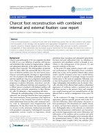
Figure 1 a) Pre-operative clinical photograph showing a large swelling over the right iliac region. b) Plain radiographs of right ilium showing
involvement of the iliac wing. Multiple septations could be appreciated even on plain radiographs. c) MRI scan of right iliac region showing multiple
fluid levels. d) 3D-CT reconstruction of the lesion showing a huge honeycomb appearance of lesion occupying almost whole of the right iliac wing
with extension to superior acetabulum.
Figure 2 a) Intraoperative photograph showing tumor size and
the psuedocapsule. b) Photograph after excision of lesion. Note the
exposed hip joint.
Agarwal et al. Journal of Orthopaedic Surgery and Research 2010, 5:24
/>Page 3 of 5
stability (Fig. 2b). After achieving hemostasis, the
exposed hip joint and raw posterior border of the iliac
bone was covered with abductor muscles. Histopatholog-
ical examination of the excised mass reconfirmed the
diagnosis of aneurysmal bone cyst (Fig. 3a, b). Postopera-
tively, she was advised complete bed rest for 4 weeks in
view of the involvement of the superior margin of the
acetabulum. Hip range of motion and strengthening exer-
cises were started on the second postoperative day. By 5
th
week, ambulation was initiated with crutch support. Four
weeks later, the crutches were discarded and patient was
encouraged to walk independently. At 18 months follow
up, the child is an independent walker, able to squat and
sit cross legged, and had full range of movements in the
hip joint (Fig. 4a, b). Her abductor group has a power of
4/5 with no other neurological deficits. X-rays and
enhanced CT repeated at this time showed good remod-
eling of the acetabulum and no signs of recurrence of the
lesion (Fig. 4c, d).
Discussion
Aneurysmal bone cysts typically involve the long bones of
the extremity, membranous bones of the thorax, or verte-
brae [1]. Ilium is not the site of predilection for the aneu-
rysmal bone cysts. In the series by Papagelopoulos et al
[2], the ilium bone was involved in only 8% out of 289
patients. Cottalorda et al series on 156 patients had pelvic
aneurysmal bone cyst in just 9% cases [3]. Capanna
detailed aneurysmal bone cysts of pelvis and mentioned
four cysts that extended into ilium [4]. Other authors
have mentioned involvement of iliac bone largely as case
reports [1,5,6]. The only reported cases of iliac aneurys-
mal bone cyst in paediatric age appear mainly as part of
large series of pelvic aneurysmal bone cysts or case
reports [2,7,8]. Thus, a review of literature indicates that
occurrence of a symptomatic aneurysmal bone cyst of
ilium in pediatric age group is distinctly rare.
The method of treatment of aneurysmal bone cyst of
the pelvis must be individualized depending on the loca-
tion, aggressiveness and extent of the lesion. Treatment
options include complete resection of the lesion, simple
curettage, curettage and bone grafting, selective arterial
embolization (primary treatment or preoperative adju-
vant therapy) and percutaneous injection of fibrosing
agent [2]. Yildirim et al [9] reported their experiences
with aneurysmal bone cyst of the adult pelvis. Lesions
less than 5 cm that exhibit minimal destruction or expan-
sion of cortical bone and don't threaten the integrity of
acetabulum or the sacroiliac joint are best treated with
intralesional curettage, with or without bone graft. Lesion
greater than 5 cm exhibiting large areas of destruction or
major expansion of cortical bone and threatening the
integrity of the acetabulum or the sacroiliac joint require
more aggressive treatment with the use of the excision or
curettage technique. Schwering et al described successful
management of large iliac aneurysmal bone cyst using
cystoscopic controlled curettage [8]. Chemical cauteriza-
tion with phenol is recommended for relatively large pri-
mary lesion to kill any surface tumor cells of the curetted
cavity [2,7,10]. Cryotherapy has also been proposed as an
adjuvant therapy with surgical treatment to achieve local
control [9]. Radiation is used in inaccessible sites where
no surgical options are available but has high recurrence
rates. Recently, percutaneous injection of fibrosing agent
has been employed in the treatment of aneurysmal bone
cysts. This technique is often associated with high com-
plication rate and is expensive [9]. Selective arterial
embolization is currently recommended as procedure of
choice for lesions whose site or size makes other types of
treatment difficult or dangerous [2]. It is especially useful
for managing huge lesions posing surgical risk due to
intraoperative bleeding and surrounding neural struc-
tures. The cost and availability, however, precludes its use
in developing countries.
Treatment of pelvic aneurysmal bone cyst in a growing
child is a challenging therapeutic problem because of the
open physis, relative inaccessibility of the lesion, associ-
ated intraoperative bleeding, proximity of the lesion to
neurovascular structures and the vulnerability of the
acetabulum or sacroiliac joint. Stability of the hip joint
was a major concern in our case, in view of the socio-cul-
tural aspect of squatting and sitting crossed legged in the
Indian setting and young age of the patient. Arthrodesis
of hip joint was not acceptable to the patient's family.
Marginal resection involving acetabulum would had
compromised the integrity of the acetabulum and hip
joint stability, hence only excision curettage of the lesion
was done and sealed with surrounding muscular flaps.
The integrity of the posterior ilium border and the sacro-
iliac joint was ensured to provide a stable hip and sacroil-
iac joint. Other authors have described use of autogenous
tricortical iliac crest bone graft to restore the structural
integrity of a compromised acetabulum [2]. Large bone
defects may require reconstruction with structural
allograft [2]. In few cases, where the integrity of the hip
Figure 3 a) Gross: The excised cyst. b) Histopathology: Blood filled
cystic spaces lined by cellular fibrous tissue lacking endothelial lining
(40×; H & E staining).
Agarwal et al. Journal of Orthopaedic Surgery and Research 2010, 5:24
/>Page 4 of 5
joint and the sacroiliac joint could not be preserved, dras-
tic step of hip or sacroiliac joint fusion have been
reported in the literature [2]. Adjuvant chemical cauter-
ization was not used in our case in view of exposed hip
cartilage (Fig. 2b). We could achieve excellent postopera-
tive range of motion and a stable, pain free hip joint by
preserving the acetabular roof. Cottalorda et al also
expressed similar views from their experience of series of
15 pelvic aneurysmal bone cysts in children. They indi-
cated that despite less aggressive surgical treatment in
form of (intralesional) curettage, the recurrence rates are
low [7].
Most of the reported recurrence of the lesion occurs
within 18 months after the primary treatment [3,10].
Figure 4 a, b) Follow up 18 months: comfortable cross legged sitting and squatting. c) Plain radiographs and d) CT showing good remodeling
and no involvement of the hip joint.
Agarwal et al. Journal of Orthopaedic Surgery and Research 2010, 5:24
/>Page 5 of 5
Capanna et al in a review of 23 aneurysmal bone cysts of
the pelvis treated with surgical intervention, noted a
recurrence rate of 13% over a 7 years period [6]. Cot-
talorda et al and Papagelopoulos et al reported recur-
rence rate of 13% and 14% respectively [2,7]. In our case,
no recurrence was noted at 18 months follow up and the
iliac bone and superior margin of acetabulum had remod-
eled well (Fig. 4).
Iliac aneurysmal bone cysts are distinctly rare in pedi-
atric age. The present case was a large lesion and associ-
ated with an atypical symptom of numbness along the
femoral nerve distribution. Hip stability and range of
movements were major concern in this patient. In our
case, we performed excision curettage of the lesion with
good outcome.
Consent
Written informed consent was obtained from the patient
for publication of this case report and any accompanying
images. A copy of the written consent is available for
review by the Editor-in-Chief of this journal.
Competing interests
The authors declare that they have no competing interests.
Authors' contributions
AA and SAK carried out planning and executed surgical procedure. PG, NAQ,
PK participated in case follow up and drafted the manuscript. PK, PG carried
out literature search. All authors read and approved the final manuscript.
Author Details
Department of Orthopedics, Chacha Nehru Bal Chikitsalaya, Geeta colony,
Delhi, India
References
1. Huang TL, Chen WM, Chen WY, Chen TH: Huge aneurysmal bone cyst of
iliac bone in a mid-aged female. J Chin Med Assoc 2004, 67:99-103.
2. Papagelopoulos PJ, Choudhury SN, Frassica FJ, Bond JR, Unni KK, Slim FH:
Treatment of aneurysmal bone cyst of pelvis and sacrum. J Bone Joint
Surg 2001, 83-A:1674-1681.
3. Cottalorda J, Kohler R, Sales de Gauzy J, Chotel F, Mazda K, Lefort G,
Louahem D, Bourelle S, Dimeglio A: Epidemiology of aneurysmal bone
cysts in children: A multicenter study and literature review. J Pediatr
Orthop B 2007, 13:389-394.
4. Bajracharya S, Khanal GP, Sundas A, Pandey SR, Singh MP: Aneurysmal
bone cyst of the pelvis: a challenge in treatment review of the
literature. Internet J Orthop Surg 2008, 8:.
5. Choe JG, Kim SH, Eoh W: Aneurysmal bone cyst arising from iliac bone
mimicking liposarcoma. Kor J Spine 2008, 5:234-236.
6. Capanna R, Bertoni F, Present D, Biaginil R, Ruggieri P, Mancini I,
Campanacci M: Aneurysmal bone cysts of pelvis. Arch Orthop Trauma
Surg 1986, 105:279-284.
7. Cottalorda J, Chotel F, Kohler R, Sales de Gauzy J, Louahem D, Lefort G,
Dimeglio A, Bourelle S: Aneurysmal bone cysts of the pelvis in children.
A multicenter study and literature review. J Pediatr Orthop 2005,
25:471-475.
8. Schwering L, Uhl M, Herget GW: Iliac aneurysmal bone cyst treated by
cystoscopic controlled curettage. SICOT Online reports E054 .
9. Yildirim E, Ersözlü S, Kirbas I, Özgür AF, Akkaya T, Karadeli E: Treatment of
pelvic aneurysmal bone cysts in two children: selective arterial
embolization as an adjunct to curettage and bone grafting. Diagn
Interv Radiol 2007, 13:49-52.
10. Campanacci M, Capanna R, Picci P: Unicameral and aneurysmal bone
cyst. Clin Orthop Relat Res 1986, 204:25-36.
doi: 10.1186/1749-799X-5-24
Cite this article as: Agarwal et al., Large aneurysmal bone cyst of iliac bone
in a female child: a case report Journal of Orthopaedic Surgery and Research
2010, 5:24
Received: 3 November 2009 Accepted: 7 April 2010
Published: 7 April 2010
This article is available from : http://www.j osr-online.com/ content/5/1/24© 2010 Agarwal et al; licensee BioMed Central Ltd. This is an Open Access article distributed under the terms of the Creative Commons Attribution License ( ), which permits unrestricted use, distribution, and reproduction in any medium, provided the original work is properly cited.Journal of Orthopaedic Surgery and Research 2010, 5:24