báo cáo hóa học:" Kyphoplasty in osteoporotic vertebral compression fractures - Guidelines and technical considerations" pptx
Bạn đang xem bản rút gọn của tài liệu. Xem và tải ngay bản đầy đủ của tài liệu tại đây (496 KB, 8 trang )
REVIEW Open Access
Kyphoplasty in osteoporotic vertebral
compression fractures - Guidelines and technical
considerations
Yohan Robinson
1*
, Christoph E Heyde
2
, Peter Försth
1
and Claes Olerud
1
Abstract
Osteoporotic vertebral compression fractures are a menace to the elderly generation causing diminished quality of
life due to pain and deformity. At first, conservative treatment still is the method of choice. In case of resulting
deformity, sintering and persistent pain vertebral cement augmentation techniques today are widely used. Open
correction of resulting deformity by different types of osteotomies addresses sagittal balance, but has comparably
high morbidity.
Besides conventional vertebral cement augmentation techniques balloon kyphopl asty has become a popular tool
to address painful thoracic and lumbar compression fractures. It showed improved pain reduction and lower
complication rates compared to standard vertebroplasty. Intere stingly the results of two placebo-controlled
vertebroplasty studies question the value of cement augmentation, if compared to a sham operation. Even though
there exists now favourable dat a for kyphoplasty from one randomised controlled trial, the absence of a sham
group leaves the placebo effect unaddressed. Technically kyphoplasty can be performed with a transpedicular or
extrapedicular access. Polymethyl methacrylate (PMMA)-cement should be favoured, since calcium phosphate
cement showed inferior biomechanical properties and less effect on pain reduction especially in less stable burst
fractures. Common complications of kyphoplasty are cement leakage and adjacent segment fractures. Rare
complications are toxic PMMA-monomer reactions, cement embolisation, and infection.
Keywords: Kyphoplasty, vertebroplasty, osteoporosis, spinal fractures
Introduction
Osteoporosis and pathological osteoporotic fractures are
common findings in the elderly population. The age-
standardised annual incidence of vertebral compression
fractures (VCF) is 10.7/1000 in women and 5.7/1000 in
men, increasing markedly with age [1]. At the age of 7 5
to 79 the annual incidence was 29.3/1000 in women and
13.6/1000 in men. Due to the continued aging of our
population, VCF represent a major cause of disability
and are a burden to the na tional healthcare budgets [2].
Non-surgical management with pain co ntrol and physi-
cal therapy-assisted mobilization has for a long time
been the only treatment option in VCF. Unfortunatelty
a great number of patients remain functionally impaired
after V CF, and some of them are severely handicapped
due to chronic back pain [3]. The functional and physi-
cal consequences of VCF lead to anxiety, depression,
and have devastating impact on interpersonal relation-
ships and social roles [4]. It is therefore no surprise that
untreated VCF c ontribute significantly to shorter life-
expectancy both in women (mortality ratio 1.66, p <
0.01) and even greater in men (mortality ratio 2.38, p <
0.0001) within one year after onset of symptoms [5].
Indications for cement augmentation
while medical therapy of osteoporosis improves dramati-
cally, the restoration of quality of life is still a major
issue in VCF treatment. Osteoporotic kyphotic compres-
sion fractures often lead to a anterior shift of the sagittal
plumb line and increased load of the anterior vertebral
colu mn, which may cause further compression fractures
[6]. This cascade of sequential compression fractures is
* Correspondence:
1
Uppsala University Hospital, Institute for Surgical Sciences, Department of
Orthopaedics, Uppsala, Sweden
Full list of author information is available at the end of the article
Robinson et al. Journal of Orthopaedic Surgery and Research 2011, 6:43
/>© 2011 Robinson et al; licensee BioMed Central Ltd. This is an Open Access article d istributed under the terms of the Creative
Commons Attribut ion License ( /by/2.0), which permits unrestricted use, distribution, and
reproduction in any medium, provided the original work is properly cited.
eventually causing the typical hump of the elderly, with
significant thoracic kyphosis and low pelvic incidence,
forcing the patient to bend hips and knees to maintain
sagittal balance [7].
Galibert et al [8] presented the first cases of successful
vertebral augmentation by intravertebral injection (ver-
tebroplasty) of polymethyl methacrylate (PMMA) in
patients with vertebral haemagiomas. Later, vertebro-
plasty was successfully introduced for the management
of osteoporotic compression fractures [9]. The primary
goal of vertebroplasty is pain relief by stabilization of
the VCF, improving indirectly pulmonary function and
patient quality of life [10]. The biomechanical under-
standing of increasing anterior column load with pro-
gressing kyphosis leading to subsequent VCF established
the basic rationale for kyphoplasty. With this technique,
partial reduction of VCF is possible by transpedicular
intracorporal balloon expansion and retention by
PMMA cement augmentation [11,12]. The results of
one multicenter randomised controlled trial found shor-
tened and improved functional recovery after kypho-
plasty with a low rate of complications if compared to
non-surgical treatment [13].
Despite the advances in percutaneous augmentation
techniques the conservative medical therapy cannot be
replaced. VCF without initial kyphosis, no consecutive
sintering and a satisfactory and quick response to con-
servative treatment should be treated conse rvatively.
Furthermore, since lack of reimbursement in most
countries kyphoplasty causes an economic burden, many
patients are not willing to take. Beyond that, it has to
me emphasised, that it remains unclear whether the
benefits of kyphoplasty outweigh its complications.
Two placebo-controlled vertebroplasty-studies have
sob ering results with regard to pain and functional out-
come after cement augmentation with vertebrop lasty, if
compared to a sham-operation [14,15]. In both studies
the sham procedure included percutaneus needle inser-
tion and opening of PMMA-monomer mixture to simu-
late the specific odour. The sham-controlled trial by
Buchbinder et al [14] in 78 patients with MRI-con-
firmed, fresh and painful VCF found no beneficial effect
of vertebroplasty when compared to a sham procedure.
A very similar study by Kallmes et al [15] investigating
131 patients found similar results. This study had
already after 3 month significant higher cross-over of
43% in the control-group (p < 0.001) diminishing the
quality of this study. Furthermore only outpatients were
included in this study, which means that no patients
being hospitalised due to acute VCF entered the study.
The randomised c ontrolled trial by Rousing et al [16]
found no greater improvement in bac k pain in patients
treated with vertebroplasty when compared to medical
therapy. Interestingly they found a significant improve-
ment in the Barthel-score after 12 month (p < 0.02)
indicating improved function, [17]. As a result to the
above-mentioned results several authors abandoned the
use of verteb roplasty [18-20] while others are hesitant
and question the quality of the sham-controlled v erteb-
roplasty trials [17,21]. It is unclear whether the results
of the multicenter randomised kyphoplasty trial could
be reproduced if sham-controlled [13]. In table 1 the
authors present clinically proven guidelines for indica-
tions and contraindications of kyphoplasty.
Due to the increased demand in cement augmentation
techniques, procedures similar to kyphopla sty have be en
developed. One competitor is Vesselplasty
®
(A-Spine),
where a porous balloon is inflated within the fractured
vertebral body and filled with cement without removing
the balloon, thus reducing the risk of cement leakage
[22]. Another new product is the Sky
®
bone expander
(Disc-O-Tech), an expandable polymer bone tamp aban-
doning the use of cement, which had favourable results
in clinical case series [23]. Then there is the StaXx
®
FX
system (Spine Wave) where a VCF is reduced percuta-
neously by gradual insertion of stacked PEEK-chips into
the vertebral body to reduce and stabilise the fracture
[24].
Indications for combined cement augmentation and
posterior instrumentation
Lately kyphoplasty has been discussed as an alternative
therapy even of burst fractures in elderly patients. This
is especi ally true in ca se of AO type A3.1 fractures,
where it could be applied instead of a posterior-only
or 360 degrees stabilisation [25]. In many of these
Table 1 Guidelines for indications and contraindications for kyphoplasty
Indications for
kyphoplasty
- Radiologically confirmed fresh compression fracture (AO type A1) (MRI shows oedema or X-ray/CT-scan
proven fracture not older than 3 months)
- Failure of 2 - 6 weeks of conservative treatment including pain medication and physiotherapy (Pain on
visual analogous scale (VAS) above 4 of 10)
Contraindications for
kyphoplasty - Burst-fractures (in some A3.1-fractures possible)
- Flexion-/distraction and rotational injuries (AO type B and C)
- Medical contraindications (bleeding disorders, sepsis, etc)
- PMMA-allergy
Robinson et al. Journal of Orthopaedic Surgery and Research 2011, 6:43
/>Page 2 of 8
cases further sintering of the fractured vertebra with
posterior dislocation of an instable fragment with
spinal stenosis is a feared complication [26,27]. Thus
several surgeons perform posterior instrumentation of
the adjacent vertebrae to protect the posterior wall and
to improve the sagittal profile [28]. This can be done
using percutaneous posterior instrumentation or with a
conventional open technique [28-30]. Possible disad-
vantages of this technique are due to segmental fusion
an increased load of the adjacent segments with degen-
eration, and possible implant loosening with loss of
correction due to low bone quality. Cement augmenta-
tion of the impla nted pedicle screws can reduce the
complication rate regarding the latter mentioned pro-
blem [ 31].
Limitations of kyphoplasty and indications for open
reduction and stabilisation
If multiple VCF lead to kyphosis with fixed sagittal
imbalance, cement augmentation will address the frac-
ture pain but not global imbalance. Major spinal imbal-
ance can be a cause of significant functional disability
leading to reduced quality of life. Increased kyphosis
may additionally cause subsequent VCF due to an
increased anterior load [32]. The anterior location of the
sagittal plumb line in fixed sagittal imbalance will lead
to falls with possible further fractures and morbidity.
Therefore the indication for correction of glob al sagittal
imbalance may be given in severe cases. As both open
and closing wedge procedures are associated with com-
plications leading to disabling morbidity surgeon are
often hesitant to perform these operations in patients
with multipl e comorbidities [33]. Due to the osteopenic
bone quality often long instrumentations are required.
Unfortunately these an increased risk of adjacent VCF
and pedicle fractures [34]. With regard to open sagittal
corrections there is growing evidence that the posterior-
only pedicle subtraction osteotomy is superior to multi-
ple Smith-Petersen osteotomies, allowing greater correc-
tion with lesser operation time [35,36].
Operation technique of kyphoplasty
Percutaneous bilateral transpedicular kyphoplasty
The bilateral transpedicular approach is the standard
kyphoplasty access for the thoracolumbar spine,
enabling a symmetric reposition and augmentation of
the VCF. Firstly, the fracture is reduced under fluoro-
scopic control by p ositioni ng and traction. Then biopsy
needles are used to enter the fractured vertebra on both
sides through the pedicle (figure 1). If fluoroscopy con-
firms correct transpedicular placement of the needles in
both planes, a K-wire is placed through the Jamshidi
biopsy needle into the vertebral body close to the ante-
rior wall. Then over the K-wire the access is widened
with the osteointroducer. Then empty bone-fillers are
used to form a cavity for the safe placement of the bal-
loons. Under fluoroscopic control two balloons are posi-
tioned anteriorly within the vertebral body, and then the
balloons are inflated under manometric control. In fresh
fractures up to 150 psi balloon pressure are mostly
enough, but sometimes up to 300 psi are necessary to
reduce a partially healed compression fracture. One has
to be careful not to fracture the endplates or the poster-
ior wall, which could lead to cement leakage. After suc-
cessful reduction the cavity is filled with bone-cement
from both pedicles. This step has to be performed with
careful fluoroscopic control. Normally the cement
should have a tooth-paste-like viscosity and should not
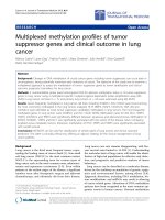
Figure 1 Transpedicular approach for ba lloon kyphoplasty. After entry in the craniolateral pedicle (red cross) in the p-a-projection (a),the
medial cortex of the pedicle is first breached when the vertebral body is entered in the lateral projection (blue cross) (b). After preparation of
the working channel a balloon can be placed in the vertebral body.
Robinson et al. Journal of Orthopaedic Surgery and Research 2011, 6:43
/>Page 3 of 8
stick to the surgeons’ gloves when testing the viscosity.
Finally the introducers are removed.
Percutaneous unilateral extrapedicular kyphoplasty
Due to the anatomical characteristics of the thoracic
spine c entral placement of the balloons can be difficult.
Firstly, the introducer often does not fit in the narrow
pedicles of the thoracic spine. Then the low angulation
of the pedicles does not allow a central placement of
the balloons disabling adequate reduction in some cases.
Thus in the thoracic spine extrapedicular accesses gain
increasing popularity, avoiding pedicle perforation with
possible neurological damage or intraspinal cement leak-
age. Most surgeon prefer the transcostovertebral access
from far lateral (figure 2), guided to the collum costae
into the costotransversary space to the cranio-posterior
wall of the fractured vertebra, the Jamshidi needle with
the tip just penetrating the lateral pedicle at its base
[37,38]. In the view from posterior the needle passes
above of the transverse process and meets the pedicle at
the craniolateral circumference. The lateral view con-
firms the placement of the tip of the needle close to the
base of the pedicle. In an axial v iew the needle is see n
to pass through the costovertebral gap, between the
neck of the rib and the lateral pedicle circumference,
towards the base of the pedicle. Then the posterolateral
cortical wall is opened with a Jamshidi cannula and
widened as described above with K-wire and osteo-
introducer. To allow central placement of the balloon in
the vertebral body a greater angle than in the transpedi-
cular placement has to be sought. This often requires a
7 to 10 cm off-midline percutaneus approach. A single
balloon is then used for reduction and the cavity is then
filled with cement as described above.
Open unilateral interlaminary kyphoplasty
Open interlaminary kyphopla sty should be reserved for
cases where an open approach has to be performed to
decompress neurological structures, and the spinal canal
has to be accessed anyway [39]. After open decompres-
sion the dural sac is retracted medially and the posterior
wall of the fractured vertebra exposed. Now kyphoplasty
can be performed with a single balloon positioned
under fluoroscopical guidance in the centre of the ver-
tebral body. After kyphoplasty the spinal canal has to be
investigated for cement leakage. This method must be
restricted to levels below the conus medullaris to avoid
myelopathy due to manipulation within the spinal canal.
Open anterior kyphoplasty
In rare cases kyphoplasty may be performed using an
anterior access, too [40]. Through a minimally-invasive
anterior access the biopsy needle may be placed directly
on the anterior wall of the fractured vertebra and a si n-
gle balloon be placed into the vertebral body. Then
under fluoroscopical control the fracture is reduced and
cement is applied.
Technical considerations
Operation room setup
Both general an local anesthesia have bee n successfully
applied for the pro cedure [41], but many surgeons
favour general anestesia allowing closed reduction in a
relaxed patient. By patient positioning o nly, more than
70% of vertebral height restoration can be achieved. Pla-
cing the patient in prone position lordosating the f rac-
tured segment by pillows or by bending the table will
lead to reduction of the fracture with ligamentotaxis
[42].
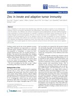
Figure 2 Unilateral extrapedicular costotransversary approach f or balloon kyphoplasty. Following the cranioposterior part of the
respective rib into the costotransversary space (c) allows extrapedicular access to the vertebral body in the thoracic spine. Due to the far lateral
approach a single balloon is placed in the middle of the vertebral body (a, b).
Robinson et al. Journal of Orthopaedic Surgery and Research 2011, 6:43
/>Page 4 of 8
As in most percutaneus surgical techniques implant
positioning and accuracy is controlled with fluoroscopic
image intensifiers. Correct positioning of the image
intensifier will lead to much lesser radiation dose for the
surgeon. Placement of x-ray tube in the image intensifier
on the opposite side of the surgeon will ca uses up to 10
times less radiation exposure [43].
Navigation
Balloon placement accuracy can be significantly
improved and the radiation exposure during kyphoplasty
can be reduced by as much as 76%, if computer-assisted
fluoroscopi c navigation is applied [44,45]. While relying
on the navigator during the transpedicular balloon pla-
cement, balloon inflation and cement injection have to
be performed under fluoroscopic control to minimise
endplate fractures and cement leakage.
Eggshell procedure
An eggshell-procedure may avoid cement leakage in
VCF suscpicious for endplate or posterior wall damage
[46]. After reduction with the kyphoplasty balloon a
small amount of doughy cement is injected into the cav-
ity, and then the balloon reinserted and reinflated. Once
the cement hardens the cavity can be filled with cement
within the “eggshell”, preventing cement leakage.
Choice of cement
Most vertebroplasty and kyphoplasty procedures have
been performed using polymethylmethacrylate
(PMMA) cement to augment the fractured vertebra.
The increasing availability of injectable calcium phos-
phate (CaP) cement led to its application in the aug-
mentation of compression fractures as an alternative to
PMMA. Advantages are high biocompatibility, no
Table 2 Overview on comparative clinical trials of kyphoplasty
Author Year Design Level of
evidence
Control
Group
Control
n
(levels)
Kyphoplasty
n (levels)
Follow-
up
Outcome
Weisskopf et
al. [54]
2003 Retrospective IIIb non-surgical 20 (35) 22 (37) 10 days Improvement in VAS (p < 0.001) Reduced days
in hospital (p < 0.01)
Fourney et
al. [55]
2003 Retrrospective IIIb vertebroplasty 34 (65) 15 (32) 4,5
months
No significant differences in VAS and ODI
Improvement of kyphosis with kyphoplasty
(p < 0.01)
Komp et al.
[56]
2004 Prospective IIb non-surgical 19(19) 21(21) 6
months
Improvement of VAS and ODI with
kyophoplasty (p < 0.01)
Kasperk et al
[57]
2005 Prospective IIb non-surgical 20 (33) 40 (72) 12
months
Improvement of VAS (p < 0.01) and
improvement of kyphosis (p < 0.001) with
kyphoplasty
Grohs et al.
[58]
2005 Prospective IIb vertebroplasty 23 (29) 28 (35) 24
months
No significant difference in ODI, but
improvement of VAS with kyphoplasty
(p < 0.05). No signi ficant improvement of kypho sis
Masala et al.
[59]
2005 Retrospective IIIb vertebroplasty 26 (33) 7 (7) 6
months
No significant difference in VAS.
Pflugmacher
et al [30]
2005 Prospective IIb vertebroplasty 20 (32) 22 (35) 12
months
No significant difference in VAS and ODI.
Improvement of kyphosis with kyphoplasty
(p < 0.05)
De Negri et
al. [60]
2007 Prospective IIb vertebroplasty 10 (18) 11 (15) 6
months
No significant difference in VAS and ODI.
Zhou et al.
[61]
2008 Prospective IIIb vertebroplasty 42 56 12
months
No significant differences in VAS, operation time
and blood loss. Improved vertebral height
restoration with kyphoplasty (p < 0.01).
Wardlaw et
al. [13]
2009 Randomised Ib non-surgical 149 151 12
months
Significant improvement in EQ-5D (p < 0.05),
RMDQ (p < 0.001) VAS (p < 0.0001).
Schmelzer-
Schmied et
al. [51]
2009 Prospective IIb non-surgical 20 20 12
months
Significant greater improvement of VAS
(p < 0.05) with kyphoplasty, which was lost after
3 months, and vertebral height preservation
after 12 months (p < 0.01)
Schofer et al.
[62]
2009 Prospective IIIb vertebroplasty 30 30 13
months
No significant differences in VAS and SF-36.
Greater improvement of kyphotic angle with
kyphoplasty (p < 0.001)
Li X et al
[63]
2011 Prospective IIIb vertebroplasty 40 45 12
months
No significant differences in VAS and ODI.
Significantly greater improvement of kyphotic
angle with kyphoplasty (p < 0.01)
VAS: Visual Analogous Scale, ODI: Oswestry Disability Index, EQ-5D: EuroQoL, RMDQ: Roland Morris Disability Questionnaire, SF-36: Short Form Health Survey.
Robinson et al. Journal of Orthopaedic Surgery and Research 2011, 6:43
/>Page 5 of 8
systemic toxic m onomers, osteoinductive capacity, and
close to isothermal cristallinisation. Disadvantages are
besides less clinical long-term experience, lesser com-
pressive s trength than PMMA [47], and the risk o f
early resorption, leading to defects prone to re-frac-
tures [48-50]. The available data does not encourage
the clinical use of CaP-cement in b urst-fractures, flex-
ion-distraction injuries, or rotational instable fractures
[48,51].
Results and complications of kyphoplasty
Fourteen years after the first vertebroplasty was per-
formed in 1984, balloon kyphoplasty challenged the con-
ventional augmentation procedures promising less
complications and sagittal reconstructive ability. Until
now several non-randomized prospective controlled
trials have been published comparing kyphoplasty to
non-surgical treatment and vertebroplasty (Table 2).
Besides pain improvement and quality of life, correction
of deformi ty and intra- and postoperative complications
were investigated. The recently presented preliminary 1-
year-results of the multicentrical randomized controlled
Fracture Reduction Evaluation (FREE) study present in
the kyphoplasty group a significant improvement of the
quality of life (EQ-5D (EuroQoL), p < 0.001), pain (VAS,
p < 0.0001), and function (SF-36 (Short form Health
Survey), p < 0.0001, ODI, p < 0.0001) after 1 month (n
= 149) controlled against non-surgical treatment (n =
151) [ 13]. These treatment-effects diminished dramati-
cally until the 12-month follow -up, but wer e still signifi-
cantly better than non-surgical treatment for quality of
life as measured with EQ-5D (p < 0.05).
The comprehensive meta-analysis of L ee et al [52]
summarized all published kyphoplasty c omplications.
Cement leakages oc curred in 1 4% of all cases, but onl y
0.01% were symptomatic. New vertebral fractures
occurred in 17%. Taylor et al [53] found in the ir metaa-
nalysis furthermore spinal stenosis with spinal cord
compression occurred 0.16% of all cases. Radiculopathy
was found in 0.17% of all cases. Furthermore there are
anecdotal reports of infections after kyphoplasty [26].
The overall mortality was 4.4%, and the perioperative
mortality was 0.13% [53].
Conclusions
Kyphoplasty is - in the hands of an experienced spine
surgeon or radiological interventionalist - an effective
tool to treat pain caused by thoracolumbar vertebral
compression fractures, but the severity of pulmonary
PMMA cement embolism and the urgent need of
immediate decompression in relevant spinal stenosis
aft er cement leak age, require an anaesthesiologist and a
spi nal surgeon on call. The comp lication rat e of kypho-
plasty is dramatically lower than in alternative open
instrumented procedures, a nd the immediate pain
reduction is significantly greater in kyph oplasty com-
pared to conservative treatment. Therefore its applica-
tion remains a pillar in VCF treatment.
Author details
1
Uppsala University Hospital, Institute for Surgical Sciences, Department of
Orthopaedics, Uppsala, Sweden.
2
Leipzig University Hospital, Department of
Orthopaedic Surgery, Spine Surgery, Leipzig, Germany.
Authors’ contributions
YR wrote the manuscript, and CEH, PF and CO revised it critically. All authors
read and approved the final manuscript.
Competing interests
YR, CEH, and PF were clinical investigators in the FREE trial, and YR and PF
were Clinical Investigators in the CAFE trial, both initiated by Kyphon Inc.
(now Medtronic Spine LLC, Sunnyvale, CA, USA). YR, CEH, PF and CO
received travel assistance by Medtronic, DePuy Spine (Johnson & Johnson)
and Synthes.
Received: 5 February 2011 Accepted: 19 August 2011
Published: 19 August 2011
References
1. Felsenberg D, Silman AJ, Lunt M, Armbrecht G, Ismail AA, Finn JD,
Cockerill WC, Banzer D, Benevolenskaya LI, Bhalla A, et al: Incidence of
vertebral fracture in europe: results from the European Prospective
Osteoporosis Study (EPOS). J Bone Miner Res 2002, 17:716-724.
2. Lad SP, Patil CG, Lad EM, Boakye M: Trends in pathological vertebral
fractures in the United States: 1993 to 2004. J Neurosurg Spine 2007,
7:305-310.
3. Pluijm SM, Tromp AM, Smit JH, Deeg DJ, Lips P: Consequences of
vertebral deformities in older men and women. J Bone Miner Res 2000,
15:1564-1572.
4. Gold DT: The clinical impact of vertebral fractures: quality of life in
women with osteoporosis. Bone 1996, 18:185S-189S.
5. Center JR, Nguyen TV, Schneider D, Sambrook PN, Eisman JA: Mortality
after all major types of osteoporotic fracture in men and women: an
observational study. Lancet 1999, 353:878-882.
6. Rohlmann A, Klockner C, Bergmann G: [The biomechanics of kyphosis].
Orthopade 2001, 30:915-918.
7. Schwab F, Lafage V, Patel A, Farcy JP: Sagittal plane considerations and
the pelvis in the adult patient. Spine (Phila Pa 1976) 2009, 34:1828-1833.
8. Galibert P, Deramond H, Rosat P, Le Gars D: [Preliminary note on the
treatment of vertebral angioma by percutaneous acrylic vertebroplasty].
Neurochirurgie 1987, 33:166-168.
9. Deramond H, Depriester C, Galibert P, Le Gars D: Percutaneous
vertebroplasty with polymethylmethacrylate. Technique, indications, and
results. Radiol Clin North Am 1998, 36:533-546.
10. Lee JS, Kim KW, Ha KY: The Effect of Vertebroplasty on Pulmonary
Function in Patients With Osteoporotic Compression Fractures of the
Thoracic Spine. J Spinal Disord Tech .
11. Voggenreiter G: Balloon kyphoplasty is effective in deformity correction
of osteoporotic vertebral compression fractures. Spine (Phila Pa 1976)
2005, 30:2806-2812.
12. Luo J, Bertram W, Sangar D, Adams MA, Annesley-Williams DJ, Dolan P: Is
kyphoplasty better than vertebroplasty in restoring normal mechanical
function to an injured spine? Bone 46:1050-1057.
13. Wardlaw D, Cummings SR, Van Meirhaeghe J, Bastian L, Tillman JB,
Ranstam J, Eastell R, Shabe P, Talmadge K, Boonen S: Efficacy and safety of
balloon kyphoplasty compared with non-surgical care for vertebral
compression fracture (FREE): a randomised controlled trial. Lancet 2009,
373:1016-1024.
14. Buchbinder R, Osborne RH, Ebeling PR, Wark JD, Mitchell P, Wriedt C,
Graves S, Staples MP, Murphy B: A randomized trial of vertebroplasty for
painful osteoporotic vertebral fractures. N Engl J Med 2009, 361:557-568.
15. Kallmes DF, Comstock BA, Heagerty PJ, Turner JA, Wilson DJ, Diamond TH,
Edwards R, Gray LA, Stout L, Owen S, et
al: A randomized trial of
Robinson et al. Journal of Orthopaedic Surgery and Research 2011, 6:43
/>Page 6 of 8
vertebroplasty for osteoporotic spinal fractures. N Engl J Med 2009,
361:569-579.
16. Rousing R, Andersen MO, Jespersen SM, Thomsen K, Lauritsen J:
Percutaneous vertebroplasty compared to conservative treatment in
patients with painful acute or subacute osteoporotic vertebral fractures:
three-months follow-up in a clinical randomized study. Spine (Phila Pa
1976) 2009, 34:1349-1354.
17. Fisher CG, Vaccaro AR: The highest level of evidence in a high impact
journal: is this the final verdict? Spine (Phila Pa 1976) 35:E676-677.
18. Karlsson MK, Ohlin A, Hasserius R: Could vertebroplasty and kyphoplasty
be regarded as evidence-based treatment of osteoporotic vertebral
fractures? Acta Radiol 51:828-831.
19. Hasserius R, Ohlin A, Karlsson MK: Vertebroplasty and kyphoplasty–
evidence-based methods? Acta Orthop 81:521-523.
20. Miller FG, Kallmes DF: The case of vertebroplasty trials: promoting a
culture of evidence-based procedural medicine. Spine (Phila Pa 1976)
35:2023-2026.
21. Heini PF: [Vertebroplasty: an update: value of percutaneous cement
augmentation after randomized, placebo-controlled trials]. Orthopade
39:658-664.
22. Flors L, Lonjedo E, Leiva-Salinas C, Marti-Bonmati L, Martinez-Rodrigo JJ,
Lopez-Perez E, Figueres G, Raoli I: Vesselplasty: a new technical approach
to treat symptomatic vertebral compression fractures. AJR Am J
Roentgenol 2009, 193:218-226.
23. Xiong J, Dang Y, Jiang BG, Fu ZG, Zhang DY: Treatment of osteoporotic
compression fracture of thoracic/lumbar vertebrae by kyphoplasty with
SKY bone expander system. Chin J Traumatol 13:270-274.
24. Renner S: Restoration of intervertebral disc mechanics after endplate
deformity reduction using structural kyphoplasty. J NeuroIntervent Surg
2009, 1:76.
25. Stoffel M, Wolf I, Ringel F, Stuer C, Urbach H, Meyer B: Treatment of
painful osteoporotic compression and burst fractures using kyphoplasty:
a prospective observational design. J Neurosurg Spine 2007, 6:313-319.
26. Robinson Y, Tschoke SK, Stahel PF, Kayser R, Heyde CE: Complications and
safety aspects of kyphoplasty for osteoporotic vertebral fractures: a
prospective follow-up study in 102 consecutive patients. Patient Saf Surg
2008, 2:2.
27. Patel AA, Vaccaro AR, Martyak GG, Harrop JS, Albert TJ, Ludwig SC,
Youssef JA, Gelb DE, Mathews HH, Chapman JR, et al: Neurologic deficit
following percutaneous vertebral stabilization. Spine (Phila Pa 1976) 2007,
32:1728-1734.
28. Noldge G, DaFonseca K, Grafe I, Libicher M, Hillmeier J, Meeder PJ,
Kauffmann GW, Kasperk C: [Balloon kyphoplasty in the treatment of back
pain]. Radiologe 2006, 46:506-512.
29. Verlaan JJ, Dhert WJ, Verbout AJ, Oner FC: Balloon vertebroplasty in
combination with pedicle screw instrumentation: a novel technique to
treat
thoracic and lumbar burst fractures. Spine (Phila Pa 1976) 2005, 30:
E73-79.
30. Pflugmacher R, Kandziora F, Schroder R, Schleicher P, Scholz M, Schnake K,
Haas N, Khodadadyan-Klostermann C: [Vertebroplasty and kyphoplasty in
osteoporotic fractures of vertebral bodies – a prospective 1-year follow-
up analysis]. Rofo 2005, 177:1670-1676.
31. Aydogan M, Ozturk C, Karatoprak O, Tezer M, Aksu N, Hamzaoglu A: The
pedicle screw fixation with vertebroplasty augmentation in the surgical
treatment of the severe osteoporotic spines. J Spinal Disord Tech 2009,
22:444-447.
32. Hato T, Kawahara N, Tomita K, Murakami H, Akamaru T, Tawara D,
Sakamoto J, Oda J, Tanaka S: Finite-element analysis on closing-opening
correction osteotomy for angular kyphosis of osteoporotic vertebral
fractures. J Orthop Sci 2007, 12:354-360.
33. Daubs MD, Lenke LG, Cheh G, Stobbs G, Bridwell KH: Adult spinal
deformity surgery: complications and outcomes in patients over age 60.
Spine (Phila Pa 1976) 2007, 32:2238-2244.
34. DeWald CJ, Stanley T: Instrumentation-related complications of multilevel
fusions for adult spinal deformity patients over age 65: surgical
considerations and treatment options in patients with poor bone
quality. Spine (Phila Pa 1976) 2006, 31:S144-151.
35. Cho KJ, Bridwell KH, Lenke LG, Berra A, Baldus C: Comparison of Smith-
Petersen versus pedicle subtraction osteotomy for the correction of
fixed sagittal imbalance. Spine (Phila Pa 1976) 2005, 30:2030-2037,
discussion 2038.
36. Kim YJ, Bridwell KH, Lenke LG, Cheh G, Baldus C: Results of lumbar pedicle
subtraction osteotomies for fixed sagittal imbalance: a minimum 5-year
follow-up study. Spine (Phila Pa 1976) 2007, 32:2189-2197.
37. Boszczyk BM, Bierschneider M, Hauck S, Beisse R, Potulski M, Jaksche H:
Transcostovertebral kyphoplasty of the mid and high thoracic spine. Eur
Spine J 2005, 14:992-999.
38. Ryu KS, Park CK, Kim MK, Kim DH: Single balloon kyphoplasty using far-
lateral extrapedicular approach: technical note and preliminary results. J
Spinal Disord Tech 2007, 20:392-398.
39. Boszczyk BM, Bierschneider M, Schmid K, Grillhosl A, Robert B, Jaksche H:
Microsurgical interlaminary vertebro- and kyphoplasty for severe
osteoporotic fractures. J Neurosurg 2004, 100:32-37.
40. Boszczyk B, Bierschneider M, Potulski M, Robert B, Vastmans J, Jaksche H:
[Extended kyphoplasty indications for stabilization of osteoporotic
vertebral compression fractures]. Unfallchirurg 2002, 105:952-957.
41. Cagli S, Isik HS, Zileli M: Vertebroplasty and kyphoplasty under local
anesthesia: review of 91 patients. Turk Neurosurg 20:464-469.
42. Cawley DT, Sexton P, Murphy T, McCabe JP: Optimal patient positioning
for ligamentotaxis during balloon kyphoplasty of the thoracolumbar and
lumbar spine. J Clin Neurosci .
43. Choi HC: Fluoroscopic
Radiation Exposure during Percutaneous
Kyphoplasty. J Korean Neurosurg Soc 49:37-42.
44. Kang JD, An H, Boden S, Phillips F, Foley K, Abdu W: Cement
augmentation of osteoporotic compression fractures and intraoperative
navigation: summary statement. Spine (Phila Pa 1976) 2003, 28:S62-63.
45. Ohnsorge JA, Siebert CH, Schkommodau E, Mahnken AH, Prescher A,
Weisskopf M: [Minimally-invasive computer-assisted fluoroscopic
navigation for kyphoplasty]. Z Orthop Ihre Grenzgeb 2005, 143:195-203.
46. Greene DL, Isaac R, Neuwirth M, Bitan FD: The eggshell technique for
prevention of cement leakage during kyphoplasty. J Spinal Disord Tech
2007, 20:229-232.
47. Wilke HJ, Mehnert U, Claes LE, Bierschneider MM, Jaksche H, Boszczyk BM:
Biomechanical evaluation of vertebroplasty and kyphoplasty with
polymethyl methacrylate or calcium phosphate cement under cyclic
loading. Spine (Phila Pa 1976) 2006, 31:2934-2941.
48. Blattert TR, Jestaedt L, Weckbach A: Suitability of a calcium phosphate
cement in osteoporotic vertebral body fracture augmentation: a
controlled, randomized, clinical trial of balloon kyphoplasty comparing
calcium phosphate versus polymethylmethacrylate. Spine (Phila Pa 1976)
2009, 34:108-114.
49. Heo HD, Cho YJ, Sheen SH, Kuh SU, Cho SM, Oh SM: Morphological
changes of injected calcium phosphate cement in osteoporotic
compressed vertebral bodies. Osteoporos Int 2009, 20:2063-2070.
50. Heo DH, Chin DK, Yoon YS, Kuh SU: Recollapse of previous vertebral
compression fracture after percutaneous vertebroplasty. Osteoporos Int
2009, 20:473-480.
51. Schmelzer-Schmied N, Cartens C, Meeder PJ, Dafonseca K: Comparison of
kyphoplasty with use of a calcium phosphate cement and non-
operative therapy in patients with traumatic non-osteoporotic vertebral
fractures. Eur Spine J 2009, 18:624-629.
52. Lee MJ, Dumonski M, Cahill P, Stanley T, Park D, Singh K: Percutaneous
treatment of vertebral compression fractures: a meta-analysis of
complications. Spine (Phila Pa 1976) 2009, 34:1228-1232.
53. Taylor RS, Fritzell P, Taylor RJ: Balloon kyphoplasty in the management of
vertebral compression fractures: an updated systematic review and
meta-analysis. Eur Spine J 2007, 16:1085-1100.
54. Weisskopf M, Herlein S, Birnbaum K, Siebert C, Stanzel S, Wirtz DC:
[Kyphoplasty - a new minimally invasive treatment for repositioning and
stabilising vertebral bodies]. Z Orthop Ihre Grenzgeb 2003, 141:406-411.
55. Fourney DR, Schomer DF, Nader R, Chlan-Fourney J, Suki D, Ahrar K,
Rhines LD, Gokaslan ZL: Percutaneous vertebroplasty and kyphoplasty for
painful vertebral body fractures in cancer patients. J Neurosurg 2003,
98:21-30.
56. Komp M, Ruetten S, Godolias G: [Minimally-invasive therapy for
functionally unstable osteoporotic vertebral fractures by means of
kyphoplasty: prospective comparative study of 19 surgically and 17
conservatively treated patients.]. J Miner Stoffwechs 2004, 11:604-612.
57. Kasperk C, Hillmeier J, Noldge G, Grafe IA, Dafonseca K, Raupp D,
Bardenheuer H, Libicher M, Liegibel UM, Sommer U, et
al: Treatment of
painful vertebral fractures by kyphoplasty in patients with primary
Robinson et al. Journal of Orthopaedic Surgery and Research 2011, 6:43
/>Page 7 of 8
osteoporosis: a prospective nonrandomized controlled study. J Bone
Miner Res 2005, 20:604-612.
58. Grohs JG, Matzner M, Trieb K, Krepler P: Minimal invasive stabilization of
osteoporotic vertebral fractures: a prospective nonrandomized
comparison of vertebroplasty and balloon kyphoplasty. J Spinal Disord
Tech 2005, 18:238-242.
59. Masala S, Lunardi P, Fiori R, Liccardo G, Massari F, Ursone A, Simonetti G:
Vertebroplasty and kyphoplasty in the treatment of malignant vertebral
fractures. J Chemother 2004, 16(Suppl 5):30-33.
60. De Negri P, Tirri T, Paternoster G, Modano P: Treatment of painful
osteoporotic or traumatic vertebral compression fractures by
percutaneous vertebral augmentation procedures: a nonrandomized
comparison between vertebroplasty and kyphoplasty. Clin J Pain 2007,
23:425-430.
61. Zhou JL, Liu SQ, Ming JH, Peng H, Qiu B: Comparison of therapeutic effect
between percutaneous vertebroplasty and kyphoplasty on vertebral
compression fracture. Chin J Traumatol 2008, 11:42-44.
62. Schofer MD, Efe T, Timmesfeld N, Kortmann HR, Quante M: Comparison of
kyphoplasty and vertebroplasty in the treatment of fresh vertebral
compression fractures. Arch Orthop Trauma Surg 2009, 129:1391-1399.
63. Li X, Yang H, Tang T, Qian Z, Chen L, Zhang Z: Comparison of Kyphoplasty
and Vertebroplasty for Treatment of Painful Osteoporotic Vertebral
Compression Fractures: Twelve-month Follow-up in a Prospective
Nonrandomized Comparative Study. J Spinal Disord Tech .
doi:10.1186/1749-799X-6-43
Cite this article as: Robinson et al.: Kyphoplasty in osteoporotic
vertebral compression fractures - Guidelines and technical
considerations. Journal of Orthopaedic Surgery and Research 2011 6:43.
Submit your next manuscript to BioMed Central
and take full advantage of:
• Convenient online submission
• Thorough peer review
• No space constraints or color figure charges
• Immediate publication on acceptance
• Inclusion in PubMed, CAS, Scopus and Google Scholar
• Research which is freely available for redistribution
Submit your manuscript at
www.biomedcentral.com/submit
Robinson et al. Journal of Orthopaedic Surgery and Research 2011, 6:43
/>Page 8 of 8