báo cáo hóa học:" Oxford unicompartmental knee arthroplasty: medial pain and functional outcome in the medium term" pptx
Bạn đang xem bản rút gọn của tài liệu. Xem và tải ngay bản đầy đủ của tài liệu tại đây (1.49 MB, 7 trang )
RESEARC H ARTIC L E Open Access
Oxford unicompartmental knee arthroplasty:
medial pain and functional outcome in the
medium term
Mark C Edmondson
*
, David Isaac, Malin Wijeratna, Sean Brink, Paul Gibb and Paul Skinner
Abstract
Background: In our experience results of the Oxford unicompartmental knee replacement have not been as good
as had been expected. A common post operative complaint is of persistent medi al knee discomfort, it is not clear
why this phenomenon occurs and we have attempted to address this in our study.
Methods: 48 patients were retrospectively identified at a mean of 4.5 years (range = 3 to 6 years) following
consecutive Oxford medial Unicompartmental Knee arthroplasties for varus anteromedial osteoarthritis. The mean
age at implantation was 67 years (range 57-86). Of these 48 patients, 4 had died, 4 had undergone revision of their
unicompartmental knee replacements and 2 had been lost to follow up leaving 38 patients with 40 replaced knees
available for analysis using the ‘new Oxford Knee Score’ questionnaire. During assessment patients were asked
specifically whether or not they still experienced medial knee discomfort or pain.
Results: The mean ‘Oxf ord score’ was only 32.7 (range = 16 to 48) and 22 of the 40 knees were uncomfortable or
painful medially.
The accuracy of component positioning was recorded, using standard post operative xrays, by summing the
angulation or displacement of each component in two planes from the ideal position (according to the ‘Oxford
knee system radiographic criteria’). No corre lation was demonstrated between the radiographic scores and the
‘Oxford scores’, or with the presence or absence of medial knee discomfort or pain.
Conclusion: In our hands the functional outcome following Oxford Unicompartmental kne e replacement was
variable, with a high incidence of medial knee discomfort which did not correlate with the postoperative
radiographic scores, pre-op arthritis and positioning of the prosthesis.
Background
There have been impressive survivorship studies, from
both originator and non originator data, for the Oxford
Unicompartmental Kn ee prosthesis, with rates of 94-
100% at 10 years, and 95% at 14 years [1-5] and 90% at
15 years [6]. There are fewer studies describing the func-
tional outcomes of this prosthesis [7-9]. Van Isaker et al
found that 79% rated as ‘ excellent’ or ‘good’, with 10.5%
moderate and 10.5% poor results following replacement
with an Oxford prosthesis in 65knees (using the HSS
score, average score 164). Cottenie et al demonstrated
80% excellent, 10% good, 4% fair, 6% poor results in 69
knees (mean HSS score 178).
In our experience the results of the Oxford medial uni-
compartmental knee arthroplasty have been variable.
Although the incidence of persistent medial knee pain
post Oxford unicompartmental replacement has been
quoted as approximately 1% [10], we found this to be a
common complaint in our patients with poorer results.
We hypothesise d that this may be due to malpositioning
of the tibial tray and particularly excessive medial
overhang.
We studied patient satisfaction in the medium term.
We also investigated whether functional scores and med-
ial pain correlated with the positioning and alignment of
the prosthesis when assesse d radiographically (us ing the
postoperative radiographic criteria listed in the Oxford
* Correspondence:
Kent and Sussex Hospital, Mount Ephraim Rd, Tunbridge Wells, Kent, TN4
8AT, UK
Edmondson et al. Journal of Orthopaedic Surgery and Research 2011, 6:52
/>© 2011 Edmondson et al; licensee BioMed Central Ltd. This is an Open Access article distributed under the terms of the Cre ativ e
Commons Attribution License ( es/by/2. 0), which permits unrestrict ed use, distribution, and
reproduction in any medium, provided the original work is properly cited.
unicompartmental knee replacement surgical technique
manual) [11].
Methods
Our study took place in a b usy district hospital Or tho-
paedic department which performs on average 180 TKRs
a year, with good published outcomes [12]. Between
August 2000 and August 2004 48 Oxford Unicompart-
mental Knee Arthroplasties were performe d, and these
were identified at a mean of 4.5y (range 3-6y) following
surgery. (These were the ‘ Phase III’ -usingoldstyle
numeric tibial trays and standard bracket non anatomic
meniscal bearings through an MIS approach). Very strict
inclusion criteria were adhered to in the selection of the
patients for UKA, as set out by Goodfellow et al [13], and
in addition patients with significant patellofemoral
osteoarthritis were excluded.
All patients that underwent Unicompartmental knee
replacement had significant anteromedial Osteoarthritis,
of these 30 of the 48 had radiographic Grade 4 (bone on
bone) arthritis, the remaining 18 had grade 3 OA.
Of the 48 patients, four had undergone revision, four
had died since implantation and 2 could not be traced.
The remaining 38 patients responded to a postal and tel-
ephone enquiry using the Oxf ord Knee Score functional
questionnaire [14] - where 0 is the worst score and 48 the
best. Scores of 0-19 as ‘poor’,20-29as‘moderate’,30-40as
‘good’ and 40-48 are perceived as ‘excellent’ (Figure 1).
Patients were specifically asked about the presence or
absence of medial knee discomfort or pain. This was done
in the postal enquiry by showing a diagram of a knee and
asking patients to report where (if at all) they experienced
persistent pain or discomfort by placing a cross on the dia-
gram at the area o f maximal discomf ort. During the te le-
phone assessment patients were asked - “which part of
your knee is painful (if at all)?” Patients then described the
area of discomfort, which was recorded.
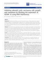
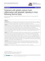
All patients had their postoperative radiographs com-
pared to the radiographic crit eria listed in the ‘Biomet
surgical technique’ manual (Figure 2). Each angle was
recorded together with the degree of overhang of the
prosthe sis in millimeters and th e presence or absence of
posterior osteophyte.
Each prosthesis was then score d radiographically by
summing the degree of deviation of the implant from per-
fect alignment in two planes, and adding the overhang in
mm and the presence of posterior osteophyte (present = 1,
absent = 0). For example a tibial tray with varus alignment
of 6 degrees, a 2 mm medial overhang and posterior osteo-
phyte would achieve a score of 9.
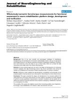
The radiographic scores are plotted against the func-
tional scores in Figure 3.
Correlation coefficients were calculated for each pros-
thesis comparing the oxford score and the xray score
(Where a poor correlation = 0.1-0.3, medium correlation
0.3-0.5, and a good correlation = 0.5-1) [15].
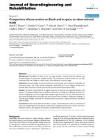
Results
38 patients with 40 Oxford knees were available for analy-
sis. Their mea n Oxford Functional Score was 32.7, range
16-48, (Figure 4). 17/40 replaced knees (42.5%) s cored
‘excellent’, 13/40 (32.5%) scored ‘good’, 7/40 (17.5%) ‘mod-
erate’ and 3/40 (7.5%) were ‘poor’. Twenty two of the forty
knees exhibited medial knee discomfort or pain (55%) and
this symptom was present in 22 of the 24 patients with
oxford scores lower than 37 (91.6%) Figure 5.
Themeanradiographicscorewas25.3(range7-43),
where 0 would signify a perfect radiograph. 6 implants
were malpositioned according to the limits for component
alignme nt as suggested in the surgical technique manual.
It was noted that the majority of abnormal X-ray criteria
arose from apparent varus or valgus placement of the tibial
tray or femoral component, and less commonly flexion of
the femoral component or posterior tilt of tibial tray. We
found no obvio us relationship between Xray scores and
presence of medial knee pain or discomfort (Figure 6).
Excessive medial overhang of the tibial component (more
than 2 mm) was seen in 4/40 knees and did not seem to
correlate with poor Oxford scores or medial knee discom-
fort (correlation coefficient = 0.18). In fact the 3 cases with
excessive medial overhang of 3 mm, 3 mm and 6 mm had
Oxford scores of 45, 43, and 42 respectively.
We found a poor correlation between Oxford Knee
Scores and the overall X-ray scores (see Figure 2). For
example, patient 1 achieved an Oxford knee score of 48
(best achievable) and scored 30 on X-ray criteria (poor),
while another patient achieved 16 on Oxford score
(poor), and 16 on X-ray (good alignment). correlation
coefficient was 0.107. The closest correlation we found
statistically, was a medium correlation, between the
varus/valgus positioning o f the femo ral component and
the Oxford score (0.38). Examples of good and poorly
positioned prosthesis can be seen in Figures 7 and 8.
Grading for the Oxford Knee Score
Score 0 to 19
Poor
Score 20 to 29
Moderate
Score 30 to 39
Good
Score 40 to 48
Excellent
Figure 1 Oxford knee score.
Edmondson et al. Journal of Orthopaedic Surgery and Research 2011, 6:52
/>Page 2 of 7
We could find no correlation between preoperative
arthritis and post operative Oxford scores (correlation
coefficient 0.12) or pre op arthritis and Medial knee dis-
comfort (correlation coefficient 0.08).
Discussion
Several authors have reported good success rates using
the Oxford Unicompartmental knee replacement system
[14,16]. It has been suggested that results are compar-
able to that of Total Knee Arthroplasty (TKA) [3].
In our small and retrospective study, 4 of the 48
Oxford unicompartmental kn ee replacements had been
revisedwithinthe4.5yearfollowupperiodandour
outcomes in the surviving knees were disappointing
compared with other studies [3,4,7,14,16-19], with 7.5%
of our patients achieving ‘poor’ results according to the
Radiographic criteria
Position and size of components
Femoral component
A/A Varus/Valgus angle <10
o
Varus- <10
o
Valgus
B/B Flexion/Extension angle <5
o
Flexion-<5
o
Extension
C/C Medial/Lateral placement Central
D Posterior fit Flush / <2mm overhang
Tibial Component (relative to tibia)
E/E Varus/Valgus <10
o
varus -<10
o
valgus
F/F Posteroinferior tilt 7
o
+/- 5
o
G Medial fit Flush or <2mm overhang
H Posterior fit Flush or <2mm overhang
J Anterior fit Flush or <3mm overhang
K Lateral fit Flush, no gap
Meniscal bearing (relative to tibial component)
L Xray marker central, and parallel with tibial
component
Bone interfaces
M Posterior Femoral Parallel surfaces cement OK
N Tibial Parallel surfaces cement OK
Other
Posterior Osteophytes None visible
Depth of Tibial saw cuts Minimal ingress of cement
Intact posterior cortex No extruded cement posteriorly
No anterior impingement Adequate bone removed no cement
Figure 2 Radiographic criteria for optimal positioning of the Oxford Unicompartmental Knee replacement.
Edmondson et al. Journal of Orthopaedic Surgery and Research 2011, 6:52
/>Page 3 of 7
‘Oxford Knee Scoring’ system. Having said t his although
we were disappointed with our average Oxford core of
33, the average Oxford score following Total knee repla-
cement has been quoted as 34.82 at two years in a
recent large study [20].
Our results are similar to those reported by Van Isaker
et al, who demonstrated functional results to be poor in
10% of their followed up knees [8], and C ottenie et al [9]
in which 6% had poor and 4% fair functional ratings.
Both of these studies used the ‘Hospital for Special Sur-
gery’ score, not the Oxford functional rating system that
we used.
In our study four UKAs required revision: two were
revised for pain secondary to progressive lateral tibiofe-
moral compartment degenerative change, one was revised
after avascular necrosis developed within the lateral
femoral condyle, and one was revised because of persitent
and unexplained medial pain, in all cases symptoms
resolved with conversion to TKA.
We found little correlation between component mal-
positioning and poor oxford scores. This is in keeping
with very recent work by the Oxford group who con-
cluded that because of the spherical femoral component,
the Oxford UKR is tolerant to femoral mal-alignment of
10° and tibial mal-alignment of 5° [21].
We feel medial knee pain is problematic in this pros-
thesis. There are several possible aetiologies for medial
discomfort including: impi ngement; medial overhang of
the tibial component; ceme nting errors; aseptic loosening
of femur or tibia; soft tissue irritation (MCL, Pes Anseri-
nus); and neuroma formation. Unfortunately there are a
group of patients that ge t unexplained medial pain wh ich
is no t attributable t o any of these f actors. Of t hose with
unexplained pain occasionally these will often settle after
1-2y, however it is our experience that an unacceptable
number (22/40) persist beyond this time. Our study
included only patients of > 3y post op and therefore
those ‘early settlers’ are excluded automatically.
Patients reporting medial knee pain had poorer
Oxford scores (Figure 4). 91.6% (22/24) of those with
medial pain had scores of 37 or less, as far as we are
aware this close correlation has not been previously
reported. It is noteworthy that we found a relatively
high incidence of medial knee pain despite the fact that
phase III Unicompartmental replacements were used.
Although excessive medial overhang of the tibial com-
ponent (more than 2 mm) was seen in 4/40 knees this
0
10
20
30
40
50
60
01020304050
Xray score
Oxford score
Figure 3 Scatterplot showing Oxford scores against
Postoperative Xray scores.
Figure 4 Distribution of scores in our series.
0
10
20
30
40
50
60
1 4 7 1013161922252831343740
Patient
Score
Oxford score
Presence of Medial Pain
Figure 5 Plot of Oxford scores against the pr esence of medial
knee pain in each patient.
0
5
10
15
20
25
30
35
40
45
50
1 4 7 1013161922252831343740
Patient
Score
Xray score
Presence of medial knee pain
Figure 6 Plot of Radiographic scores against the presence of
medial knee pain for each patient.
Edmondson et al. Journal of Orthopaedic Surgery and Research 2011, 6:52
/>Page 4 of 7
did not seem to correlate with poor Oxford scores or
medial knee disc omfort. This is in keeping with the
most recent results reported by Murray et al [22]. They
reported that medial overhang o f < 3 mm and did not
worsen Oxford scores when compared with an overhang
of > 3 mm which did have a negative impact on the
scores, they did not report an association with medial
jointdiscomfortorpain.Itshouldbenotedthatin
Figure 8 the Radiograph is rotated so the overhang visi-
ble is likely to be mostly posteromedial, which could be
less problematic than direct or anteromedial overhang.
This may have some bearing on the lack of correlation
between overhang and medial pain as some reported
overhangs could have been the less signific ant ‘postero-
medial’ type. This, however, still does not help in our
understanding of why medial pain occurs in high num-
bers of patients (in our study) following Oxford unicom-
partmental knee replacement.
A large proportion of our patients experienced medial
knee pain (more than half). We believe that this medial
dis comfort does correlate with poorer results, as none of
those with scores > 37 complained of the symptom and
all those with scores below that did. However it is not the
single most important determinant of poor functional
results as several patients (18/22 complaining of medial
pain) had outcomes which were ‘moderate’ to ‘good’.Isit
possible that the presence of medial knee pain is irrele-
vant to the outcome of these k nees? Certainly we do not
believe th is to be the case as we h ave found that medial
joint disco mfor t was a common reason for patient dissa-
tisfaction with the Oxford UKA, with one patient requir-
ing revision to TKR (With successful outcome).
There are suggestions that patients with lesser degrees
of osteoarthritis preoperatively do not achieve such
good results with arthoplasty as those with greater wear.
Within our small sample we did not find this to be the
case, and furthermore, we did not note a correlation
between severity of preoperative osteoarthritis and pre-
sence of post op persistent medial discomfort.
There are limitations to our study including being a
retrospective review of a small cohort. Due to the fact
that we excluded all patients with significant patellofe-
moral a rthritis, we performed very few UKAs (48) when
compared with TKAs (around 740) during the period
studied and this may, of course, have a significant bearing
on our results. It has been suggested that as the Oxford
unicompartmental arthroplasty is a demanding proce-
dure that the outcomes are better in units where the
operation is being performed frequently [18,23-25].
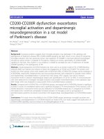
Figure 7 An example of a knee with a good radiographic
score.
Figure 8 An example of a knee with a poor radiographic score.
Edmondson et al. Journal of Orthopaedic Surgery and Research 2011, 6:52
/>Page 5 of 7
When the cause for revision of Knee replacement was
studied from the New Zealand Joint registry data, it was
noted that the early revision rate for the Oxford unicom-
partmental knee was 2.9 times greater than that for Total
knee replacement. However, higher-use surgeons (i.e.
those performing one/month or more) had a revision
rate comparable to TKA. Those performing > 12 per year
had a revision rate of 0.99%, those performing 8-11 per
year had revision rates of 4%, those performing 2-7 per
year 6.4% and those performing 1 per y had an 8% revi-
sion rate [26].
We used standard post operative Xrays to score align-
ment of prosthes es, rather than ‘screened’ radiographs,
and we accept this may affect the calculation of the
radiographic scores.
Conclusion
Our small study demonstrated disappointing medium
term results with the ‘Oxford Unicom partmental Knee
Arthroplasty’,7.5%achieving‘ poor’ Oxford scores, and
around 9% requiring revision within 5 years. We accept
that these poor results could be attributable to the rela-
tively low numbers performed in our unit. We also accept
that performing unicompartmental replacements more
frequently could improve our results, this could be done
by extending our indications and ignoring the presence of
patellofemoral arthritis (if not clinically symptomatic) as
suggested in the new guidelines by the Oxford group.
The v ast majority of those patients in our study report-
ing medial knee pain recorded Oxford scores of < 37, and
we feel that the presence of medial knee pain is asso-
ciated with poorer functional results. Furthermore, it is
our experience that this symptom is a common com-
plaint when following up t hese patients, regardless of the
alignment of the prosthesis. Although not formally
ass essed in this study, we find our patients exhibited sig-
nificant dissatisfaction with the persistence of medial
knee pain post operatively. We also noted no significant
correlation between grade of p reoperative arthritis and
post operative Oxford score or medial knee pain.
Finally, we note that despite current interest in optimis-
ing the positioning of UKA to improve functional results,
our study failed to demonstrated a correlation between the
radiographic alignment of the prosthesis and the patients
functional Oxford score.
Acknowledgements
No funding was received for this study. All contributors were fully involved with
the preparation and analysis of the results of this study. I would like to
acknowledge the help of Matthew Hankins of the Brighton and Sussex University
Department of Statistics, for his advice and statistical analysis of the results.
Authors’ contributions
All authors were involved with the assessment and subsequent follow up of
these patients, and all authors have read and approved the manuscript.
Competing interests
The authors declare that they have no competing interests.
Received: 7 August 2009 Accepted: 10 October 2011
Published: 10 October 2011
References
1. Murray DW, Goodfellow JW, O’Connor JJ: The Oxford medial
unicompartmental arthroplasty: a ten year survival study. Journal of Bone
and Joint surgery (B) 1998, 80(6):983-9.
2. Keys GW, Ul-Abbiddin Z, Toh EM: Analysis of first forty Oxford medial
unicompartmental knee replacement from a small district general
hospital in UK. Knee 2004, 11(5):375-7.
3. Rajasekhar C, Das S, Smith A: Unicompartmental knee arthroplasty 2-12
year results in a community hospital. Journal of Bone and Joint surgery (B)
2004, 86(7):983-5.
4. Svard UC, Price AJ: Oxford medial unicompartmental knee arthroplasty. A
survival analysis of an independent series. Journal of Bone and Joint
surgery (B) 2001, 83(2):191-4.
5. Verdonk R, Cottenie D, Almqvist KF, Vorlat P: The Oxford
unicompartmental knee prosthesis: a 2-14 year follow-up. Knee Surg
Sports Traumatol Arthros 2005, 13:163-166.
6. Newman J, Pydissiti R, Ackroyd C: Unicompartmental or total knee
replacement. The 15 year results of a prospective randomised controlled
trial. Journal of Bone and Joint Surgery 2009, 91-B(1):52-57.
7. Volpin G, Schachar R, Shtarkjer H: Functional outcome after
unicompartmental knee arthroplasty in patients with osteoarthritis of
the medial compartment. Journal of Bone and Joint Surgery 2006, 88-
B(SUPP_II):335.
8. Van Isacker T, Cottenie D, Vorlat P, Verdonk R: The oxford
unicompartmental knee replacement an independent 10 year follow up.
Journal of Bone and Joint Surgery 2004, 86-B(SUPP_III):290.
9. Cottenie Dominique, Vorlat P, Byn P, Almqvist KF, Verdonk R: The oxford
unicompartmental knee replacement a 2-14 year follow up. Journal of
Bone and Joint Surgery 2004, 86-B(SUPP_III):308.
10. Kim KT, Lee S, Park H, Cho K, Kim K: A Prospective Analysis of Oxford
Phase 3 Unicompartmental Knee Arthroplasty. Orthopedics 2007,
30(5):15-18.
11. Oxford Unicompartmental knee replacement. Biomet Surgical technique
manual [ />FLK089 Oxford Knee Surgical Technique(screen res).pdf], Appendix pp34-35.
12. Chana R, Shenava Y, Nicholl A, Lusted F, Skinner P, Gibb P: Five to 8 year
results of the uncemented Duracon total knee arthroplasty system. The
Journal of Arthroplasty 2008, 23(5):677-682.
13. White SH, Goodfellow JW, O’connor JJ: Anteromedial Osteoarthritis of the
Knee. Journal of Bone and Joint Surgery (Br) 1991, , 73-8: 582-86.
14. Dawson J, Fitzpatrick R, Murray D, Carr A: Questionnaire on the
perceptions of patients about total knee replacement. Journal
of Bone
Joint Surgery (Br) 1998, 80(1):63-9.
15. Cohen J: Statistical power analysis for the behavioral sciences (2nd ed.).
Hillsdale, NJ: Lawrence Erlbaum Associates; 1988, ISBN 0-8058-0283-5.
16. Cartier P, Sanouiller JL, Greisamer RP: The Oxford Unicompartmental,
Arthroplasty - a ten year survival study. Journal of Bone and Joint Surgery
(Br) 1998, , 80-B: 983-9.
17. Palacious BF, Montes SF: Unicompartmental knee arthroplasty with an
Oxford prosthesis. Acta Ortop Mex 2007, 21(2):49-54.
18. Robertson O, Knutson K, Lewold S, Lidgren L: The routine of surgical
management reduces failure after unicompartmental knee implant.
Journal of Bone and Joint Surgery 2001, , 83B: 45-9.
19. Emerson RH Jr, Hansborough T, Reitman RD, Rosenfeldt W, Higgins LL:
Comparison of mobile and fixed bearing unicompartmental knee
implant. Clinical Orthopaedics 2002, 404:62-70.
20. The KAT trial group: The knee arthroplasty trial Design features, Baseline
characteristics and two year functional outcomes after alternative
approaches to knee replacement. J Bone Joint Surg Am 2009, 91:134-141.
21. Gulati A, Chua R, Simpson DJ, Dodd CAF, Murray DW: Influence of
component alignment on Unicompartmental Knee replacement. The
Knee 2009, 16(3):196-199.
22. Murray D, Simpson D, Dodd C, Gill H, Beard D, Pandit H, Chau R: An
acceptable limit of tibial component overhang in the Oxford
Edmondson et al. Journal of Orthopaedic Surgery and Research 2011, 6:52
/>Page 6 of 7
unicompartmental knee arthroplasty. British Association for Surgery of the
Knee, free paper session ‘Technical aspects of UKA’, Bournemouth 2008.
23. Gleeson RE, Evans R, Ackroyd CE, Webb J, Newman J: Fixed or mobile
bearing unicompartmental knee replacement? A comparative cohort
study. The Knee 2004, 11:379-384.
24. Murray D: Mobile bearing Unicompartmental knee replacement.
Orthopaedics 2005, 28(9):985-87.
25. Kasodekar VB, Yeo SJ, Othman S: Clinical outcome of unicompartmental
knee arthroplasty and influence of alignment on prosthesis survival rate.
Singapore Medical Journal 2006, 47(9):796-802.
26. Tregonning R, Rothwell A, Hobbs T, Hartnett N: Early failure of the Oxford
phase 3 cemented medial uni-compartmental knee joint arthroplasty. J
Bone Joint Surg Br 2009, 91-B(Supp II):339.
doi:10.1186/1749-799X-6-52
Cite this article as: Edmondson et al.: Oxford unicompartmental knee
arthroplasty: medial pain and functional outcome in the medium term.
Journal of Orthopaedic Surgery and Research 2011 6:52.
Submit your next manuscript to BioMed Central
and take full advantage of:
• Convenient online submission
• Thorough peer review
• No space constraints or color figure charges
• Immediate publication on acceptance
• Inclusion in PubMed, CAS, Scopus and Google Scholar
• Research which is freely available for redistribution
Submit your manuscript at
www.biomedcentral.com/submit
Edmondson et al. Journal of Orthopaedic Surgery and Research 2011, 6:52
/>Page 7 of 7