Báo cáo hóa học: "Inhibiting adenoid cystic carcinoma cells growth and metastasis by blocking the expression of ADAM 10 using RNA interference" pdf
Bạn đang xem bản rút gọn của tài liệu. Xem và tải ngay bản đầy đủ của tài liệu tại đây (1.06 MB, 10 trang )
RESEARC H Open Access
Inhibiting adenoid cystic carcinoma cells growth
and metastasis by blocking the expression of
ADAM 10 using RNA interference
Qin Xu, Xiuming Liu, Wantao Chen, Zhiyuan Zhang
*
Abstract
Background: Adenoid cystic carcinoma is one of the most common types of salivary gland cancers. The poor
long-term prognosis for patients with adenoid cystic carcinoma is mainly due to local recurrence and distant
metastasis. Disintegrin and metalloprotease 10 (ADAM 10) is a transmembrane protein associated with metastasis
in a number of dive rse of cancers. The aim of this study was to analyze the relationship between ADAM 10 and
the invasive and metastatic potentials as well as the proliferation capability of adenoid cystic carcinoma cells
in vitro and in vivo.
Methods: Immunohistochemistry and Western blot analysis were applied to detect ADAM 10 expression levels in
metastatic cancer tissues , corresponding primary adenoid cystic carcinoma tissues, adenoid cystic carcinoma cell
lines with high metastatic potential, and adenoid cystic carcinoma cell lines with low metastatic potential. RNA
interference was used to knockdown ADAM 10 expression in adenoid cystic carcinoma cell lines with high
metastatic potential. Furthermore, the invasive and metastatic potentials as well as the proliferation capability of
the treated cells were observed in vitro and in vivo.
Results: It was observed that ADAM 10 was expressed at a significantly higher level in metastatic cancer tissues
and in adenoid cystic carcinoma cell lines with high metastatic potential than in corresponding primary adenoid
cystic carcinomas and adenoid cystic carcinoma cell lines with low metastatic potential. Additionally, silencing of
ADAM 10 resulted in inhibition of cell growth and invasion in vitro as well as inhibition of cancer metastasis in an
experimental murine model of lung metastases in vivo.
Conclusions: These studies suggested that ADAM 10 plays an important role in regulating proliferation and
metastasis of adenoid cystic carcinoma cells. ADAM 10 is potentially an important therapeutic target for the
prevention of tumor metastases in adenoid cystic carcinoma.
Background
Adenoid cystic carcinoma is one of the most common
types of salivary gland cancers, characterized by hetero-
geneous phenotypic features and persistently progressive
biological behavior. The poor long-term prognosis for
patients with adenoid cystic carcinoma is mainly due to
local recurrence related to perineural invasion and
delayed onset of distant metastasis, particularly to the
lungs [1,2]. In-depth studies on its invasion and
metastasis mechanisms are of great significance for the
prognosis, evaluation, and selection of treatment
protocols.
The ADAM (A disintegrin and metalloprotease) family
is a class of type I transmembrane proteins that partici-
pate in a wide range of physiological functions. This
family of proteins is named because they have two main
structural domains, the disintegrin domain and the
matrix metalloproteinase domain. They can degrade the
extracellular matrix (ECM) and control cell adhesion
and movement through regulation of intercellular adhe-
sion, protease activity and cell activities that are closely
related to the metastasis of human tumors [3,4]. Among
the members of the ADAM family, some ADAMs, such
* Correspondence: zhang.zhiyuan2010@hotm ail.com
Department of Oral and Maxillofacial Surgery, Ninth People’s Hosp ital,
Shanghai Jiao Tong University School of Medicine, Shanghai Key Labor atory
of Stomatology, Shanghai 200011, China
Xu et al. Journal of Translational Medicine 2010, 8:136
/>© 2010 Xu et al; lice nsee BioMed Central Ltd. This is an Open Access article d istributed under the terms of the Creative Commons
Attribution License ( which permits unrestricted use, distribution, and reproduction in
any medium, provided the original work is properly cited.
as ADAM 9, 10, 17, are closely involved in the tumori-
genesis, development, and metastasis of tumors [5-7].
Recently, ADAM 10 has bee n reported to play impor-
tant roles in cell migration, tumor development, and
metastasis by proteolytic shedding of cell surface pro-
teins. It has been demonstrated that ADAM 10 can
cleave collagen type IV of the basement membrane and
is relevant to tumor metastasis [8]. In another study, it
was shown that the cleavage of CD44 catalyzed by
ADAM 10 contributed to the migration and invasion of
glioblastoma tumor cells [9]. In addition, our previous
study found that ADAM 10 expression in adenoid cystic
carcinoma cells with high metastatic potential was sig-
nificantly higher than t hat in adenoid cystic carcinoma
cells with low metastatic potential based on gene chip
analysis [10]. These findings strongly suggest that
ADAM 10 plays an essential role in tumor metastases.
The a im of this study was to analyze the relationship
between the expression of ADAM 10 and the invasive
and metastatic potentials as well as the proliferation
capability of adenoid cystic carcinoma cells in vitro and
in vivo. In the present study, the expression level of
ADAM 10 was examined both in primary tumor sec-
tions and corresponding metastatic lymph nodes from
patients with adenoid cystic carcinoma. RNA interfer-
ence (RNAi) was applied to inhibit the expression of
ADAM 10 i n an adenoid cystic carcinoma cell line with
high metastatic potential, and the changes in biological
behaviors such as cell proliferation and metastasis were
observed both in vitro and in vivo.
Materials and methods
Cell lines and specimens
Adenoid cystic carcinoma cells with high metastatic
potential (SACC-LM) and low metastatic potential
(SACC-83) wer e provided bythePekingUniversity
School of Stomatology [11]. Both cell lines were cul-
tured in RPMI 1640 complete medium with 10% inacti-
vated FBS, 200000 u/L penicillin, and 200000 u/L
streptomycin at 37°C. Paraffin specimens of primary foci
and metasta tic lymph nodes from 15 patient s with ade-
noid cystic carcinoma and cervical lymph node metasta-
sis and paraffin specimens of primary foci of adenoid
cystic carcinoma from 20 patients without cervical
lymph node metastasis were pro vided by the Depart-
ment of Oral Pathology, Ninth People’s Hospital, Shang-
hai Jiao Tong University Scho ol of Medicine. T he
metastatic lymph node tissues were histopathologically
graded using a specific three-tier grading system, origin-
ally proposed by Szanto et al [12].
Immunohistochemistry
Immunohistochemistry for ADAM 10 was performed
using standard methods. Endogenous peroxidase activity
was blocked by treatment with 3% hydrogen peroxide in
PBS for 30 min. The specimens were rinsed in PBS. The
tissue sections were stained with a mouse monoclonal
anti-ADAM 10 antibody (R&D Systems, Minneapolis,
MN, USA). The sections were incubated overnight at
4°C (1:50 dilution of primary antibodies). The bound
antibody was detected with a secondary biotinylated
antibody for 30 min at room temperature and visualized
using diaminobenzidine as a chromogenic substrate.
The sections were then counterstained with hemat oxy-
lin. Immunostaining was defined as positive when more
than 30% of tumor cells stained positive. The level of
immunostaining was quantified using a semi-automated
computerized image analysis system (Image Pro Plus
6.0; Media Cybernetics, Bethesda, FL, USA), which has
been successfully applied to analyze histological sections
and d escribed in previous reports [13-15]. In brief, the
integrated optical density (IOD; IOD = area × average
optical density) o f positive staining was calculated for
each tissue section. The average IOD scores were calcu-
lated from triplicate values from each section. The
image analysis was performed by three pathologists
blinded to the treatment group.
Preparation of plasmid based ADAM 10 shRNA vector
The ADAM 10 small interfering RNA (siRNA) sequence
(CAGUGUGCAUUCAAGUCAA) was designed using
the software siRNA Target Designer (Promega, Madison,
WI, USA). The preparation of the RNAi vector expres-
sing the human ADAM 10 short hairpin RNA (shRNA)
was performed using the pSuper siRNA expression plas-
mid with the U6 promoter (Oligoengine, Seattle, WA,
USA) [16].
Construction of stable silencing cell lines
SACC-LM cells were transduced with the specific ADAM
10 shRNA vector or an empty plasmid using Lipofecta-
mine 2000 transfection reagent. G418 (300 μg/ml)
was used to screen stably transfected clones. The
expression of ADAM 10 was examined by real time
RT-PCR and Western blotting with an antibody against
ADAM 10 (these experiments we re repeated three
times) to validate the silencing efficiency of the target
gene after RNAi. The cell line with stable transfection
and effective inhibition of the ADAM 10 gene was
named SACC-ADAM 10-RNAi, and the c ell line with
stable transfection of the control plasmid was named
SACC-Mock.
Quantitative RT-PCR
Quantitative RT-PCR (qRT-PCR) for ADAM 10 tran-
scripts in adenoid carcinoma cell lines was carried out
using the PrimeScript RT reagent kit following the man-
ufacturer’ s instructions (TaKaRa Bio, Shiga, Japa).
Xu et al. Journal of Translational Medicine 2010, 8:136
/>Page 2 of 10
ADAM 10 gene-specific amplification was confirmed by
PCR with specific primers (5’ -C TGCCCAGCATCT-
GACCCTAA-3’ and 5’ -TTGCCATCAGAACTGGCA-
CAC-3’ ) and subjected to melting curve analysis.
GAPDH was used as an internal control for standardiza-
tion. All qRT-PCR tests were performed in triplicate.
ThedatawereanalyzedusingthecomparativeCt
method.
Western blot analysis
Cells were washed twice with cold phosphate-buffered
saline (PBS; 137 mM NaCl, 2.7 mM KCl, 10 mM
sodium phosphate dibasic, 2 mM potassium phosphate
monobasic, pH 7.4) and lysed on ice in buffer (150 mM
NaCl,50mMTris-Hcl,2mMEDTA,1%NP-40,
pH 7.4) containing protease inhibitors. Equal amounts
of protein (20 μg/lane) from the cell lysates were elec-
trophoresed under nonreducing conditions on 10% acry-
lamide gels. After SDS-PAGE, proteins were transferred
to a polyvinylidene difluoride membrane. The mem-
brane was incubated for 2 h in PBS plus 0.1% Tween-20
and 5% nonfat skim milk to block nonspecific binding.
Subsequently, the membrane was incubated for 2 h
with an antibody against ADAM 10 (R&D Systems,
Minneapolis, MN, USA). After washing, proteins were
visualized using an ECL detection kit with the appropri-
ate HRP-conjugated secondary antibody (Amersham
Pharmacia Biotech, Piscataway, NJ, USA). The mem-
branes were stripped a nd probed with monoclonal anti-
bodies for GAPDH for loading control as per standard
protocols.
Proliferation assay
The MTT (3-[4,5-dimethylth iazol-2-yl]-2, 5-diphenylte-
trazolium bromide) colorimetric assay was used to
screen for cell proliferation. Briefly, cells were seeded in
8 wells of 96-well plates at a density of 2 × 10
3
cells/
well. One plate was taken out at the same time every
day after the cells had adhered to the wall. Twenty
microliters of MTT (5 mg/ml) were added into each
well, and the cell culture was continued fo r 4 h. After
aspiration of the medium, the cells were lysed with
DMSO. The absorbance was measured using a micro-
plate reader at a wavelength of 490 nm. The measure-
ment was carried out for 8 consecutive days, and the
cell growth curve was plotted with OD values as ordi-
nate against tim e as abscissa. The experiment was
repeated three times.
In vitro invasion assay
Cell invasive behavior was evaluated using 24-well trans-
well units with 8-μm porosity polycarbonate filters. The
filters were coated with 50 μl of 8 mg/ml reconstituted
basement membrane substance (Matrigel; BD Biosciences,
San Diego, CA, USA). The coated filters were air-dried at
4°C prior to the addition of the cells. The basement mem-
brane was hydrated with 50 μl serum-fre e RPMI 1640
medium 30 min before use. The cells were digested with
trypsin, and the cell density was adjusted to 1 × 10
6
/ml
using serum-free RPMI 1640 medium. A total of 200 μlof
cell suspension was a dded into each upper Transwe ll
chamber, and 600 μl of RPMI 1640 me dium containing
5% fetal bovine serum was added into the lower chamber.
There were three duplicates for each cell group. Then, the
cells were incubated for 24 h in a humidified atmosphere
of 5% CO
2
at 37°C. Cells were fixed with methanol and
stained with Giemsa. Cells on the upper surface of the fil-
ter were removed by wiping with a cotton swab, and inva-
sion was determi ned by cou nting the cells that migrated
to the lower side of the filter with optical microscopy at
400×. A total of five visual fields at the center and in the
surrounding areas were counted, and the average was cal-
culated [17]. The experiment was repeated three times.
Analysis of lung metastasis in vivo
Four-week-o ld female BALB/c nu/nu nude mice were
raised under specific patho gen free conditions. All ani-
mal experiments were carried out according to the stan-
dards of animal care a s outlined in the Guide for the
Care and Use of Experimental Animals of the Medical
College of Shanghai Jiaotong University. The study pro-
tocol was approved by the hospital ethical committee.
As an experimental lung metastasis model, 0.2 ml sin-
gle-cell suspensions (10
6
cells) were injected via the
mouse tail vein. There were seven mice in each group.
The mice were sacrificed 40 days after inoculation, and
bilateral lung tissues were removed. Pathological sec-
tions of lung tissues with the maximum cross-sectional
area were prepared. Tumor burden was determined by
weighing the lungs of the animals as described in pre-
vious reports [18-20].
Statistical analysis
AFisher’ s exact test was perf ormed to compare differ-
ences in ADAM 10 expression levels between primary
tumors and corresponding metastatic lymph node
groups. Normally distributed, continuous variables were
compared using one-way analysis of variance (ANOVA).
When ANOVA produced a significant difference
between groups, mult iple comparisons of gro up means
were performed using the Bonferroni procedure with a
type I error adjustment. Repeated measure analyse s
were performed to assess the group effect s on prolifera-
tive capacity over the time course. Data are presented as
mean ± standard deviation. All statistical assessments
were two-sided and evaluated at the 0.05 significance
level. All statistical analyses were performed using SPSS
13.0 statistics software (SPSS, Chicago, IL, USA).
Xu et al. Journal of Translational Medicine 2010, 8:136
/>Page 3 of 10
Results
ADAM 10 expression in primary and metastasized
adenoid cystic carcinoma tissue samples
First, ADAM 10 expression was examined by immunos-
taining of 15 paired tissues from patients with oral adenoid
cystic carcinoma and cervical lymph node metastasis. For
each pair of tissues, primary tumor sections and corre-
sponding metastatic lymph nodes were examined. ADAM
10 was only detected in 26.7% of primary tumors (4/15;
Figure 1A), whereas 80% of corresponding metastatic
lymph nodes showed positive ADAM 10 staining (12/15;
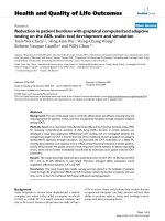
Figure 1B). Table 1 shows the overall ADAM 10 expres-
sion in metastatic lymph nodes according to the histologic
grade, which indicated that the ADAM 10 immuno-
reaction was stronger with a higher histologic grade. The
Fisher’s exact test indicated that the expression levels of
ADAM 10 in corresponding metastatic lymph nodes were
statistically higher than those in the primary tumors (p =
0.004). The IOD value of ADAM 10 staining for metastatic
lymph nodes was also significantly higher than the ADAM
10 staining for primary tumors (p < 0.001; Figure 1D), sug-
gesting that ADAM 10 expression is closely related to
tumor metastasis. Next, ADAM 10 expression in 20 pri-
mary foci tissues without cervical lymph node metas tasis
were detected. In these cases, 30% of primary tumors (6/
20) showed positive staining (Figure 1C), which indicated
a similar expression rate in primary foci.
ADAM 10 expression in adenoid cystic carcinoma cells
with different metastatic potentials
The metastatic potential of SACC-LM and SACC-83
cells was investigated using a matrigel invasion assay and
experimental lung metastasis tests. The invasion assay
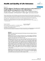
results indicated that SACC-LM cells had a significantly
higher ability to pass through the basement membrane
compared to SACC-83 cells (p < 0.001; Figure 2A, B, E).
Similarly, the experime ntal lung metastasis results (n = 7
mice per group) showed the lung weight derived from
SACC-LM group was 0.61 ± 0.15 g, compared to 0.24 ±
0.06 g from the SACC-83 group (p < 0.001; Figure 2C, D,
F). These results verified the difference in metastasis
potential of SACC-LM and SACC-83 bothin vitro and
in vivo.
Subsequently, both ADAM 10 mRNA and protein
levels were examined in adenoid cystic carcinoma cells
with either high (SACC-LM) or low (SACC-83)
Figure 1 Immunohistochemical staining for ADAM 10 on paired primary adenoid cystic carcinoma (a) and corresponding metastatic
lymph nodes (b) and in 20 primary foci tissues without cervical lymph node metastasis (c). Scale bar = 100 μm. (d) The IOD value of
ADAM 10 staining (mean ± SD) in metastatic lymph nodes was significantly higher than that in primary tumors (*p < 0.001).
Table 1 ADAM 10 expression in metastatic lymph nodes
according to the histologic grade
ADAM 10 expression
Grade Negative No. (%) Positive No. (%) Total
I0 0 0 0 0
II 1 33.3% 3 25% 26.7%
III 2 66.7% 9 75% 73.3%
Xu et al. Journal of Translational Medicine 2010, 8:136
/>Page 4 of 10
Figure 2 Detection of the meta static potential of SACC-LM and SACC-83 cells. (a), (b) A Matrigel transwell invasion assay was used to test
the ability of SACC-LM and SACC-83 cells to invade the filter membrane. (c), (d) Overview of lung tissues from mice injected with SACC-LM and
SACC-83 cells (scale bar = 0.5 cm). Tumors are indicated by black arrows. (e) Values represent the cell number (mean ± SD) per visible field (*p <
0.001). (f) Lung weight
(*p < 0.001).
Xu et al. Journal of Translational Medicine 2010, 8:136
/>Page 5 of 10
metastatic potential. ADAM 10 was more a bundant at
both the mRNA and protein level (about 2 .6 fold) in
SACC-LM cells when compared to SACC-83 (Figure 3A
and 3B), which corroborated the tumor tissue results
and indicated that ADAM 10 overexpression might cor-
relate with cancer metastasis.
Abolished ADAM 10 expression in SACC-LM cells
To investigate whether ADAM 10 expression was essen-
tial for the metastatic capability of SACC-LM cells,
stable ADAM 10 RNAi transfected cells (SACC-
ADAM10-RNAi) and a mock-transfected control cell
line (SACC-Mock) were established as described above.
Three cellular clones with stable ADAM 10 RNAi trans-
fection, SACC-ADAM10-RNAi ( 1), (2), and (3), were
selected for further evaluation. Compared t o parental
(SACC-LM) and mock-transfected (SACC-Mock) cells,
both m RNA and protein expression of ADAM 10 were
significantly reduced in SACC-ADAM10-R NAi (1), (2),
and (3) cells (all, p < 0.001; Figure 4A, B).
Gene silencing of ADAM 10 reduces cell proliferation and
migration in SACC-LM cells
To examine w he ther the knockdown A D AM 10 expression
had any effect on cell growth, an MTT cell proliferation
assay was performed. Compared to parental (SACC-LM)
and mock-transfected (SACC-Mock) cells, ADAM 10-
RNAi cells showed decreased cell proliferation, supporting
theroleofADAM10incellgrowthinSACC-LMcells
(Figure 5 C). In addition, the affect of gene silencing
of ADAM 10 on the cell migration ability of SACC-LM
cells was also investigated by transwell invasion assay
(Figure 5A). The results indicated that ADAM 10-RNAi
cells had a significantly reduced ability to pass through the
basement membrane when compared to the parental and
mock-transfected cells (all, p < 0.00 1; Figure 5B). These
data supported the notion that ADAM 10 expression is
essential for both cell proliferation and migration.
Gene silencing of ADAM 10 reduces tumor metastasis in v ivo
To evaluate if ADAM 10 expression was essential for
the metastatic potential of SACC-LM cells in vivo,par-
ental (SACC-LM), mock-transfected SACC-LM cells
(SACC-Mock), or ADAM 10-RNAi SACC-LM cells-
SACC-ADAM 10-RNAi (1), (2), and (3)-were injected
into BALB/c nude mice (n = 7 mice per group). Mice
Figure 3 ADAM 10 expression levels in SACC-83 and SACC-LM
cell lines. (a) Quantitative RT-PCR showing relative ADAM 10 mRNA
levels (mean ± SD) in SACC-83 cells (low metastatic potential)
compared with SACC-LM cells (high metastatic potential) (*p <
0.001). (b) Western blot analysis showing ADAM 10 protein
expression in SACC-83 and SACC-LM cell lines. GAPDH served as a
loading control.
Figure 4 Abolishment of ADAM 10 expression in SACC-LM
cells. (a) ADAM 10 mRNA levels were determined by qRT-PCR.
Relative fold induction for the ADAM 10 mRNA (mean ± SD) in
mock- and ADAM 10 siRNA-transfected cells is presented relative to
the expression in parental SACC-LM cells (*p < 0.001 compared with
SACC-LM). (b) Western blot analysis for ADAM 10 protein expression
in the indicated cell lines. GAPDH was used as a loading control.
SACC-LM (high metastatic potential control); SACC-Mock (mock
transfection control); SACC-ADAM10-RNAi (1), (2), and (3) represent
the three different clones, respectively.
Xu et al. Journal of Translational Medicine 2010, 8:136
/>Page 6 of 10
were sacrificed 40 days after inoculation, and their bilat-
eral lung tissues were removed and subjected to histolo-
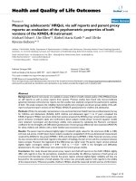
gical examination (Figure 6A). The lung weights derived
from parental and mock-transfected SACC-LM cells
were 0.57 ± 0.19 g and 0.60 ± 0.17 g, respectively, com-
pared to 0.23 ± 0.08 g, 0.21 ± 0.07 g, and 0.24 ± 0.07 g
for the SACC-ADAM 10-RNAi (1), (2), and (3) groups.
The lung weight test revealed a significant reduction of
tumor burden in ADAM 10-RNAi cells as compared to
parental or mock-transfected SACC-LM cells (p < 0.001;
Figure 6C). Next, ADAM 10 expression in the se tumors
was examined. As expected, ADAM 10 expression was
severely reduced in tumors der ived from ADAM
10-RNAi cells compared to tumors derived from paren-
tal or mock-transfected cells (Figure 6B, D). These data
again supported the argument that ADAM 10 is essen-
tial for metastasis in adenoid cystic carcinoma.
Discussion
A variety of ADAMs including ADAM 10 have been
shown to be overexpressed in cancers, and it has been
hypothesized that the downregulation of ADAM 10 may
suppress tumor growth and metastasis in adenoid cystic
carcinoma. However, previous reports that may relate to
this hypothesis are very limited. T he purpose of this
study was to analyze the relationship between the gene
silencing o f ADAM 10 and the invasive and metastatic
potentials as well as the proliferation capability of ade-
noid cystic carcinoma cells in vitro and in vivo.
In this study, we have characterized the expression of
ADAM 10 in adenoid cystic carcinoma tissues. Immu-
nohistochemical analysis indicated that ADAM 10
expression was significantly elevated in metastatic lymph
nodes compared with corresponding primary tumors,
and ADAM 10 immunoreactivity was stronger w ith a
higher histologic grade in metastatic lymph nodes. In
addition, both mRNA and protein levels of ADAM 10
were more abundant in an adenoid cystic carcinoma cell
line with high metastatic potential (SACC-LM) than in a
cell line with low metastatic potential (SACC-83). This
result indicated that high ADAM 10 expression tends to
occur in metastatic tumor tissues and overexpress ion of
ADAM 10 might be a potential p rognostic sign of high
metastatic risk, which is consistent with prior studies.
Lee et al. reported that ADAM 10 was upregulated in
melanoma metastases compared with primary melano-
mas [21]. In another study, Gavert et al. reported that
the expression of ADAM 10 was detected at the invasive
front of human colorectal tumor tissues [22]. Based on
these data, it is reasonable to speculate that ADAM 10
may play a role in tumor invasion and metastasis.
To provide evidence supporting this supposition, we
investigated the effects of ADAM 10 silencing on
in vitro cell invasion as well as in vivo cancer metastasis
in an experimental murine model of lung metastasis.
The expression of ADAM 10 was specifically knocked
down in human adenoid cystic carcinoma cell lines with
high metastatic potential using RNAi. Downregulation
Figure 5 Gene silencing of ADAM 10 reduces cell proliferation and migration in SACC-LM cells. (a) A Matrigel transwell invasion assay
was used to test the ability of the indicated cell lines to invade the filter membrane. (b) Values represent the cell number (mean ± SD) per
visible field (*p < 0.001 compared with SACC-LM). (c) Cell proliferation was analyzed using the MTT assay. Cells were monitored for 8 days and
the average OD490 (± SD) for each cell line is shown. Cells transfected with ADAM 10 siRNA showed reduced cell growth relative to parental
and mock-transfected cells. SACC-LM (high metastatic potential control); SACC-Mock (mock transfection control); SACC-ADAM10-RNAi (1), (2), and
(3) represent the three different clones, respectively.
Xu et al. Journal of Translational Medicine 2010, 8:136
/>Page 7 of 10
of ADAM 10 re sulted in a suppression of tumor cell
invasion in vitro and decreased experimental lung
metastasis in vivo, which strongly supported that
ADAM 10 is involved in the process of tumor metasta-
sis. Our finding is in agreement with previous reports
on the functional roles of ADAM 10. As we know, to
metastasize, malignant cells must first detach from the
dense, cross-linked collagen network of the ECM and
migrate through the host vasculature before extravasat-
ing the vasculature and infiltrating the host tissues
[23]. Therefore, tumor metastasis is dependent on the
tumor’ s ability to degrade the surrounding ECM and
reduced cell adhesion. A number of studies have
demonstrated that the metalloprotease domain of
ADAM 10 can cleave and remodel ECM proteins such
as type-IV collagen and CD44 [24] and influence cell-
cell signaling, including the Notch pathway [25,26].
The disintegrin domain of ADAM 10 can also interact
with matrix adhesion molecules. Hence, ADAM 10 is
able to modulate a variety of cell-cell and cell-ECM
interactions and consequently digest the basement
membrane, facilitate cell migration, and promote
tumor m etastasis. However, the detailed mechanism by
which ADAM 10 interacts with ECM proteins is not
very clear. Further studies are required to determine
these exact mechanisms. Moreover, in our study,
downregulation of ADAM 10 expression significantly
inhibited experimental lung metastasis, which sug-
gested this therapy might be a novel and promising
treatment strategy for metastasis.
In addition, in the present study, the transf ection of
ADAM 10 siRNA resulted in a significant reduction of
cellular growth of adenoid cystic carcinoma cells. Our
data are in line with previous reports showing that
Figure 6 Gene silencing of ADAM 10 reduces tumor metastasis in vivo. (a) Overview of lung tissues from mice injected with the indicated
cell lines (scale bar = 0.5 cm). Tumors are indicated by black arrows. (b) Immunohistochemical staining of ADAM 10 from tumors derived from
injected SACC-LM cells (scale bar = 50 μm). (c) Lung weight. (d) Quantification of immunohistochemical staining of ADAM 10 from b using
Image Pro Plus software (*p < 0.001 compared with SACC-LM). SACC-LM (high metastatic potential control); SACC-mock (mock transfection
control); SACC-scrambled RNA (scrambled siRNA control); SACC-ADAM 10-RNAi (1), (2), and (3) represent the three different clones, respectively.
Xu et al. Journal of Translational Medicine 2010, 8:136
/>Page 8 of 10
ADAM 10 expression is correlated with the proliferation
of tumor cells. Lee et al. demonstrated that the expres-
sion of ADAM 10 correlated with increased melanoma
cell proliferation [18]. Simi larl y, Ko et al. conf irmed the
effects of ADAM 10 on the growth of oral squamous
cell carcinoma cells [27]. In another study, results indi-
cated that suppression of ADAM 10 expression leads to
a significant decrease in prostate cell growth [28].
This effect on growth promotion might also be related
to its protease activity. It has been demonstrated that
ADAM 10 can cleave amyloid precursor protein [29-31],
a critical transmembrane molecule related to the growth
of several types of cells [32-34], which suggests that
ADAM 10 may influence the proliferation of adenoid
cystic carcinoma cells via amyloid precursor protein
shedding. Furthermore, Ko et al. reported that ADAM
10 could inhibit oral squamous cell carcinoma cell
growth through its a-secretase activity [27]. Jin et al.
have indicated that A DAM 10 can active Notch signal-
ing by suppressing ectodomain shedding of delta-1,
which subsequently leads to a strong inhibitory effect on
tumor cell proliferation [35]. These studies reveal that
different mechanisms seem to be involved in the anti-
proliferative effects of ADAM 10 against tumor cells.
Importantly, in the present study, we discovered a sig-
nificant growth inhibition of adenoid cystic carcinoma
cells following downregulation of ADAM10 via ADAM
10-specific siRNA, which suggested that ADAM 10 is a
promising new therapeutic target for the treatment of
adenoid cystic carcinoma.
Conclusions
Collectively, our data suggested that ADAM 10 expres-
sion is closely associated with adenoid cystic carcinoma
metastasis. Reduced ADAM 10 expression not only
impacted cell proliferation, b ut it also decreased the
metastatic potential of adenoid cystic carcinoma cells.
Thus, ADAM 10 i s a potential therapeutic target for the
treatment of adenoid cystic carcinoma.
Acknowledgements
This work was supported by the Chinese National Natural Science
Foundation of China (Grant Number 30600715, 81070845), Shanghai Leading
Academic Discipline Project (Project Number S30206).
Authors’ contributions
QX participated in the design of the study, carried out the
immunohistochemistry, Western blot analysis, performed the statistical
analysis, and drafted the manuscript. XL participated in animal sacrifice. WC
carried out proliferation and invasive analyses. ZZ conceived the study and
participated in its design. All authors have read and approved the final
manuscript.
Competing interests
The authors declare that they have no competing interests.
Received: 8 August 2010 Accepted: 20 December 2010
Published: 20 December 2010
References
1. Van der Wal JE, Becking AG, Snow GB, van der Waal I: Distant metastases
of adenoid cystic carcinoma of the salivary glands and the value of
diagnostic examinations during follow-up. Head Neck 2002, 24:779-83.
2. Ramer N, Wu H, Sabo E, Ramer Y, Emanuel P, Orta L, Burstein DE:
Prognostic value of quantitative p63 immunostaining in adenoid cystic
carcinoma of salivary gland assessed by computerized image analysis.
Cancer 2010, 116:77-83.
3. Murphy G: The ADAMs: signalling scissors in the tumour
microenvironment. Nat Rev Cancer 2008, 8:929-41.
4. Lu X, Lu D, Scully M, Kakkar V: ADAM proteins - therapeutic potential in
cancer. Curr Cancer Drug Targets 2008, 8:720-32.
5. Wu K, Liao M, Liu B, Deng Z: ADAM-17 over-expression in gallbladder
carcinoma correlates with poor prognosis of patients. Med Oncol .
6. Zubel A, Flechtenmacher C, Edler L, Alonso A: Expression of ADAM9 in
CIN3 lesions and squamous cell carcinomas of the cervix. Gynecol Oncol
2009, 114:332-6.
7. McCulloch DR, Harvey M, Herington AC: The expression of the ADAMs
proteases in prostate cancer cell lines and their regulation by
dihydrotestosterone. Mol Cell Endocrinol 2000, 167:11-21.
8. Endres K, Fahrenholz F: Upregulation of the alpha-secretase ADAM10–risk
or reason for hope? FEBS J 2010, 277:1585-96.
9. Murai T, Miyazaki Y, Nishinakamura H, Sugahara KN, Miyauchi T, Sako Y,
Yanagida T, Miyasaka M: Engagement of CD44 promotes Rac activation
and CD44 cleavage during tumor cell migration. J Biol Chem 2004,
279:4541-50.
10. Huang D, Chen W, Zhang Z, Zhang P, He R, Zhou X, Qiu W: Identification
of genes with consistent expression alteration pattern in ACC-2 and
ACC-M cells by cDNA array. Chin Med J (Engl) 2003, 116:448-52.
11. Hu K, Li SL, Gan YH, Wang CY, Yu GY: Epiregulin promotes migration and
invasion of salivary adenoid cystic carcinoma cell line SACC-83 through
activation of ERK and Akt. Oral Oncol 2009, 45:156-63.
12. Szanto PA, Luna MA, Tortoledo ME, White RA: H istologic grading of
adenoid cys tic carcinoma of the salivary gl ands . Cancer 1984,
54:1062-9.
13. Xu Q, Zhang Z, Zhang P, Chen W: Antisense oligonucleotides and all-
trans retinoic acid have a synergistic anti-tumor effect on oral squamous
cell carcinoma. BMC Cancer 2008, 8:159.
14. Wang-Tilz Y, Tilz C, Wang B, Tilz GP, Stefan H: Influence of lamotrigine and
topiramate on MDR1 expression in difficult-to-treat temporal lobe
epilepsy. Epilepsia 2006, 47:233-9.
15. van Holten J, Smeets TJ, Blankert P, Tak PP:
Expression of interferon beta
in synovial tissue from patients with rheumatoid arthritis: comparison
with patients with osteoarthritis and reactive arthritis. Ann Rheum Dis
2005, 64:1780-2.
16. Brummelkamp TR, Bernards R, Agami R: A system for stable expression of
short interfering RNAs in mammalian cells. Science 2002, 296:550-3.
17. Yu Y, Chen W, Zhang Y, Hamburger AW, Pan H, Zhang Z: Suppression of
salivary adenoid cystic carcinoma growth and metastasis by ErbB3
binding protein Ebp1 gene transfer. Int J Cancer 2007, 120:1909-13.
18. Cuneo KC, Fu A, Osusky KL, Geng L: Effects of vascular endothelial growth
factor receptor inhibitor SU5416 and prostacyclin on murine lung
metastasis. Anticancer Drugs 2007, 18:349-55.
19. Nakashima Y, Yano M, Kobayashi Y, Moriyama S, Sasaki H, Toyama T,
Yamashita H, Fukai I, Iwase H, Yamakawa Y, et al: Endostatin gene therapy
on murine lung metastases model utilizing cationic vector-mediated
intravenous gene delivery. Gene Ther 2003, 10:123-30.
20. Walser TC, Rifat S, Ma X, Kundu N, Ward C, Goloubeva O, Johnson MG,
Medina JC, Collins TL, Fulton AM: Antagonism of CXCR3 inhibits lung
metastasis in a murine model of metastatic breast cancer. Cancer Res
2006, 66:7701-7.
21. Lee SB, Schramme A, Doberstein K, Dummer R, Abdel-Bakky MS, Keller S,
Altevogt P, Oh ST, Reichrath J, Oxmann D, et al: ADAM10 is upregulated in
melanoma metastasis compared with primary melanoma. J Invest
Dermatol 2010, 130:763-73.
Xu et al. Journal of Translational Medicine 2010, 8:136
/>Page 9 of 10
22. Gavert N, Conacci-Sorrell M, Gast D, Schneider A, Altevogt P, Brabletz T,
Ben-Ze’ev A: L1, a novel target of beta-catenin signaling, transforms cells
and is expressed at the invasive front of colon cancers. J Cell Biol 2005,
168:633-42.
23. Makale M: Cellular mechanobiology and cancer metastasis. Birth Defects
Res C Embryo Today 2007, 81:329-43.
24. Anderegg U, Eichenberg T, Parthaune T, Haiduk C, Saalbach A, Milkova L,
Ludwig A, Grosche J, Averbeck M, Gebhardt C, et al: ADAM10 is the
constitutive functional sheddase of CD44 in human melanoma cells. J
Invest Dermatol 2009, 129:1471-82.
25. Tian L, Wu X, Chi C, Han M, Xu T, Zhuang Y: ADAM10 is essential for
proteolytic activation of Notch during thymocyte development. Int
Immunol 2008, 20:1181-7.
26. Dyczynska E, Sun D, Yi H, Sehara-Fujisawa A, Blobel CP, Zolkiewska A:
Proteolytic processing of delta-like 1 by ADAM proteases. J Biol Chem
2007, 282:436-44.
27. Ko SY, Lin SC, Wong YK, Liu CJ, Chang KW, Liu TY: Increase of disintergin
metalloprotease 10 (ADAM10) expression in oral squamous cell
carcinoma. Cancer Lett 2007, 245:33-43.
28. Arima T, Enokida H, Kubo H, Kagara I, Matsuda R, Toki K, Nishimura H,
Chiyomaru T, Tatarano S, Idesako T, et al: Nuclear translocation of ADAM-
10 contributes to the pathogenesis and progression of human prostate
cancer. Cancer Sci 2007, 98:1720-6.
29. Allinson TM, Parkin ET, Turner AJ, Hooper NM: ADAMs family members as
amyloid precursor protein alpha-secretases. J Neurosci Res 2003,
74:342-52.
30. Jorissen E, Prox J, Bernreuther C, Weber S, Schwanbeck R, Serneels L,
Snellinx A, Craessaerts K, Thathiah A, Tesseur I, et al: The disintegrin/
metalloproteinase ADAM10 is essential for the establishment of the
brain cortex. J Neurosci 2010, 30:4833-44.
31. Jacobsen KT, Adlerz L, Multhaup G, Iverfeldt K: Insulin-like growth factor-1
(IGF-1)-induced processing of amyloid-beta precursor protein (APP) and
APP-like protein 2 is mediated by different metalloproteinases. J Biol
Chem 2010, 285:10223-31.
32. Fan X, Liu Y, Jiang J, Ma Z, Wu H, Liu T, Liu M, Li X, Tang H: miR-20a
promotes proliferation and invasion by targeting APP in human ovarian
cancer cells. Acta Biochim Biophys Sin (Shanghai) 2010, 42:318-24.
33. Venkataramani V, Rossner C, Iffland L, Schweyer S, Tamboli IY, Walter J,
Wirths O, Bayer TA: Histone deacetylase inhibitor valproic acid inhibits
cancer cell proliferation via down-regulation of the alzheimer amyloid
precursor protein. J Biol Chem 2010, 285:10678-89.
34. Zhao H, Zhu J, Cui K, Xu X, O
’Brien M, Wong KK, Kesari S, Xia W, Wong ST:
Bioluminescence imaging reveals inhibition of tumor cell proliferation
by Alzheimer’s amyloid beta protein. Cancer Cell Int 2009, 9:15.
35. Jin EJ, Choi YA, Sonn JK, Kang SS: Suppression of ADAM 10-induced
Delta-1 shedding inhibits cell proliferation during the chondro-inhibitory
action of TGF-beta3. Mol Cells 2007, 24:139-47.
doi:10.1186/1479-5876-8-136
Cite this article as: Xu et al.: Inhibiting adenoid cystic carcinoma cells
growth and metastasis by blocking the expression of ADAM 10 using
RNA interference. Journal of Translational Medicine 2010 8:136.
Submit your next manuscript to BioMed Central
and take full advantage of:
• Convenient online submission
• Thorough peer review
• No space constraints or color figure charges
• Immediate publication on acceptance
• Inclusion in PubMed, CAS, Scopus and Google Scholar
• Research which is freely available for redistribution
Submit your manuscript at
www.biomedcentral.com/submit
Xu et al. Journal of Translational Medicine 2010, 8:136
/>Page 10 of 10