báo cáo hóa học:" Curcumin induces chemo/radio-sensitization in ovarian cancer cells and curcumin nanoparticles inhibit ovarian cancer cell growth" pot
Bạn đang xem bản rút gọn của tài liệu. Xem và tải ngay bản đầy đủ của tài liệu tại đây (1.97 MB, 12 trang )
Yallapu et al. Journal of Ovarian Research 2010, 3:11
/>Open Access
RESEARCH
BioMed Central
© 2010 Yallapu et al; licensee BioMed Central Ltd. This is an Open Access article distributed under the terms of the Creative Commons
Attribution License ( which permits unrestricted use, distribution, and reproduction in
any medium, provided the original work is properly cited.
Research
Curcumin induces chemo/radio-sensitization in
ovarian cancer cells and curcumin nanoparticles
inhibit ovarian cancer cell growth
Murali M Yallapu
†1
, Diane M Maher
†1
, Vasudha Sundram
1
, Maria C Bell
2
, Meena Jaggi
1,2
and Subhash C Chauhan*
1,2
Abstract
Background: Chemo/radio-resistance is a major obstacle in treating advanced ovarian cancer. The efficacy of current
treatments may be improved by increasing the sensitivity of cancer cells to chemo/radiation therapies. Curcumin is a
naturally occurring compound with anti-cancer activity in multiple cancers; however, its chemo/radio-sensitizing
potential is not well studied in ovarian cancer. Herein, we demonstrate the effectiveness of a curcumin pre-treatment
strategy for chemo/radio-sensitizing cisplatin resistant ovarian cancer cells. To improve the efficacy and specificity of
curcumin induced chemo/radio sensitization, we developed a curcumin nanoparticle formulation conjugated with a
monoclonal antibody specific for cancer cells.
Methods: Cisplatin resistant A2780CP ovarian cancer cells were pre-treated with curcumin followed by exposure to
cisplatin or radiation and the effect on cell growth was determined by MTS and colony formation assays. The effect of
curcumin pre-treatment on the expression of apoptosis related proteins and β-catenin was determined by Western
blotting or Flow Cytometry. A luciferase reporter assay was used to determine the effect of curcumin on β-catenin
transcription activity. The poly(lactic acid-co-glycolic acid) (PLGA) nanoparticle formulation of curcumin (Nano-CUR)
was developed by a modified nano-precipitation method and physico-chemical characterization was performed by
transmission electron microscopy and dynamic light scattering methods.
Results: Curcumin pre-treatment considerably reduced the dose of cisplatin and radiation required to inhibit the
growth of cisplatin resistant ovarian cancer cells. During the 6 hr pre-treatment, curcumin down regulated the
expression of Bcl-X
L
and Mcl-1 pro-survival proteins. Curcumin pre-treatment followed by exposure to low doses of
cisplatin increased apoptosis as indicated by annexin V staining and cleavage of caspase 9 and PARP. Additionally,
curcumin pre-treatment lowered β-catenin expression and transcriptional activity. Nano-CUR was successfully
generated and physico-chemical characterization of Nano-CUR indicated an average particle size of ~70 nm, steady
and prolonged release of curcumin, antibody conjugation capability and effective inhibition of ovarian cancer cell
growth.
Conclusion: Curcumin pre-treatment enhances chemo/radio-sensitization in A2780CP ovarian cancer cells through
multiple molecular mechanisms. Therefore, curcumin pre-treatment may effectively improve ovarian cancer
therapeutics. A targeted PLGA nanoparticle formulation of curcumin is feasible and may improve the in vivo
therapeutic efficacy of curcumin.
Background
Ovarian cancer is the most lethal gynecological cancer
and the fifth most common cause of cancer mortality in
women in the United States: in 2009 it is estimated that
21,550 women will be diagnosed with ovarian cancer and
14,600 women will die due to this disease [1]. A high per-
cent of women with ovarian cancer are diagnosed at an
advanced stage (67%) and have a 5 year survival rate of
only 46% [1]. The usual treatment modality involves sur-
gical cytoreduction followed by treatment with a combi-
* Correspondence:
1
Cancer Biology Research Center, Sanford Research/University of South
Dakota, Sioux Falls, SD 57105, USA
†
Contributed equally
Full list of author information is available at the end of the article
Yallapu et al. Journal of Ovarian Research 2010, 3:11
/>Page 2 of 12
nation of platinum (cisplatin or carboplatin) and taxane
based therapies. This is effective in 60-80% of patients;
however, the majority of women with advanced disease
will have cancer recurrence [2,3]. Unfortunately, almost
all relapsing ovarian cancers eventually develop platinum
resistance and patients with resistant tumors have a
median survival time of 6 months, with only 27% living
longer than 12 months [4]. In addition to improving diag-
nosis of ovarian cancer, there is an urgent need to develop
effective therapeutic modalities for advanced stage drug
resistant ovarian cancer.
Although the mechanism of resistance to cisplatin has
been widely studied in vitro, the actual reasons underly-
ing cisplatin resistance in vivo is still not well understood.
Cisplatin functions primarily by forming DNA adducts
that inhibit cell replication and induce apoptosis if the
DNA damage is not repaired by the cell. Recently, it has
been suggested that while initial sensitivity to cisplatin is
via nonfunctional DNA repair genes (i.e. BRCA1/2), cis-
platin resistance may be acquired through a gain of func-
tion in BRCA1/2 [5]. Independent of the mechanism of
resistance, inhibition of cell death via apoptosis is an
important event leading to cisplatin resistance. Another
important aspect limiting the use of cisplatin is the nega-
tive side effects which accumulate in severity with multi-
ple cisplatin treatments and include gastrointestinal
distress, kidney and nerve damage, hearing loss, and bone
marrow suppression [2,3,6]. Additionally, treatment of
ovarian cancer with radiation is limited due to gastroin-
testinal toxicity [6]. While significant progress has been
made in developing targeted radioimmunotherapy (RIT),
current drawbacks to this therapy include toxicity and
resistance to radiation [7,8].
One strategy to improve the effectiveness and limit the
toxicity of cisplatin and/or radiation therapy is to induce
chemo/radio-sensitization in cancer cells. A number of
natural dietary phytochemicals, such as curcumin, quer-
cetin, xanthorrhizol, ginger, green tea, genistein, etc., are
candidates for inducing chemo/radio-sensitization of
cancer cells [9-11]. Among these agents, curcumin (difer-
uloyl methane), a polyphenol derived from the rhizomes
of tumeric, Curcuma longa, has received considerable
attention due to its beneficial chemopreventive and che-
motherapeutic activity via influencing multiple signaling
pathways, including those involved in survival, growth,
metastasis and angiogenesis in various cancers [12-15].
Importantly, curcumin has demonstrated no toxicity to
healthy organs at doses as high as 8 grams/day [16]. How-
ever, the low bioavailability and poor pharmacokinetics of
curcumin limits its effectiveness in vivo [17]; therefore,
we have developed a PLGA nanoparticle formulation of
curcumin (Nano-CUR) to provide increased bioavailabil-
ity as well as antibody conjugation abilities for targeted
delivery of curcumin into tumors.
Given the need for therapies to treat cisplatin resistant
ovarian cancer, we investigated the effect of curcumin
pre-treatment on a cisplatin resistant ovarian cancer cell
line model. We demonstrate, for the first time, that cur-
cumin pre-treatment sensitizes A2780CP cells (which are
cisplatin resistant) to cisplatin and radiation treatment.
Curcumin pre-treatment dramatically inhibits prolifera-
tion and clonogenic potential of cisplatin resistant cells in
the presence of low levels of cisplatin or radiation. We
also identified molecular pathways involved in curcumin
mediated sensitization to cisplatin/radiation induced
apoptosis. This study advances the understanding regard-
ing the molecular mechanisms involved in curcumin
mediated chemo/radio-sensitization in ovarian cancer
cells.
Materials and methods
Cell culture and drugs
A2780 and A2780CP (resistant to cisplatin) paired cells
[18] were generously provided by Dr. Stephen Howell,
University of California, San Diego. These cells were
maintained as monolayer cultures in RPMI-1640 medium
(HyClone Laboratories, Inc. Logan, UT) supplemented
with 10% fetal bovine serum (Atlanta Biologicals, Law-
renceville, GA) and 1% penicillin-streptomycin (Gibco
BRL, Grand Island, NY) at 37°C in a humidified atmo-
sphere (5% CO
2
). Curcumin (≥ 95% purity, (E, E)-1,7-
bis(4-Hydroxy-3-methoxyphenyl)-1,6-heptadiene-3,5-
dione, Sigma, St. Louis, MO) was stored at -20°C as 10
mM stock solution in DMSO and protected from light.
Cisplatin (cis-Diammineplatinum(II) dichloride, Sigma)
was stored at 4°C as 10 mM stock solution in 0.9% saline.
Cell growth and viability
Cells were seeded at 5,000 per well in 96-well plates,
allowed to attach overnight and different concentrations
(2.5-40 μM) of curcumin or cisplatin diluted in medium
were added. DMSO and PBS containing medium served
as the respective controls. In another set, cells were
treated for 6 hrs with 10 or 20 μM curcumin in medium
and followed by cisplatin treatment (2.5-40 μM). DMSO-
PBS medium was used as a control. The anti-proliferative
effect of these drugs was determined at 2 days with a
MTS based colorimetric assay (CellTiter 96 AQ
eous
One
Solution Cell Proliferation Assay, Promega, Madison,
WI). The reagent (20 μL/well) was added to each well and
plates were incubated for 2 hrs at 37°C. The color inten-
sity was measured at 492 nm using a microplate reader
(BioMate 3 UV-Vis spectrophotometer, Thermo Electron
Corporation, Waltham, MA). The anti-proliferative effect
of each treatment was calculated as a percentage of cell
growth with respect to the appropriate controls after sub-
tracting intensity values for curcumin, DMSO, PBS and
DMSO-PBS in medium without cells. Phase contrast
Yallapu et al. Journal of Ovarian Research 2010, 3:11
/>Page 3 of 12
microscope cell images were taken on an Olympus BX 41
microscope (Olympus, Center Valley, PA).
Colony formation assay
For this assay, cells were seeded at 500 cells per 100 mm
culture dish and allowed to attach overnight. The cells
were treated with curcumin or cisplatin or with a pre-
treatment of curcumin followed by cisplatin treatment
and maintained under standard cell culture conditions at
37°C and 5% CO
2
in a humid environment. After 8 days,
the dishes were washed twice in PBS, fixed with metha-
nol, stained with hematoxylin (Fisher Scientific, Pitts-
burgh, PA), washed with water and air dried. The number
of colonies was determined by imaging with a Multim-
age™ Cabinet (Alpha Innotech Corporation, San Leandro,
CA) and using AlphaEase Fc software. The percent of col-
onies was calculated using the number of colonies
formed in treatment divided by number of colonies
formed in DMSO or PBS or DMSO-PBS control.
Radiation
Cells were seeded at 200 per well in 6 well plates and
allowed to attach overnight. These cells were treated with
different concentrations of curcumin for 6 hrs and
exposed to 1-5 Gy dose of radiation. A 1060 kV industrial
RS-2000 Biological X-ray irradiator (Radiation Source,
Alpharetta, GA) was used to irradiate the cultures at
room temperature. The machine was operated at 25 mA.
The dose rate with a 2 mm Al and 1 mm Be filter was
~1.72 Gy/min at a focus surface distance of 15 cm. Cells
treated with different concentrations of curcumin or radi-
ation alone was used as controls. These cells were main-
tained under standard cell culture conditions at 37°C and
5% CO
2
in a humid environment. After 8 days, the colo-
nies were counted as described earlier.
Immunoblot assay
Following treatment, cells were processed for protein
extraction and Western blotting using standard proce-
dures as described earlier [19]. Briefly, 800,000 cells per
100 mm cell culture dish were plated, allowed to attach
overnight and treated with curcumin or cisplatin or pre-
treated with curcumin followed by cisplatin. After 48 hrs
cells were washed twice with PBS, lysed in SDS buffer
(Santa Cruz Biotechnology, Santa Cruz, CA) and kept at
4°C for 30 min. Cell lysates were passed through one
freeze-thaw cycle and sonicated on ice for 30 sec (Sonic
Dismembrator Model 100, Fisher Scientific) and the pro-
tein concentration was normalized using SYPRO Orange
(Invitrogen, Carlsbad, CA). The cell lysates were heated
at 95°C for 5 min, cooled down to 4°C, centrifuged at
14,000 rpm for 3 min and the supernatants were col-
lected. SDS-PAGE (4-20%) gel electrophoresis was per-
formed and the resolved proteins were transferred onto
PVDF membrane. After rinsing in PBS, membranes were
blocked in 5% nonfat dry milk in TBS-T (Tris buffered
saline containing 0.05% Tween-20) for 1 hr and incubated
with Bcl-X
L
, Mcl-1, Caspase 3, 7 and 9, Poly (ADP-ribose)
polymerase (PARP), β-catenin, c-Myc and β-actin specific
primary antibodies (Cell Signaling, Danvers, MA) over-
night at 4°C. The membranes were washed (4 × 10 min)
in TBS-T at room temperature and then probed with
1:2000 diluted horseradish peroxidase-conjugated goat
anti-mouse or goat anti-rabbit secondary antibody (Pro-
mega) for 1 hr at room temperature and washed (5 × 10
min) with TBS-T. The signal was detected with the Lumi-
Light detection kit (Roche, Nutley, NJ) and a BioRad Gel
Doc (BioRad, Hercules, CA).
Annexin V staining
Cells were plated, allowed to attach overnight and treated
with cisplatin or curcumin alone or pre-treated with cur-
cumin for 6 hrs and followed by cisplatin treatment for an
additional 42 hrs. Both adherent and floating cells were
collected, washed with PBS, suspended in Annexin V
binding buffer, stained with Annexin V-PE (BD Biosci-
ences, San Diego, CA) and analyzed by flow cytometry
using an Acuri C6 flow cytometer (Accuri Cytometers,
Inc., Ann Arbor, MI).
TOPFlash reporter assay
The β-catenin-TCF transcription activity was measured
using a luminescence reporter assay as described earlier
[20]. In short, 200,000 cells were plated per well in a 12
well plate for 16 hrs prior to transient transfection with
reporter construct TOPFlash or FOPFlash (Gift from Dr.
R. Moon, Washington University) and cotransfected with
Renilla luciferase (pRL-TK, Promega). After 3 hrs of
transfection, the wells were treated with either 20 μM
curcumin, 5 μM cisplatin or a 6 hr pre-treatment with 20
μM curcumin followed by treatment with 5 μM cisplatin.
After a 24 hr incubation, the cells were harvested in
luciferase lysis buffer and the luciferase activity was
assayed using Dual-Glo luciferase assay system with a
GLOMAX™ 96 microplate luminometer (Promega).
Curcumin-PLGA Nanoparticles (Nano-CUR)
PLGA nanoparticles (PLGA NPs) containing curcumin
were prepared from curcumin and PLGA (50:50 lactide-
glycolide ratio; inherent viscosity 1.32 dL/g in at 30°C)
(Birmingham Polymers, Pelham, AL) using modified
nano-precipitation technique [21]. In brief, 90 mg of
PLGA was dissolved in 10 mL of acetone over a period of
3 hrs and 1 mg of curcumin was added to get a uniform
PLGA-curcumin solution. This solution was drop wise
added to 20 mL of aqueous solution containing 2% (wt./
v.) poly(vinyl alcohol) (PVA) (M.W. 30,000-70,000) and 10
mg of poly-L-lysine (M.W. 30,000-70,000) (PLL), over a
Yallapu et al. Journal of Ovarian Research 2010, 3:11
/>Page 4 of 12
period of 10 min on a magnetic stir plate operated at 800
rpm. Within a few minutes precipitation can be observed
in the aqueous layer. This suspension was stirred at room
temperature for ~24 hrs to completely evaporate the ace-
tone. Unentrapped curcumin was removed by centrifuga-
tion at 5,000 rpm on an Eppendorf Centrifuge 5810 R
(Eppendorf AG, Hamburg, Germany) for 10 min. PLGA
NPs with entrapped curcumin were recovered by ultra-
centrifugation at 30,000 rpm using Rotor 30.50 on an
Avanti J-30I Centrifuge (Beckman Coulter, Fullerton, CA)
and were subsequently lyophilized using a freeze dry sys-
tem (-48°C, 133 × 10
-3
mBar Freeze zone
®
, Labconco, Kan-
sas City, MO) and stored at 4°C until further use.
Curcumin loading and release was estimated at 450 nm
using Biomate 3 UV-vis spectrophotometer (Thermo
Electron) as described earlier [22].
Internalization of PLGA NPs
Cellular uptake of PLGA NPs was determined with nano-
particles prepared as described above but with 500 μg of
fluorescein-5
'
-isothiocyanate (FITC) used in place of cur-
cumin. The FITC loading in PLGA NPs was determined
using UV-vis spectrophotometer [23] at 490 nm after
extracting FITC for 1 day in acetone. FITC standards (1-
10 μg/ml) were used for estimation of FITC in PLGA
NPs. To determine the PLGA NPs uptake in A2780CP
cells, 50,000 cells were plated in 4 well chamber slides and
after 24 hrs the media was replaced with PLGA NPs (20
μg of FITC) diluted in media. After 6 hrs incubation with
FITC-PLGA NPs, cells were washed twice in PBS, fixed
with ice cold methanol for 10 min, washed with PBS and
stained with DAPI (1:1000 dilution) (Invitrogen) to label
the nucleus of the cells. Fluorescence microscope images
were taken on an Olympus BX 51 microscope (Olympus,
Center Valley, PA) equipped with an X-cite series (ExFo,
Quebec Canada) excitation source and an Olympus DP71
camera.
Anti-TAG-72 MAb conjugation to PLGA NPs
The feasibility of antibody conjugation was determined
with PLGA NPs. The conjugation reaction was per-
formed with anti-TAG-72 MAb (CC49) and PLGA NPs
utilizing conjugation chemistry employing a reactive di-
functional cross-linker, NANOCS NHS-PEG-NHS (MW
5,000) (NANOCS, New York, NY) at a ratio 1:20 (anti-
body to NPs), as shown in Figure 6F. Unconjugated anti-
TAG-72 MAb was removed by ultra centrifugation. The
antibody conjugation was confirmed by immunoblotting.
The samples (5 μg of anti-TAG-72 MAb conjugated
PLGA NPs, PLGA NPs or 2 μg free anti-TAG-72 MAb)
were heated at 95°C for 5 min, cooled down to 4°C and
centrifuged at 14,000 rpm for 3 min and supernatants
were collected. Following gel electrophoresis and protein
transfer, membranes were probed with a horseradish per-
oxidase- conjugated goat anti-mouse antibody and the
signal was detected as described above.
Statistical Methods
Analysis of variance (ANOVA) was followed by the stu-
dent t-test with Bonferroni correction for multiple com-
parisons (to be considered significant, the p value must
be less than 0.017 (0.05/3 = 0.017)). Normality of distri-
bution, equal variance, ANOVA, and t-tests were per-
formed using the statistical software package, JMP 8.0
(SAS, Carry, NC).
Results
Curcumin pre-treatment induces chemo/radio-
sensitization in ovarian cancer cells
To determine if curcumin could sensitize cisplatin-resis-
tant ovarian cancer cells (A2780CP) to cisplatin treat-
ment, we designed a curcumin pre-treatment strategy
and compared individual treatments (curcumin or cispla-
tin) to a combination of treatments (curcumin and cispla-
tin) (Figure 1A). When used individually, curcumin and
cisplatin have limited dose dependent anti-proliferative
effects on A2780CP cells (Figure 1B, CUR + CIS). How-
ever, pre-treatment with 20 μM curcumin for 6 hrs fol-
lowed by treatment with 2.5-40 μM cisplatin for an
additional 42 hrs resulted in drastic cell growth inhibition
compared to each agent alone (Figure 1B, CUR + CIS).
The cisplatin sensitive ovarian cancer cell line, A2780
(the parental cell line of A2780CP), also showed
increased sensitivity to cisplatin following pre-treatment
with curcumin (data not shown). Additionally, a 6 hr pre-
treatment with curcumin was more effective than treat-
ing the cells with curcumin and cisplatin simultaneously
(data not shown). Of note, the MTS assay that is used to
determine cell proliferation does not directly distinguish
between induction of cell death or prevention of cell divi-
sion; however, the result is clear that curcumin pretreat-
ment dramatically increases the effects of cisplatin on
ovarian cancer cells. Microscopic examination of treated
cells revealed that 2.5 μM cisplatin did not change cell
number or morphology and that 20 μM curcumin had a
moderate decrease in cell number (Figure 1C). However,
when pre-treated with 20 μM curcumin, 2.5 μM cisplatin
drastically reduced the cell survival (Figure 1C).
To determine the long-term effect of chemo-sensitiza-
tion with curcumin pre-treatment, we performed colony
forming assays with cells either treated individually with
curcumin or cisplatin, or with a 6 hr pre-treatment of
curcumin followed by cisplatin treatment (Figure 2). Pre-
treatment of cells with curcumin (2 and 4 μM) followed
by cisplatin (1-3 μM) resulted in a greater inhibition of
colony formation than each agent alone (Figure 2). Due to
the prolonged incubation after drug treatment(s), it is not
surprising that lower doses of cisplatin/curcumin had sig-
Yallapu et al. Journal of Ovarian Research 2010, 3:11
/>Page 5 of 12
nificant effects compared to the 48 hr proliferation assay.
Further, we have determined the effect of curcumin pre-
treatment on ovarian cancer cell's sensitivity to radiation.
Pre-treatment of cells with curcumin (2-8 μM) followed
by radiation exposure (2-8 Gy) resulted in greater inhibi-
tion of colony formation than curcumin or radiation
alone (Figure 3). From these data, it is apparent that cur-
cumin can induce chemo/radio-sensitization in ovarian
cancer cells and may considerably lower the minimum
effective dose of cisplatin or radiation treatment.
Curcumin pre-treatment modulates the expression of pro-
survival/pro-apoptosis proteins
To examine the possible molecular mechanisms by which
curcumin induces chemo/radio-sensitization effects in
A2780CP cells, we examined the expression pattern of
Figure 1 Curcumin pre-treatment effectively lowers the cisplatin dose needed for inhibiting growth of cisplatin resistant A2780CP ovarian
cancer cells. (A) Design of treatment method for curcumin sensitization followed by cisplatin treatment. Cisplatin resistant ovarian cancer cells
(A2780CP) were either treated with curcumin or cisplatin alone for 48 hrs, or pre-treated with curcumin for 6 hrs followed by cisplatin for an additional
42 hrs. (B) Curcumin pre-treatment followed by cisplatin exposure decreases cell proliferation at lower doses of cisplatin. A2780CP cells were
treated with either 2.5-40 μM of curcumin (CUR) or cisplatin (CIS) alone for 48 hrs or pre-treated with 10 or 20 μM curcumin for 6 hrs followed by 2.5-
40 μM of cisplatin treatment for 42 hrs (CUR + CIS). Cell proliferation was determined by MTS assay and normalized to control cells treated with ap-
propriate amounts of vehicle (DMSO or DMSO-PBS). Data represent mean ± SE of 6 repeats for each treatment and the experiment was repeated three
times. (C) Phase contrast microscopic analysis reveals curcumin sensitization to cisplatin. Phase contrast images of A2780CP cells treated with
vehicle (DMSO, control), 2.5 μM CIS for 48 hrs, 20 μM CUR for 48 hrs, and 20 μM CUR for 6 hrs followed by 2.5 μM CIS for 42 hrs. Bar equals 100 microns.
Control
CUR 20 P
P
M
BC
Over night
Plate cells Add compounds
CIS (48 hrs)
CUR (48 hrs)
CUR (6 hrs)
CIS (42 hrs)
Proliferation assay
A
0 10203040
0
20
40
60
80
100
120
Treatment (
P
M)
% Proliferation
CUR
CIS
10
P
M CUR + CIS
20
P
M CUR + CIS
CUR 20
P
M + CIS 2.5
P
M
CIS 2.5
P
M
Yallapu et al. Journal of Ovarian Research 2010, 3:11
/>Page 6 of 12
pro-survival Bcl-2 family members expressed by
A2780CP cells (data shown for Bcl-X
L
and Mcl-1). Fol-
lowing a 6 hr pre-treatment with 20 μM curcumin, the
expression of Bcl-X
L
and Mcl-1 was decreased (Figure
4A), which would suggest increased sensitivity to apopto-
sis. Hence, we sought to determine if cell death was
occurring through an apoptotic pathway. Following cur-
cumin pre-treatment, both adherent and floating cells
were collected, stained with Annexin V-PE and analyzed
by flow cytometry. Curcumin pre-treatment followed by
cisplatin treatment resulted in a substantial increase in
Annexin V positive cells (Figure 4B), indicating induction
of cell death via an apoptotic pathway. We confirmed this
observation by probing for the expression of PARP and
caspases 3, 7 and 9, as proteolytic cleavage and subse-
quent activation of these molecules activate apoptotic
pathways. A2780CP cells pre-treated with curcumin and
then treated with cisplatin showed higher levels of
Figure 2 Curcumin pre-treatment followed by cisplatin exposure reduces the clonogenic potential of A2780CP cells. A2780CP cells were
treated with the indicated amounts of curcumin or cisplatin alone or pre-treated with curcumin for 6 hrs followed by cisplatin and allowed to grow
for 8 days. (A) Representative images of colony forming assays. (B) Colonies were counted and expressed as a percent of the DMSO vehicle control.
Data represent mean of 3 repeats for each treatment (Mean ± SE; * p < 0.017, compared to the same cisplatin dose without curcumin).
0123
0
20
40
60
80
100
120
*
*
*
*
Cisplatin (
P
M)
% Colonies
No CUR
2
P
M CUR
4
P
M CUR
CUR 2
P
M
CUR 4
P
M
No CUR
CIS 1
P
M CIS 2
P
M CIS 3
P
MCIS 0
P
M
BA
*
*
*
*
*
*
Figure 3 Curcumin pre-treatment sensitizes cells to radiation exposure and reduces the clonogenic potential of A2780CP cells. A2780CP
cells were treated with the indicated amounts of curcumin or radiation alone or pre-treated with curcumin for 6 hrs followed by radiation exposure
and allowed to grow for 8 days. (A) Representative images of colony forming assays. (B) Colonies were counted and expressed as a percent of each
respective dose of radiation. Data represent mean of 3 repeats for each treatment (Mean ± SE; * p < 0.017, compared to the same dose of curcumin
with no radiation exposure).
Control CUR
2 P
P
M 4
P
M 6
P
M 8
P
M
0 Gy
2 Gy
4 Gy
024
0
20
40
60
80
100
120
*
*
*
*
% Colonies
Radiation dose (Gy)
2
P
M CUR
4
P
M CUR
6
P
M CUR
8
P
M CUR
BA
Radiation dose
*
*
*
*
*
*
*
*
Yallapu et al. Journal of Ovarian Research 2010, 3:11
/>Page 7 of 12
cleaved caspase 9, in contrast to cells treated with cur-
cumin or cisplatin alone (Figure 4C). Additionally, the
expression level of full-length caspase 3 and 7 was
decreased, suggesting cleavage and activation of the cas-
pase pathway; however, cleaved products of caspase 3 or
7 were not detectable (data not shown). Furthermore, we
also assessed treated cells for cleavage of PARP, a classic
marker for apoptotic cells. Pre-treatment with curcumin
followed by cisplatin exposure resulted in increased
PARP cleavage in a dose dependent manner, while cispla-
tin alone was unable to induce PARP cleavage even at the
highest dose (Figure 4C and 4D). We detected an increase
in full length PARP after 20 μM cisplatin treatment (Fig-
ure 4C), which could be an indication of the cancer cell's
attempt to survive cisplatin induced DNA damage by
increasing DNA repair proteins, such as PARP. However,
in curcumin pre-treated cells, cisplatin exposure resulted
in a significant (p < 0.05) increase in PARP cleavage, indi-
cating the induction of apoptosis.
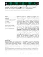
Curcumin suppresses β-catenin activity
Inappropriate activation of β-catenin is linked with the
development of a wide variety of cancers, including mela-
noma, colorectal and prostate cancer [24,25]. Addition-
ally, deregulation of the Wnt/β-catenin pathway has also
been shown in ovarian cancer [26,27]. As a modulator of
the Wnt signaling pathway, β-catenin functions as a tran-
scription factor that is translocated into the nucleus
Figure 4 Curcumin treatment alters the expression of pro-survival and pro-apoptosis related proteins. (A) Curcumin decreases the expres-
sion of Bcl2 family of pro-survival proteins during 6 hr pre-treatment. A2780CP cells were treated with 20 μM curcumin for 6 hrs and protein
lysates were collected and analyzed by immunoblotting for Bcl-xl, Mcl-1 and β-actin. Appropriate bands were quantified by densitometry, normalized
to β-actin, scaled to the DMSO control and expressed as relative expression levels (number beneath the blots). (B) Curcumin pre-treatment fol-
lowed by low dose cisplatin increases percent of Annexin V positive cells. A2780CP cells treated as indicated with vehicle (DMSO), curcumin (20
μM) or cisplatin (5 μM) only or pre-treated with curcumin (20 μM) followed by cisplatin (5 μM) treatment. After 48 hrs, adherent and attached cells
were stained with Annexin V-PE and analyzed by Flow Cytometry. Representative histograms are shown for 1 of 3 similar experiments. (C and D) Cur-
cumin pre-treatment promotes the induction of apoptosis by cisplatin. A2780CP cells were treated as indicated for a total of 48 hrs and protein
lysates were collected and analyzed by immunoblotting for caspase 9 and PARP. (D) Bands for full length and cleaved PARP were quantified by den-
sitometry, normalized to β-actin and calculated as a ratio of cleaved PARP to full length PARP. Data represent mean of 3 repeats for each treatment
(Mean ± SE, * p < 0.017, compared to curcumin only).
0
1
2
3
4
5
6
7
8
9
002.51020
C
ɴ-actin (42 kDa)
PARP (116 kDa)
Cleaved (89 kDa)
Caspase 9 (47 kDa)
Cleaved (35/37 kDa)
2 .5 10 20
0
CIS (
P
M)
CUR + CIS (
P
M)
0 2 .5 10 20
B
D
ɴ-actin (42 kDa)
Bcl-X
L
(30 kDa)
Mcl-1 (40 kDa)
DMSO CUR 20 μM
A
1.0 0.65
1.0 0.25
CIS (μM)
20 μM CUR
Cleaved PARP/PARP
*
*
Yallapu et al. Journal of Ovarian Research 2010, 3:11
/>Page 8 of 12
where it binds with the TCF transcription factor and up-
regulates the expression of cell survival genes such as c-
Myc and c-Jun, which as a result, enhances cell prolifera-
tion in cancer cells. It has also been shown that β-catenin
activity can also inhibit apoptosis in cancer cells [28-31].
Therefore, we sought to investigate the effects of cur-
cumin treatment on nuclear β-catenin function in cispla-
tin resistant ovarian cancer cells using TOPFlash reporter
assay. The cells were treated with either curcumin, cispla-
tin or a 6 hr pre-treatment with curcumin followed by
treatment with cisplatin. After 24 hrs of incubation, cell
lysates were collected and analyzed for β-catenin tran-
scription activity. While treatment of the cells with cispl-
atin caused no change in the β-catenin activity, curcumin
treatment repressed the β-catenin mediated transcription
activity by 60% (Figure 5A). The combination of cur-
cumin and cisplatin also reduced β-catenin activity to
similar levels as when treated with curcumin (there is not
a significant difference between curcumin only and com-
bination treatment with curcumin and cisplatin). To fur-
ther investigate curcumin mediated repression of β-
catenin activity, we analyzed the overall expression of β-
catenin levels and the expression of a downstream target
of nuclear β-catenin signaling (c-Myc) by Western blot-
ting. Curcumin treatment leads to ~50% reduction in β-
catenin and c-Myc protein levels (Figure 5B). This data
suggest that curcumin treatment attenuates nuclear β-
catenin signaling, which is known to play a significant
role in cancer cell proliferation.
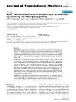
PLGA nanoparticle formulation of curcumin (Nano-CUR)
effectively inhibits ovarian cancer cells growth
While we have shown that curcumin has effective chemo/
radio sensitization effects in ovarian cancer cells, low
water solubility and poor pharmac okinetics greatly ham-
per curcumin's in vivo therapeutic efficacy. Therefore, we
decided to synthesize a PLGA nanoparticle (NP) formu-
lation of curcumin, which is expected to improve bio-
availability in vivo [32,33]. Following synthesis, Nano-
CUR was physically characterized by both dynamic light
scattering (DLS) and transmission electron microscopy
(TEM). The average size of Nano-CUR was observed to
be ~72 nm by DLS (Figure 6A) and 70 ± 3.9 nm by TEM
(Figure 6B). Additionally, curcumin is released from
PLGA NPs in a controlled fashion, which may be useful
for sustained and long term delivery of curcumin for
ovarian cancer treatment (Figure 6C). Following particle
characterization, we examined the in vitro therapeutic
efficacy of Nano-CUR and found that Nano-CUR treat-
ment effectively inhibited proliferation of ovarian cancer
cells (Figure 6D). Additionally, PLGA NPs are efficiently
internalized by A2780CP cells (Figure 6E). Further, to
verify that these nanoparticles are capable of antibody
conjugation for targeted delivery specifically to ovarian
cancer cells, we conjugated nanoparticles with anti-TAG-
72 monoclonal antibody (MAb) (Figure 6F). TAG-72, a
tumor-associated glycoprotein, is over-expressed in vari-
ous tumors, including ovarian cancer [34]. Western blot
analysis of conjugated PLGA NPs revealed that anti-
TAG-72 MAb was effectively conjugated to PLGA NPs
Figure 5 Curcumin inhibits nuclear β-catenin signaling. (A)Curcumin inhibits β-catenin transcription activity. A2780CP cells were transiently
transfected with TOPFlash or FOPFlash and co-transfected with Renilla luciferase to determine β-catenin/TCF transcription activity. The cells were
treated with 20 μM curcumin, 5 μM cisplatin or a 6 hr pre-treatment with 20 μM curcumin followed by treatment with 5 μM cisplatin. After 24 hrs of
incubation, cell lysates were collected and probed for luciferase activity. Treatment of A2780CP cell line with 20 μM curcumin resulted in over a 60%
reduction in β-catenin activity (Mean ± SE, n = 3, *p value p < 0.017, compared to control). (B) Curcumin treatment reduces overall β-catenin and
c-Myc protein levels. A2780CP cell lines were treated as in (A), protein lysates were collected and analyzed by immunoblotting for β-catenin, c-Myc
and β-actin. Protein bands were quantified by densitometry, normalized to β-actin, scaled to the DMSO control and expressed as relative expression
levels (number beneath the blots). Curcumin treatment caused a 50% reduction in β-catenin and c-Myc levels.
Control Cisplatin Curcumin Curcumin +
Cisplatin
Relative luminescence units
A
ɴ-catenin (94kDa)
B
C
CIS CUR
CUR+
CIS
c-Myc (65kDa)
ɴ-actin (42kDa)
*
*
1.0 0.6 0.4 0.2
1.0 0.6 0.5 0.2
Yallapu et al. Journal of Ovarian Research 2010, 3:11
/>Page 9 of 12
(Figure 6G). These data suggest that, in the future, tar-
geted delivery of curcumin specifically to tumors will be
possible. This strategy will improve the therapeutic effi-
cacy of curcumin and will be useful for specific chemo/
radio-sensitization of cancer cells.
Discussion
Most ovarian cancers initially respond well to current
treatment modalities, but the majority of patients will
experience recurrence. Unfortunately, almost all recur-
rent ovarian cancers eventually develop resistance to
platinum based treatment. Tumors with intrinsic or
acquired resistance may have various altered characteris-
tics, including: (a) altered membrane transport proper-
ties, (b) altered expression of target enzymes, (c)
promotion of DNA repair, (d) degradation of drug mole-
cules, and (e) generalized resistance to apoptosis [35-37].
A promising strategy for improving current ovarian can-
cer therapy is to employ a chemo/radio-sensitizer along
with chemo/radiation therapies.
Curcumin is an excellent candidate as a chemo/radio
sensitizer and has been shown to have in vitro chemo-
sensitization effects for cervical cancer and radio-sensi-
tizing effects for prostate cancer [38,39]. However, cur-
cumin's utility for ovarian cancer treatment has not been
fully explored [40-42]. Chirnomas et al. reported that a
functional Fanconi anemia (FA)/BRCA pathway limits
sensitivity to cisplatin and that curcumin can inhibit this
pathway, leading to increased sensitivity to cisplatin
treatment in ovarian cancer cells [41]. Our study shows
that a 6 hr pre-treatment with curcumin effectively sensi-
tized cisplatin resistant ovarian cancer cells to the cyto-
toxic effects of cisplatin, at doses at least 10 times lower
compared to cisplatin treatment alone. Using clonogenic
assays, we assessed the long term effects of curcumin pre-
treatment along with cisplatin treatment or radiation
exposure. We found that curcumin pre-treatment fol-
lowed by cisplatin or radiation exposure dramatically
reduced colony formation compared to either treatment
alone. Curcumin pre-treatment clearly lowers the dose of
cisplatin and radiation treatment needed to suppress the
growth of ovarian cancer cells.
Apoptosis is normally a carefully balanced system of
checks and balances. In cancer cells, often the balance has
been tilted to be more resistant to the initiation of apop-
tosis. Over-expression of pro-survival Bcl2 family mem-
bers is common in many types of cancer and has been
correlated with decreased sensitivity to chemotherapy
Figure 6 Characterization of PLGA-nanoparticle (NP) containing curcumin (Nano-CUR) and its in vitro therapeutic efficacy. (A and B) Nano-
CUR particles are an appropriate size of ~70 nm. Nano-CUR size was determined by (A) dynamic light scattering (DLS) and (B) transmission elec-
tron microscopy (TEM). (C) Nano-CUR formulation demonstrates sustained release of curcumin. Cumulative release of curcumin from PLGA NPs
was determined by UV spectrophotometer at 450 nm over a period of 18 days. (D) Nano-CUR effectively inhibits the growth of cisplatin resistant
ovarian cancer cells. A2780CP cells were treated with Nano-CUR (5-80 μM) or PLGA NPs without curcumin (NPs control) for 48 hrs. Cell proliferation
was determined by MTS assay and normalized to control cells treated with vehicle (PBS). (E) A2780CP cells internalize PLGA-NPs. A2780CP cells
were incubated with FITC-PLGA NPs for 6 hrs and analyzed by fluorescent microscopy. Original magnifications 400×. Inset image represents PLGA NPs
no FITC. (F) Strategy used for antibody conjugation of PLGA-NP for targeted delivery of curcumin to ovarian cancer cells. (G) PLGA-NPs can
be conjugated with anti-TAG-72 MAb (CC49). PLGA-NPs were incubated with anti-TAG-72 MAb. Nano-immunoconjugates were run on 10% SDS-
PAGE, transferred to the PVDF membrane and were probed with an anti-mouse secondary antibody as indicated.
048121620
0
20
40
60
% Cumulative release
Day
200 nm
10 20 30 40
0
20
40
60
80
100
% Proliferation
Concentration (
P
M)
Nano-CUR
NPs control
-CC49
-NPs
-NPs-CC49
A
B
C
D
E
F
G
F
G
Yallapu et al. Journal of Ovarian Research 2010, 3:11
/>Page 10 of 12
and radiation [43]. We found that curcumin pre-treat-
ment reduced the expression of two pro-survival pro-
teins, Bcl-X
L
and Mcl-1, potentially allowing curcumin
treated cells to undergo apoptosis upon cisplatin treat-
ment. Indeed, pre-treatment with curcumin followed by
cisplatin increased the percent of Annexin V positive cells
and increased the amount of cleaved caspase 9 and PARP,
as compared to cisplatin or curcumin alone, indicating
that curcumin pre-treatment followed by cisplatin
enhanced apoptosis.
Curcumin treatment reduced the transcriptional activ-
ity and expression level of β-catenin. The β-catenin path-
way is known to be disrupted in a variety of cancers,
including ovarian cancer. Activation of the β-catenin sig-
naling pathway leads to nuclear localization of β-catenin
which interacts with the TCF transcription factor and
modulates the expression of a wide range of proto-onco-
genes. The functions of these responsive genes are
thought to increase proliferation and recent studies have
also suggested that β-catenin signaling may also inhibit
apoptosis [28-31]. Taken together, these results suggest
that curcumin pre-treatment increases the effectiveness
of cisplatin treatment in cisplatin resistant cells by
increasing the sensitivity of cells to apoptotic pathways
and modulating nuclear β-catenin signaling.
Curcumin is in early phase clinical trials for various
types of cancers [44]. Curcumin is remarkably well toler-
ated and has no toxicity issues [45,46], but it has limited
bioavailability and poor pharamacokinetics [47,48]. To
improve curcumin's in vivo effectiveness we have devel-
oped a PLGA nanoformulation of curcumin. Nanoparti-
cles can deliver anti-cancer drugs to the site of disease
with an antibody targeting approach; however, major
drawbacks include interaction with serum proteins (caus-
ing opsonization), clearance by the reticuloendothelial
system, and non specific accumulation in organs [49]. To
counter these difficulties and to extend the circulation
time of nanoparticles in the blood, nanoparticles may be
modified with inert hydrophilic polymers, such as
poly(ethylene glycol) and poly(vinyl alcohol). In addition,
formulating a small particle size (less than 100 nm) with
high antibody conjugation efficiency will further enhance
the ability to target tumors efficiently [50]. In our current
study, we have developed PLGA nanoparticles which are
made using FDA approved polymer (PLGA) and coated
with poly(vinyl alcohol). The formulated Nano-CUR
effectively inhibits proliferation in cisplatin resistant
ovarian cancer cell lines. The size of these PLGA NPs
were formulated to ~70 nm which is an important
parameter for enhancing the circulation life time and
ensuring diffusion of particles into tumor sites. Recent
literature suggests that antibody conjugated nanoparti-
cles could efficiently deliver chemotherapeutic drugs to
the tumor site [51-53]. Accordingly, we have shown effi-
cient conjugation of anti-TAG-72 MAb to PLGA NPs
with our conjugation chemistry for targeting applica-
tions. Targeted delivery of curcumin will improve the
therapeutic efficacy of curcumin and will be useful for
specific chemo/radio-sensitization of cancer cells. Over-
all, the results of this study suggest that curcumin pre-
treatment induces chemo/radio-sensitization in ovarian
cancer cells via modulating pro-survival cellular signaling
and nanoparticle mediated curcumin delivery may fur-
ther improve the therapeutic efficacy of curcumin.
Conclusion
We report that curcumin acts as a chemo/radio-sensitizer
by modulating the expression of pro-survival proteins
and increasing apoptosis in response to a low dose of cis-
platin. Nanoparticle mediated curcumin delivery will fur-
ther improve the sensitization and therapeutic
capabilities of curcumin. This study demonstrates a novel
curcumin pre-treatment strategy that could be imple-
mented in pre-clinical animal models and in future clini-
cal trials for the effective treatment of chemo/radio-
resistant ovarian cancers.
Competing interests
The authors declare that they have no competing interests.
Authors' contributions
MMY designed and performed MTS assays, colony formation assays, Western
blotting, and synthesis of PLGA NP formulations. DM participated in the design
of the study, provided technical support and performed flow cytometry analy-
sis. DM and MMY drafted the manuscript together. VS performed and analyzed
the β-catenin assays and participated in manuscript preparation. SCC and MJ
participated in the inception of the idea, experimental design, and revision of
the manuscript. All authors read and approved the manuscript.
Acknowledgements
Authors thankfully acknowledge Cathy Christopherson for editorial assistance
and James Pottala for statistical consultation. This work was supported in part
by a Sanford Research/USD grant and Department of Defense Grants awarded
to SCC (PC073887) and MJ (PC073643).
Author Details
1
Cancer Biology Research Center, Sanford Research/University of South Dakota,
Sioux Falls, SD 57105, USA and
2
Department of Obstetrics and Gynecology,
Sanford School of Medicine, University of South Dakota, Sioux Falls, SD 57105,
USA
References
1. Jemal A, Siegel R, Ward E, Hao Y, Xu J, Thun MJ: Cancer statistics, 2009.
CA Cancer J Clin 2009, 59:225-249.
2. Armstrong DK: Relapsed ovarian cancer: challenges and management
strategies for a chronic disease. Oncologist 2002, 7(Suppl 5):20-28.
3. Markman M: Pharmaceutical management of ovarian cancer: current
status. Drugs 2008, 68:771-789.
4. Markman M, Webster K, Zanotti K, Peterson G, Kulp B, Belinson J: Survival
following the documentation of platinum and taxane resistance in
ovarian cancer: a single institution experience involving multiple
phase 2 clinical trials. Gynecol Oncol 2004, 93:699-701.
5. Borst P, Rottenberg S, Jonkers J: How do real tumors become resistant to
cisplatin? Cell Cycle 2008, 7:1353-1359.
Received: 11 February 2010 Accepted: 29 April 2010
Published: 29 April 2010
This article is available from: 2010 Yallapu et al; licensee BioMed Central Ltd. This is an Open Access article distributed under the terms of the Creative Commons Attribution License ( which permits unrestricted use, distribution, and reproduction in any medium, provided the original work is properly cited.Journa l of Ovaria n Resear ch 2010, 3:11
Yallapu et al. Journal of Ovarian Research 2010, 3:11
/>Page 11 of 12
6. Herzog TJ, Pothuri B: Ovarian cancer: a focus on management of
recurrent disease. Nat Clin Pract Oncol 2006, 3:604-611.
7. Alvarez RD, Huh WK, Khazaeli MB, Meredith RF, Partridge EE, Kilgore LC,
Grizzle WE, Shen S, Austin JM, Barnes MN, et al.: A Phase I study of
combined modality (90)Yttrium-CC49 intraperitoneal
radioimmunotherapy for ovarian cancer. Clin Cancer Res 2002,
8:2806-2811.
8. Meredith RF, Buchsbaum DJ, Alvarez RD, LoBuglio AF: Brief overview of
preclinical and clinical studies in the development of intraperitoneal
radioimmunotherapy for ovarian cancer. Clin Cancer Res 2007,
13:5643s-5645s.
9. Meenakshi Kuhar SI, Neeta Singh: Curcumin and Quercetin Combined
with Cisplatin to Induce Apoptosis in Human Laryngeal Carcinoma
Hep-2 Cells through the Mitochondrial Pathway. Journal of Cancer
Molecules 2007, 3:121-128.
10. Cheah YH, Nordin FJ, Sarip R, Tee TT, Azimahtol HL, Sirat HM, Rashid BA,
Abdullah NR, Ismail Z: Combined xanthorrhizol-curcumin exhibits
synergistic growth inhibitory activity via apoptosis induction in human
breast cancer cells MDA-MB-231. Cancer Cell Int 2009, 9:1.
11. Siddiqui IA, Malik A, Adhami VM, Asim M, Hafeez BB, Sarfaraz S, Mukhtar H:
Green tea polyphenol EGCG sensitizes human prostate carcinoma
LNCaP cells to TRAIL-mediated apoptosis and synergistically inhibits
biomarkers associated with angiogenesis and metastasis. Oncogene
2008, 27:2055-2063.
12. Bal Krishnan Jaggi SCC, Meena Jaggi : Review of Curcumin Effects on
Signaling Pathways in Cancer. Proceedings of the South Dakota Academy
of Science 2007, 86:283-293.
13. Karmakar S, Banik NL, Patel SJ, Ray SK: Curcumin activated both receptor-
mediated and mitochondria-mediated proteolytic pathways for
apoptosis in human glioblastoma T98G cells. Neurosci Lett 2006,
407:53-58.
14. Shishodia S, Amin HM, Lai R, Aggarwal BB: Curcumin (diferuloylmethane)
inhibits constitutive NF-kappaB activation, induces G1/S arrest,
suppresses proliferation, and induces apoptosis in mantle cell
lymphoma. Biochem Pharmacol 2005, 70:700-713.
15. Shishodia S, Chaturvedi MM, Aggarwal BB: Role of curcumin in cancer
therapy. Curr Probl Cancer 2007, 31:243-305.
16. Cheng AL, Hsu CH, Lin JK, Hsu MM, Ho YF, Shen TS, Ko JY, Lin JT, Lin BR,
Ming-Shiang W, et al.: Phase I clinical trial of curcumin, a
chemopreventive agent, in patients with high-risk or pre-malignant
lesions. Anticancer Res 2001, 21:2895-2900.
17. Garcea G, Jones DJ, Singh R, Dennison AR, Farmer PB, Sharma RA, Steward
WP, Gescher AJ, Berry DP: Detection of curcumin and its metabolites in
hepatic tissue and portal blood of patients following oral
administration. Br J Cancer 2004, 90:1011-1015.
18. Hamaguchi K, Godwin AK, Yakushiji M, O'Dwyer PJ, Ozols RF, Hamilton TC:
Cross-resistance to diverse drugs is associated with primary cisplatin
resistance in ovarian cancer cell lines. Cancer Res 1993, 53:5225-5232.
19. Chauhan SC, Vannatta K, Ebeling MC, Vinayek N, Watanabe A, Pandey KK,
Bell MC, Koch MD, Aburatani H, Lio Y, Jaggi M: Expression and functions
of transmembrane mucin MUC13 in ovarian cancer. Cancer Res 2009,
69:765-774.
20. Jaggi M, Chauhan SC, Du C, Balaji KC: Bryostatin 1 modulates beta-
catenin subcellular localization and transcription activity through
protein kinase D1 activation. Mol Cancer Ther 2008, 7:2703-2712.
21. Govender T, Stolnik S, Garnett MC, Illum L, Davis SS: PLGA nanoparticles
prepared by nanoprecipitation: drug loading and release studies of a
water soluble drug. J Control Release 1999, 57:171-185.
22. Bisht S, Feldmann G, Soni S, Ravi R, Karikar C, Maitra A, Maitra A: Polymeric
nanoparticle-encapsulated curcumin ("nanocurcumin"): a novel
strategy for human cancer therapy. J Nanobiotechnology 2007, 5:3.
23. Dong S, Roman M: Fluorescently labeled cellulose nanocrystals for
bioimaging applications. J Am Chem Soc 2007, 129:13810-13811.
24. Gavert N, Ben-Ze'ev A: beta-Catenin signaling in biological control and
cancer. J Cell Biochem 2007, 102:820-828.
25. Wheelock MJ, Johnson KR: Cadherin-mediated cellular signaling. Curr
Opin Cell Biol 2003, 15:509-514.
26. Gatcliffe TA, Monk BJ, Planutis K, Holcombe RF: Wnt signaling in ovarian
tumorigenesis. Int J Gynecol Cancer 2008, 18:954-962.
27. Sarrio D, Moreno-Bueno G, Sanchez-Estevez C, Banon-Rodriguez I,
Hernandez-Cortes G, Hardisson D, Palacios J: Expression of cadherins
and catenins correlates with distinct histologic types of ovarian
carcinomas. Hum Pathol 2006, 37:1042-1049.
28. Dehner M, Hadjihannas M, Weiske J, Huber O, Behrens J: Wnt signaling
inhibits Forkhead box O3a-induced transcription and apoptosis
through up-regulation of serum- and glucocorticoid-inducible kinase
1. J Biol Chem 2008, 283:19201-19210.
29. Huang M, Wang Y, Sun D, Zhu H, Yin Y, Zhang W, Yang S, Quan L, Bai J,
Wang S, et al.: Identification of genes regulated by Wnt/beta-catenin
pathway and involved in apoptosis via microarray analysis. BMC Cancer
2006, 6:221.
30. Yang F, Zeng Q, Yu G, Li S, Wang CY: Wnt/beta-catenin signaling inhibits
death receptor-mediated apoptosis and promotes invasive growth of
HNSCC. Cell Signal 2006, 18:679-687.
31. Liu M, Yang S, Wang Y, Zhu H, Yan S, Zhang W, Quan L, Bai J, Xu N: EB1 acts
as an oncogene via activating beta-catenin/TCF pathway to promote
cellular growth and inhibit apoptosis. Mol Carcinog 2009, 48:212-219.
32. Anand P, Nair HB, Sung B, Kunnumakkara AB, Yadav VR, Tekmal RR,
Aggarwal BB: Design of curcumin-loaded PLGA nanoparticles
formulation with enhanced cellular uptake, and increased bioactivity
in vitro and superior bioavailability in vivo. Biochem Pharmacol 2010,
79:330-338.
33. Shaikh J, Ankola DD, Beniwal V, Singh D, Kumar MN: Nanoparticle
encapsulation improves oral bioavailability of curcumin by at least 9-
fold when compared to curcumin administered with piperine as
absorption enhancer. Eur J Pharm Sci 2009, 37:223-230.
34. Ponnusamy MP, Venkatraman G, Singh AP, Chauhan SC, Johansson SL,
Jain M, Smith L, Davis JS, Remmenga SW, Batra SK: Expression of TAG-72
in ovarian cancer and its correlation with tumor stage and patient
prognosis. Cancer Lett 2007, 251:247-257.
35. Morris PG, Fornier MN: Microtubule active agents: beyond the taxane
frontier. Clin Cancer Res 2008, 14:7167-7172.
36. Perez EA: Impact, mechanisms, and novel chemotherapy strategies for
overcoming resistance to anthracyclines and taxanes in metastatic
breast cancer. Breast Cancer Res Treat 2009, 114:195-201.
37. Krishna R, Mayer LD: Multidrug resistance (MDR) in cancer. Mechanisms,
reversal using modulators of MDR and the role of MDR modulators in
influencing the pharmacokinetics of anticancer drugs. Eur J Pharm Sci
2000, 11:265-283.
38. Chendil D, Ranga RS, Meigooni D, Sathishkumar S, Ahmed MM: Curcumin
confers radiosensitizing effect in prostate cancer cell line PC-3.
Oncogene 2004, 23:1599-1607.
39. Javvadi P, Segan AT, Tuttle SW, Koumenis C: The chemopreventive agent
curcumin is a potent radiosensitizer of human cervical tumor cells via
increased reactive oxygen species production and overactivation of
the mitogen-activated protein kinase pathway. Mol Pharmacol 2008,
73:1491-1501.
40. Chan MM, Fong D, Soprano KJ, Holmes WF, Heverling H: Inhibition of
growth and sensitization to cisplatin-mediated killing of ovarian
cancer cells by polyphenolic chemopreventive agents. J Cell Physiol
2003, 194:63-70.
41. Chirnomas D, Taniguchi T, de la Vega M, Vaidya AP, Vasserman M,
Hartman AR, Kennedy R, Foster R, Mahoney J, Seiden MV, D'Andrea AD:
Chemosensitization to cisplatin by inhibitors of the Fanconi anemia/
BRCA pathway. Mol Cancer Ther 2006, 5:952-961.
42. Montopoli M, Ragazzi E, Froldi G, Caparrotta L: Cell-cycle inhibition and
apoptosis induced by curcumin and cisplatin or oxaliplatin in human
ovarian carcinoma cells. Cell Prolif 2009, 42:195-206.
43. Frenzel A, Grespi F, Chmelewskij W, Villunger A: Bcl2 family proteins in
carcinogenesis and the treatment of cancer. Apoptosis 2009,
14:584-596.
44. Lopez-Lazaro M: Anticancer and carcinogenic properties of curcumin:
considerations for its clinical development as a cancer
chemopreventive and chemotherapeutic agent. Mol Nutr Food Res
2008, 52(Suppl 1):S103-127.
45. Hsu CH, Cheng AL: Clinical studies with curcumin. Adv Exp Med Biol
2007, 595:471-480.
46. Sharma RA, Euden SA, Platton SL, Cooke DN, Shafayat A, Hewitt HR,
Marczylo TH, Morgan B, Hemingway D, Plummer SM, et al.: Phase I clinical
trial of oral curcumin: biomarkers of systemic activity and compliance.
Clin Cancer Res 2004, 10:6847-6854.
47. Ireson C, Orr S, Jones DJ, Verschoyle R, Lim CK, Luo JL, Howells L, Plummer
S, Jukes R, Williams M, et al.: Characterization of metabolites of the
Yallapu et al. Journal of Ovarian Research 2010, 3:11
/>Page 12 of 12
chemopreventive agent curcumin in human and rat hepatocytes and
in the rat in vivo, and evaluation of their ability to inhibit phorbol ester-
induced prostaglandin E2 production. Cancer Res 2001, 61:1058-1064.
48. Ireson CR, Jones DJ, Orr S, Coughtrie MW, Boocock DJ, Williams ML,
Farmer PB, Steward WP, Gescher AJ: Metabolism of the cancer
chemopreventive agent curcumin in human and rat intestine. Cancer
Epidemiol Biomarkers Prev 2002, 11:105-111.
49. Singh R, Lillard JW Jr: Nanoparticle-based targeted drug delivery. Exp
Mol Pathol 2009, 86:215-223.
50. Emerich DF, Thanos CG: Targeted nanoparticle-based drug delivery and
diagnosis. J Drug Target 2007, 15:163-183.
51. Byrne JD, Betancourt T, Brannon-Peppas L: Active targeting schemes for
nanoparticle systems in cancer therapeutics. Adv Drug Deliv Rev 2008,
60:1615-1626.
52. Davis ME, Chen ZG, Shin DM: Nanoparticle therapeutics: an emerging
treatment modality for cancer. Nat Rev Drug Discov 2008, 7:771-782.
53. Kukowska-Latallo JF, Candido KA, Cao Z, Nigavekar SS, Majoros IJ, Thomas
TP, Balogh LP, Khan MK, Baker JR Jr: Nanoparticle targeting of anticancer
drug improves therapeutic response in animal model of human
epithelial cancer. Cancer Res 2005, 65:5317-5324.
doi: 10.1186/1757-2215-3-11
Cite this article as: Yallapu et al., Curcumin induces chemo/radio-sensitiza-
tion in ovarian cancer cells and curcumin nanoparticles inhibit ovarian can-
cer cell growth Journal of Ovarian Research 2010, 3:11