báo cáo hóa học:" Hematopoietic-Prostaglandin D2 synthase through PGD2 production is involved in the adult ovarian physiology" pot
Bạn đang xem bản rút gọn của tài liệu. Xem và tải ngay bản đầy đủ của tài liệu tại đây (1.14 MB, 13 trang )
RESEARCH Open Access
Hematopoietic-Prostaglandin D2 synthase
through PGD2 production is involved in the
adult ovarian physiology
Andalib Farhat
1
, Pascal Philibert
1,2
, Charles Sultan
1,2
, Francis Poulat
1
, Brigitte Boizet-Bonhoure
1*
Abstract
Background: The prostaglandin D2 (PGD2) pathway is involved in numerous biological processes and while it has
been identified as a partner of the embryonic sex determining male cascade, the roles it plays in ovarian function
remain largely unknown. PGD2 is secreted by two prostaglandin D synthases (Pgds); the male-specific lipocalin (L)-
Pgds and the hematopoietic (H)-Pgds.
Methods: To study the expression of the Pgds in the adult ovary, in situ hybridization were performed. Then, to
evaluate the role of H-Pgds produced PGD2 in the ovarian physiology, adult female mice were treated with HQL-
79, a specific inhibitor of H-Pgds enzymatic activity. The effects on expression of the gonadotrophin receptors FshR
and LhR, steroidogenic genes Cyp11A1, StAR and on circulating progesterone and estradiol, were observed.
Results: We report the localization of H-Pgds mRNA in the granulosa cells from the primary to pre-ovulatory
follicles. We provide evidence of the role of H-Pgds-produced PGD2 signaling in the FSH signaling through
increased FshR and LhR receptor expre ssion. This leads to the activation of steroidogenic Cyp11A1 and StAR gene
expression leading to progesterone secretion, independently on other prostanoid-synthetizing mechanisms. We
also identify a role whereby H-Pgds-produced PGD2 is involved in the regulation of follicular gro wth through
inhibition of granulosa cell proliferation in the growing follicles.
Conclusions: Together, these results show PGD2 signaling to interfere with FSH action within granulosa cells, thus
identifying an important and unappreciated role for PGD2 signaling in modulating the balance of proliferation,
differentiation and steroidogenic activity of granulosa cells.
Background
Folliculogenesis is under the control o f growth factors
and two pituitary gonadotropin hormones; follicle-
stimulating hormone (FSH) a nd luteinizing hormone
(LH). These heterodimeric glycoproteins bind in the
ovary to specific G-protein coupled receptors, FshR and
LhR respectively, to facilitate the growth and differen tia-
tion of ovarian cells and also to c ontrol the production
of the two steroid hormones estradiol and progesterone,
for review see [1,2].
Amongst the several autocrine and/ or paracrine
growth factors produced by the follicle itself, prostaglan-
dins are critical for multiple stages of reproduction [3,4].
Mice lacking the cyclo-oxygenase-2 (Cox-2) gene encod-
ing the rate limiting step in prostaglandin synthesis,
show pre-implantation deficiencies throughout ovulation
and fertilization [5]. This phenotype is also seen in the
absence of prostaglandin E2 (PGE2) receptor EP2 [6].
A surge in LH levels in granulosa cells of pre-ovulatory
follicles induces expression of Cox-2 and EP2 [7], while
elevated PGE2 in turn, stimu lates cumulus expansion by
elevating cAMP [8]. It h as also bee n shown that PGE2
increases expression of the aroma tase Cyp19A1 gene,
the key gene in estrogen biosynthesis in granulosa cells
[9], as well as acting as a luteotrophic component to
stimulate luteal progesterone secretion through a
cAMP-mediated pathway in both human and ruminants
[10]. Besides PGE2, prostaglandin PGF2a secretion via
cyclo-oxygenase COX-1 expression and the action of its
receptor FP, also plays an important role in diminishing
* Correspondence:
1
Institut de Génétique Humaine, Department of Genetic and Development,
CNRS UPR1142, 141, rue de la Cardonille, 34396 Montpellier CEDEX5, France
Full list of author information is available at the end of the article
Farhat et al. Journal of Ovarian Research 2011, 4:3
/>© 2011 Farhat et al; licensee BioMed Central Ltd. This is an Open Access article distributed under the terms of the Creative Commons
Attribution License (http://creativec ommons.or g/licenses/by/2.0), which permits unrestricted use, distribution, and reproduction in
any medium, pro vided the original work is properly cited.
progesterone levels and stimulating luteolysis, a crucial
stage in inducing labor and pup delivery during parturi-
tion in human and mice [11,12]. Whereas PGE2 and
PGF2a are both involved in regulating ovulation, lutei-
nization, luteolysis and fertility [13-16], the role(s) of
PGD2 signaling in folliculogenesis and ovarian physiol-
ogy is not precisely understood.
PGD2 has been implicated as a signaling molecule in
the mediation or regulation of various biological pr o-
cesses such as platelet aggregation, broncho-constriction
and alle rgic diseases [17,18], whilst also being identif ied
as a partner of the embryonic sex-determining male cas-
cade [19,20]. Secreted PGD2 interacts with two r ecep-
tors: (i) the specific membrane-bound DP receptor
(DP1) associated with adenylcyclase and intracellular
cAMP production [21,22], and (ii) chemo attractant
receptor Th2 (CRTH2) cells (DP2) which is coupled to
Ca
2+
signaling. A metabolite of P GD2, PGJ2, has also
been shown to bind the peroxisome proliferat or-
activated receptor PPARg amemberoftheorphan
nuclear receptor superfamily implicated in k ey female
reproductory roles [23]. PGD2 is produced by two pros-
taglandin D synthases (Pgds) responsible for mediating
the final regulatory step in the biosynthetic pathway of
PGD2 production [24]: (i) the lipocalin-type Pgds (L-
Pgds), a member of the lipocalin ligand-carrier protein
family [24,25] and (ii) the hematopoietic-type Pgds (H-
Pgds) or GSH-requiring enzyme [26].
The L-Pgds transcript initially found in the brain [27],
represents one of the ten most abundant transcripts in
the cortex, hypothalamus and pituitary gland [28]. How-
ever, it is not expressed in either the embryonic or the
adult ovary [20,29,30] whereas H-Pgds is expressed in the
embryonic gonad of both sexes (submitted data). H-Pgds
is a cytosolic protein responsible for the biosynthesis of
PGD2 in immune and inflammatory cells such as mast
cells or Th2 cells, and is also expressed in the spleen, thy-
mus, skin and liver [26], in the microglia where H-Pgds-
produced PGD2 is responsible for the neuroinflammation
associated with bra in injury and neurodegenerative dis-
eases [31], as well as in trophoblasts, uterine epithelium
and endometrial glands at the implantation site of the
humandecidua[32].H-Pgdsexpressionwasalsofound
in the hypothalamus-pituitary axis of hens and has been
associated with high egg production [33]. Recently,
PGD2 produced by H-Pgds and its metabolite PGJ2 have
been shown to induce transcription of the Lhb subunit
gene in the primary culture of chicken anterior pituitary
cells, via the PPARa and PPARg signaling pathways [34].
On the other hand, a stimulatory effect of PGD2 on pro-
gesterone secretion has b een found in vitro in isolated
human corpus lutea [35]. However, the precise H-Pgds
exp ression profile and function of PGD2 signaling in the
adult ovary remain unknown.
Here, we report the characterization and ovarian loca-
lization of H-Pgds mRNA and provide evidence of a role
of H-Pgds-produced PGD2 signaling in the FSH signal-
ing via the increase of FshR and LhR receptor expres-
sion, leading to activation of steroidogenic Cyp11A1 and
StAR gene expression and progesterone secretion. We
found that in vivo inhibition of H-Pgds activity failed to
modify PGE2 and PGF2a synthesis in the ovary and
also identify a role for H-Pgds-produced PGD2 in folli-
cular growth regulation. Our results provide evidence
that PGD2 signaling is a modulator of the differentiation
and steroidogenic activity of granulosa cells.
Methods
Mouse strain and treatments
Female C57BL/6J mic e (Charles River Laboratories,
Saint Germain sur l’ Arbresle, France) were housed at
the IGH animal care facility under controlled environ-
mental conditions (12 h light/12 h darkness, tempera-
ture 21°C). Animal care and handling conformed to the
Réseau des Animaleries de Montpellier (RAM) and all
procedures were approved by the Languedoc-Roussillon
Regional Ethic committee (permit number 34-366, 2008
to BBB).
HQL-79 (4-benzhydryloxy-1-[3-(1H-tetrazol-5-
yl)-propylpiperidine) [36], an inhibitor of H-Pgds activ-
ity, was purchased from Cayman Chemical (SpiBio,
Interchim Montluçon, France). A HQL-79 solution (2.5
mg/ml) was made in methanol as recommended by the
supplier and diluted to 0.125 mg/ml in 0.6% saline solu-
tion. Daily oral administration of HQL-79 was performed
on 8 weeks old- cycling female mice for 5 to 9 days (for
ovariesanalyzisattheestrousphase)orfor16days(for
study of the length of the estrous cycle (three to four
cycles)), as mentioned in the text. According to previous
studies [36-38], 0.1, 1 or 10 mg/kg/day were admini-
strated for the first experiment and then 1 mg/kg/day
was administrated in the following experiments since the
three doses had the same impact on the expression of
ovarian markers. As a control,thesamevolumeofvehi-
cle (0.5% metha nol) was orally administrated into control
cycling mice during the same period.
Young cycling female mice (6 weeks) were treated
with 5 I. U. PMSG (pregnant mare serum gonadotropin,
Sigma-Aldrich, St Louis, MO, USA) without or with
administration of HQL-79 inhibitor (1 mg/kg/d ay).
PMSG was dissolved in 0.6% saline solution and injected
s.c. in a total volume of 0.1 ml, at the diestrous or
proestrous stages of the cycle to initiate follicular devel-
opment. Ovaries were dissected 48 h later for analysis.
Determination of estrous cycle
To determine the stages of estrous cycle, vaginal washes
were collected for 16 days (three to four cycles) from
Farhat et al. Journal of Ovarian Research 2011, 4:3
/>Page 2 of 13
five wild type (WT) and fiveHQL-79mice.Diestrous
phase was de fined by t he exclusive presence of leuko-
cytes; proestrous phas e by leukocytes and nucleated
epithelial cells; estrous phase by large and squamous-
type epithelial cells without nuclei; and m etestrous by
leukocytes and epithelial cells with translucent nuclei.
Histology, immunofluorescence and in situ hybridization
For each female mouse, one ovary was processed for
immunofluorescence and the other one was subjected to
quantitative RT-PCR. Tissues were fixed in 4% parafor-
maldehyde at 4°C overnight and then embedded in
OCT [39]. Cryosections (10 mm) were processed for
immunofluorescence, after rehydr ation. Sections were
then incubated overnigh t at room temperature with pri-
mary antibodies at the indicated dilutions: rabbit anti-
CYP11A1 (1/200 dilution, gift of Dr Nadia Cherradi,
CEA Grenoble) [40], rabbit anti-phospho-histone H3 (1/
100 dilution, sc-8656, Santa Cruz Biotechnology, Santa-
Cruz, CA, USA)), rat anti-H-Pgds (1/100 dilution, Cay-
man Chemical (SpiBio, France)), mouse anti- laminin
(1/500 dilution, Sigma Aldrich), goat anti-FOXL2 (1/100
dilution, Santa Cruz Biotechnology) and goat anti-AMH
(1/200 dilution, sc- 6886, Santa Cruz Biotechnology).
After washing, sections were incubated with appropriate
secondary antibodi es (1/800 dilution, Alexa) (Mo lecular
Probes, Invitrogen, Carlsbad, CA, USA) for 40 min.
The antisense H-Pg ds and FoxL2 RNA probes were
PCR-amplified from embryonic mouse cDNAs, cloned
in a pCRII Topo vector (Invitrogen) and sequenced
using an ABI automatic sequencer. Digoxigenin-labeled
riboprobes were synthesized using a digoxigenin RNA
labeling kit, following the manufacturer’ s instructions
(Roche Diagnostics, Indianapolis, IN, USA) and used for
in situ hybridization experiments on cryosections of WT
ovaries, as previously described [20,41].
RNA isolation and quantitative RT-PCR analysis of gene
expression
RNA isolation using the RNeasy Midi kit (Qiagen,
Valencia, CA, USA) from frozen ovaries, reverse tran-
scriptase and quantitative RT-PCR using a LightCy-
cler480 apparatus (Roche Diagnostics) were carried out
as previously described [20]. Gene expression levels
were investigated using different pairs of primers (Table 1)
and normalized to those of Gapdh or Hprt .
Hormone and prostaglandin assays
Hormone assays for estradiol and progesterone were
performed from sera, by using ELISA kits (Cayman Che-
micals, Progesterone EIA kit 582601 and Estradiol EIA
kit 582251). Mice (n = 20 for WT and n = 20 for HQL-
79-treated) at the estrous phase of their cycle, were
anesthetized and b lood was colle cted by ca rdiac punc-
ture into plastic eppendorf tubes containing heparin.
After centrifugation, the serum was ex tracted twice with
methylene chloride; after evaporation, steroid extracts
were stored at -80°C until assays were performed. Deter-
mination of the hormone concentrations was performed
in triplicate at two different dilutions according t o the
Table 1 Sequences of oligonucleotides for real time PCR
Primers Sequence 5’-3’ Primers Sequence 5’-3’
mFSHRfwd gtgcgggctactgctacact mGapdhFwd tggcaaagtggagattgttgcc
mFSHRrev caggcaatcttacggtctcg mGapdhRev aagatggtgatgggcttcccg
mLHRqFwd gatgcacagtggcaccttc mP27Fwd gagcagtgtccagggatgag
mLHRqRev cctgcaatttggtggaagag mP27Rev tctgttctgttggccctttt
mStARqFwd ttgggcatactcaacaacca mCycD2Fwd ctgtgcatttacaccgacaac
mStARqRev acttcgtccccgttctcc mCycD2Rev cactaccagttcccactccag
mSCCqFwd aagtatggccccatttacagg mCox-1Fwd cctctttccaggagctcaca
mSCCqRev tggggtccacgatgtaaact mCox-1Rev tcgatgtcaccgtacagctc
mDP1Fwd cccagtcaggctcagactaca mCox-2Fwd gctcttccgagctgtgct
mDP1Rev aagtttaaaggctccatagtacgc mCox-2Rev cggttttgacatggattgg
mDP2Fwd catcgtggttgccttcgt mPges-2Fwd cccaggaaggagacagctt
mDP2Rev gcctccagcagactgaagat mPges-2Rev aggtaggtcttgagggcactaat
mSF-1Fwd cacgaaggtgcatggtctt mHPgdsFwd cacgctggatgacttcatgt
mSF-1Rev cagttctgcagcagtgtcatc mHpgdsRev aattcattgaacatccgctctt
mCYP19Fwd cctcgggctacgtggatg mLPgdsFwd ggctcctggacactacacct
mCYP19Rev gagagcttgccaggcgttaaa mLPgdsRev atagttggcctccaccactg
mEP2Fwd tgctccttgcctttcacaat mFPFwd ctggccataatgtgcgtct
mEP2Rev ctcggaggtcccacttttc mFPRev tgcaatgttggccattgtta
hGapdhFwd gagaaggctggggctcat hHPgdsFwd gagaatggcttattggtaactctgt
hGapdhRev tgctgatgatcttgaggctg hHPgdsRev aaagaccaaaagtgtggtactgc
Farhat et al. Journal of Ovarian Research 2011, 4:3
/>Page 3 of 13
kits’manufacturer. In each case, the twenty values were
averaged.
PGD2, PGE2 and PGF2a levels were determined using
the PGD2 - MOX EIA Kit (Cayman Chemical 500151),
PGE2 express EIA kit (500141, Cayman Chemical) and
13,14-dihydro-1 5keto PGF2a (516671, Cayman Chemi-
cal), respectively. Ovaries were collected from mice trea-
ted(n=8)ornot(n=8)byHQL-79andimmediately
frozen on dry ice and then stored at -80°C. Ovaries
were lyzed and proteins were extracted with cold acet-
one on ice and lyzates were evaporated under nitrogen
flow. Prostaglandins were resuspended in 500 μlEIA
buffer and assayed as recommended by the kits supplier.
Two dilutions (1 and 1/20) were assayed for prostaglan-
dins content. The eight values for each group were aver-
aged and statistical analysis was performed using
Student’s t test, and results were considered statistically
significant at a P < 0.05.
Statistical analysis
Quantified real time RT-PCR signals were normalized to
Gapdh or Hprt levels and the hormone levels of treated
ovaries were compared to those of untreated ovaries. All
values were presented as means ± SE. Student ’ sttest
was used to determine the significance of differences in
expression and hormone data. Results were considered
significant at P < 0.05 for two-sided analysis.
Results
H-Pgds and DP2 expression in adult mouse ovaries
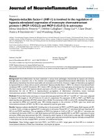
The mRNA for H-Pgds was detected by in situ hybridi-
zation in the growing follicles from the primary t o the
pre-ovulat ory stage and in the corpus luteum. Figure 1A
shows an expression of H-Pgds mRNA in the granulosa
cells of the developing follicles similar to that of the
granulosa cell marker FoxL2 whereas hybridization with
the control sense H-Pgds cRNA probe showed no signif-
icant signal (data not shown). In the antral and pre-ovu-
latory follicles, H-Pgds expression is likely abolished in
the external layers of mural granulosa cells, remaining
only in the internal layers of granulosa cells and in gran-
ulosa cells forming the cumulus in the ovulatory follicle.
H-Pgds mRNA was not detectable in the other ovarian
cell types. In order to confirm H-Pgds expression in the
granulosa cells at the protein level, we used immuno-
fluorescence with (Figure 1B, arrows) or without (Figure
1C, IgG control) a specific H-Pgds antibody. We then
showed the DP2 receptor expression in the granulosa
cells of primary, secondary, preantral (Figure 2A), antral
(Figure 2B) and preovulatory (Figure 2C) follicles using
an anti-rabbit DP2 antibody together with anti-FOXL2
(A) or anti-AMH ( B, C) antibodies, two specific granu-
losa markers. Specific expression of DP2 in the granu-
losa cells was confirmed by high magnification imaging
(Figure 2D). However, DP1 recepto r was not detected in
any cell type at any stage (data not shown). Indeed,
using real-time RT-PCR we observed significant levels
of Dp2 transcripts (Figure 2E), whereas Dp 1 expression
level remained undetectable in WT ovaries.
Prostaglandin synthesis in the ovary upon inhibition of H-
Pgds enzymatic activity
We evaluated the implication of H-Pgds mediated-PGD2
signaling within the ovarian physiology using the
H-Pgds specific inhibitor HQL-79 [36-38]. To confirm
the significance of the inhibition by HQL-79 and evalu-
ate the incidence of PGD2 depletion on pr ostaglandin
production, we measured the level of PGD2, PGE2 and
PGF2a in ovaries of HQL-79-treated mice. As expected,
the ovarian level of PGD2 was markely reduced by 65%
in the HQL-79 treated mice compared to that in the
untreated mice. However, no significant different levels
of PGE2 and PGF2a were measured (Figure 3A).
We then analyzed the PGD2 pathway components and
showed by real time RT-PCR that H-Pgds expression
was u p-regulated concomitantly to the reduced level of
PGD2 in HQL-79 treated ovaries (Figure 3B). On the
other hand, no significantly different expression of the
Dp2 and PP ARg genes (Figure 3B) was detected upon
HQL-79 treatment and no expression of L-Pgds and
Dp1 receptor genes was detect ed in the control or trea-
ted ovaries (data not shown).
To evaluate the impact of the PGD2 signaling on
other prostaglandin pathways and considering the
importance of PGE2 and PGF2a for ovarian function,
we then d etermined the mRNA contents of cyclooxy-
genases Cox1 and Cox2, prostaglandin synth ase (mem-
brane-bound) m-Pges-2, and the receptors Ep2 and Fp
by quantitative RT-PCR in ovaries from mice (in estrous
phase) treated with vehicle or HQL-79. The ovarian
Cox1, Pges and Ep2, Fp m RNA levels were not signifi-
cantly different in the untreated or HQL-79 treated
mice (Figure 3C) that were in agreement with the stable
levels of PGE2 and PGF2a. Howev er, the expre ssion of
Cox-2 was significantly increased by 10 fold in HQL-79
treated ovaries compared to control ovaries (Figure 3C).
Taken together, these results indicate that 65% of H-
Pgds activity were inhibited by HQL-79 but this treat-
ment has no effect on PGE2 and PGF2a prostaglandin
pathways in the ovary; however, the reduced level of
PGD2 induces Cox-2 gene expression that could contri-
bute to the up-regulation of H-Pgds gene expression in
order to restore the intraovarian PGD2 content.
PGD2 signaling is necessary for FSH signaling and
steroidogenesis in the mouse ovary
Folliculogenesis and synthesis of steroid hormones in
the ovary depends on the coordinated actions of FSH
Farhat et al. Journal of Ovarian Research 2011, 4:3
/>Page 4 of 13
A
H-Pgds HST
H-Pgds
GC
GC
TC
HST
B C
IgG control
AMH
AMH
GC
GC
secondaryprimary
antral ovulatory corpus luteum
preantral
Foxl2
H-PgdsFoxl2H-Pgds
GC
GC
GC
GC
GC
GC
GC
GC
GC
GC
Figure 1 Expression of H-Pgds in the mouse adult ovary.(A), In situ hybridization for H-Pgds and granulosa cell marker FoxL2 was performed
on sections from wild type adult ovaries. Primary, secondary, pre-antral, antral, ovulatory follicles and corpus luteum are represented for H-Pgds
and FoxL2 mRNA expression and expressing granulosa cells (GC) are labeled by a blue arrow. Scale bars = 50 μm. (B), H-Pgds protein expression
was detected in granulosa cells on wild type adult ovary sections, using an anti-H-Pgds antibody (in green) whereas nuclei are labeled in blue
by the Hoescht Dye (HST). The merge panel has been enlarged on the right bottom panel. Arrows indicate H-Pgds expressing granulosa cells.
TC, theca cells; GC, granulosa cells. Scale bar = 50 μm. (C), Control immunofluorescence experiment with no primary H-Pgds antibody (IgG
control) showing the specificity of the antibody. AMH staining in granulosa cells was used on the same slide. Arrows indicate granulosa cells
(GC).
Farhat et al. Journal of Ovarian Research 2011, 4:3
/>Page 5 of 13
GC
TC
GC
GC
GC
GC
GC
TC TC
GC
GC
TC
TC
A
B
C
D
FOXL2 DP2+FOXL2
DP2
DP2
DP2
DP2
DP2
AMH
AMH
+
DP2
AMH
+
DP2
AMH
+
HST
HST
E
Relative mRNA expression
Dp1 Dp2
0
1
3
2
primary
secondary
preantral
antral
preovulatory
Figure 2 Expression of PGD2-receptors in the mouse adult ovary. DP2 protein expression was detected in granulosa cells of primary,
secondary and preantral follicles (A), of antral (B) and preovulatory (C) follicles of wild type adult ovary, using immunofluorescence staining with
an anti-DP2 antibody (in red) whereas FOXL2 (A) or AMH (B,C) (in green) were used to delineate granulosa cells. Right panels are the merge
between DP2 and FOXL2 or AMH stainings. Dotted lines delineate granulosa (GC) and theca (TC) cells. Scale bars = 50 μm. (D), Control
immunofluorescence experiment using an anti-DP2 antibody with the Hoescht dye (HST) labeling nuclei. Dotted lines delineate granulosa (GC)
and theca (TC) cells within a follicle. Scale bars = 25 μm. (E), Expression levels of PGD2 receptors Dp1 and Dp2 mRNAs by real time RT-PCR. Dp2
was expressed at high levels in ovaries from adult cycling mice (n = 4) whereas Dp1 transcripts were undetectable. The values of three repeats
were averaged and normalized to Gapdh expression.
Farhat et al. Journal of Ovarian Research 2011, 4:3
/>Page 6 of 13
and LH acting through their respective receptors FshR
and LhR [2 ]. We thus evaluated the implication of H-
Pgds mediated-PGD2 signaling within the gonadotropin
pathways. Adult female mice were treated with the H-
Pgds inhibitor HQL-79 (at doses 0.1-1 or 10 mg/kg/day)
[36-38] or with vehicle for five to nine days until mice
reached the estrous phase and the resulting ovaries were
examined in terms of their expression of gonadotropin
receptors and ovarian markers. For the three doses of
HQL-79, the reduced level of H-Pgds produced PGD2
clearly impaired ovarian gonadotropin re ceptor expres-
sion, as shown by the reduction in FshR and LhR levels
by 50% and 80% respectively (data not shown for 0.1
and 10 mg/kg/day and Figure 4A, dose 1 mg/kg/day).
Induced steroidogenesis is regulated by increased StAR
(steroidogenic acute regulatory) protein expression
under the positive control of gonadotropin signaling.
StAR is the primary regulator of cholesterol transport
into the mitochondria where the steroid precursor is
then converted by CYP11A1 side-chain cleavage enzyme
(P45 0scc) to pregnenolone. We demonstrated here that,
when compared to levels in the untreated ovary, inhibi-
tion of H-Pgds enzymatic activity significantly reduced
expression of StAR and Cyp11A1 genes by 60% and 50%
respectively (Figure 4B), whereas PGD2 signaling did
not affect expression levels of SF-1, a major activator of
steroidogenesis gene expression. In contrast, expression
levels of the Cyp19A1 gene increased significantly by
30% (Figure 4B). CYP11A1 protei n expression was also
largely reduced in granulosa cells of the growing follicles
of ovaries treated by HQL-79, when compared to that
observed in WT ovaries (Figure 4C).
We next evaluated serum levels of the ovarian steroid
hormones estradiol and progesterone in twenty WT and
twenty female mice treated with HQL-79 for five to nine
days, all in the estrous period. The results showed a signif-
icant reduction of 50% in the basal level of progesterone in
themicetreatedwithHQL-79,whencomparedtothat
measured in the WT (Figure 5A). In contrast, the estradiol
level increased by 50% in the HQL-79 treated mice com-
pared to WT (Figure 5B), following the increased aroma-
tase Cyp19A1 expression described above (Figure 4B).
C
B
-HQL79 +HQL79-HQL79 +HQL79
0
10
20
30
40
50
60
ovarian PGE2 pg/ml
ovarian PGF2α pg/ml
0
2
4
6
8
10
12
ovarian PGD2 pg/ml
-HQL79 +HQL79
A
0
10
20
30
5
15
25
*
-HQL79 +HQL79
Relative mRNA expression
Dp2
0
0.2
0.4
0.6
0.8
1.0
1.2
1.4
1.6
Cox-1
Pges
Fp
R
elative mRNA expression
-HQL79 +HQL79
-HQL79 +HQL79
-HQL79 +HQL7
9
0
0.2
0.4
0.6
0.8
1.0
1.2
1.4
1.6
0
0.4
0.8
1.2
1.6
Ep2
-HQL79 +HQL79
0
0.2
0.4
0.6
0.8
1.0
1.2
1.4
0
0.2
0.4
0.6
0.8
1.0
1.2
Relative mRNA expression
-HQL79 +HQL79
0
1
2
3
0.5
1.5
2.5
3.5
H-Pgds
*
Cox-2
-HQL79 +HQL79
4
5
0
1
2
3
*
-HQL79 +HQL79
Relative mRNA expression
0
1
2
3
0.5
1.5
2.5
3.5
PPARγ
Figure 3 Prostaglandins synthesis in the ovary upon PGD2 depletion.(A), Levels of PGD2, PGE2 and PGF2a were measured using ELISA in
HQL-79 treated or not ovaries (n = 8 for each condition). Results expressed in pg of prostaglandin/ml showed that PGD2 content is significantly
decreased (P-value < 0.01) by 65% upon HQL-79 treatment whereas PGE2 and PGF2a contents were not affected; error bars indicate SD of
assays done with two dilutions of the eight samples of each group. Expression levels of H-Pgds, Dp2, PPARg (B) and Cox-1, Cox-2, mPges-2, Ep2,
Fp (C) in ovaries of HQL-79 treated (n = 8) or not (n = 8) mice. By real time RT-PCR, no significant difference of Dp2, PPARg (B) and Cox-1,
mPges-2, Ep2, Fp (C) expression level was detectable whereas a large increase of Cox-2 and H-Pgds expression level was measured upon HQL-79
treatment. All the expression level values were normalized to those of Hprt. Data are expressed as means +/- SE (columns and bars); * P < 0.05
vs control.
Farhat et al. Journal of Ovarian Research 2011, 4:3
/>Page 7 of 13
To evaluate the relationships between PGD2 signaling
and FSH action, we stimulated mice with PMSG which
mimics the function of FSH. As expected, FshR and LhR
expression was increased by 2.5 fold in PMSG-treated
versus untreated control ovaries (Figure 6A). Accord-
ingly, this stimulation was inhibited upon co-treatment
with the HQL-79 inhibitor (Figure 6A), indicating the
requirement for intact PGD2 signaling in order for
PSMG to take effect. Subsequently, inhibition of H-Pgds
activity also inhibited StAR expression induced after
PMSG treatment (Figure 6D) whereas Cyp11A 1 expres-
sion decreased after HQL-79 treatment (Figure 6C), co n-
firming that PGD2 is involved in Cyp11A1 activation. On
the other hand, SF-1 expression level remained indepen-
dent of PMSG and HQL-79 treatment (Figure 6B).
H-Pgds-produced PGD2 is implicated in the control of
granulosa cell proliferation
We assessed the length of estrous cycles in five WT and
five HQL-79-treated adult mice using vaginal smears
collected over 16 consecutive days (three to four cycles).
The WT mice (-HQL-79) had cyclical estrous cycles
lasting more than five days (5.3 days) whereas in con-
trast, HQL-79 treated (+HQL-79) mice had significantly
shorter cycles lasting less than four days (3 .8 days) (Fig-
ure 7A, P-value: 0.0097). To chara cterize the observed
changes of inactivation of H-Pgds activi ty at the cellular
level, we examined the proliferation rate of granulosa
cells (GCs) in the developing follicles. GCs partially
depleted of PGD2 signaling showe d an increased prolif-
eration upon immunostaining for mitosis marker
Lhr Fshr
Relative mRNA expression
0
10
20
30
A
C
*
*
-Hql79
+Hql79
B
0
1
3
2
0
0.4
1.2
0.8
0
0.4
0.8
0.2
0.6
Sf-1
Cyp11A1
Star
Relative mRNA expression
control
+HQL-79
control
+HQL-79
control
+HQL-79
control
+HQL-79
0
1
3
2
Cyp19A1
*
*
*
control +HQL-79
Cyp11A1 + HST
Cyp11A1 + HST
Cyp11A1
Cyp11A1
GC
GC
c
GC
GC
c
antral
preovulatory
Figure 4 PGD2 si gnaling regulates g onadotropin receptors and steroidogenic genes expression. FshR and LhR (A)andSf-1, Cyp11A1,
StAR, Cyp19A1 (B) mRNA expression levels were assessed using real time RT-PCR in ovaries from adult cycling mice treated (n = 10) or not (n =
10) using H-Pgds inhibitor HQL-79 (1 mg/kg/day). The values of at least two repeats of two different RT reactions were averaged and
normalized to Gapdh expression. Values represent mean +/- SEM and * represents significant differences P < 0.025 compared with untreated
ovaries (control). (C), CYP11A1 protein expression was detected in untreated (control) or treated (+HQL-79) ovaries (in red). Upon HQL-79
treatment, a largely decreased expression is detected in antral and preovulatory follicles. Nuclei are labeled in blue (Hoescht dye, HST). GC:
granulosa cells, c: cumulus cells. Scale bars = 50 μm.
Farhat et al. Journal of Ovarian Research 2011, 4:3
/>Page 8 of 13
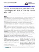
phosphohistone H3 (phosphoH3) (Figure 7B). A signifi-
cant increase of 30% in granulosa cell proliferation was
seen in the pre-antral follicles and reached 50% in the
GCs of antral follicles of HQL-79 treated ovaries, com-
pared to untreated ovaries (Figure 7C). In contrast,
apoptosis in the GCs of the growing follicles was not
modified by the lack of PGD2 signaling (data not
shown). As shown in Figure 7D, this increase in cell
proliferation is associated with a significantly decreased
expression of CDKN1B (p27) in the treated ovaries,
whereas levels of CyclinD2 expression remained unmo-
dified. Consequently, the number of corpora lutea in
HQL-79 ovaries was increased by two fold compared to
that in untreated ovaries (Figure 7E) (female mice at the
proestrous phase of their cycle), suggesting that upon
HQL-79 treatment, the number of growing and matur-
ating follicles have increased. Collectively, these results
support the hypothesis where PGD2 signaling negatively
impacts GC proliferation in vivo, thus promoting condi-
tions favoring granulosa cell differentiation and subse-
quently steroidogenesis.
Discussion
In this study, we describe the expression of H-Pgds mRNA
in the adult mouse ovary. This localization includes granu-
losa cells from growing follicles through primary to antral
and pre-ovulatory stages, a nd the corpus luteum formed
after ovulation. H-Pgds is thus the sole source of PGD2 in
the ovary since the second enzyme able to produce PGD2
(L-Pgds) is not expressed [19]. In the embryonic gonad, L-
Pgds secreted PGD2 signals through the adenylcyclase-
coupled receptor DP1 to activate expression of the Sertoli
cell differentiating gene Sox9 and contribute to the nuclear
translocation of SOX9 protein [19,30]. In the adult ovary,
the Ca
++
coupled DP2 receptor is exclusively expressed in
granulosa cells. Considering how Sertoli and granulosa
cells have common ancestor precursor cells [42], this dif-
ferential expression of both receptors and the dual func-
tional convergence between L- and H-Pgds might
constitute part of the antagonistic regulation between
male and female pathways [43,44] and be a key regulatory
step in maintaining the differentiation of both Sertoli and
granulosa cell types [45]. PGD2 is metabolized to 15d-
PGJ2, the high affinity natural ligand for the PPARg recep-
tor expressed in granulosa cells of developing follicles
[46,47]. These results thus suggest that both receptors
DP2 and PPARg might relay PGD2 signaling in the adult
ovary.
The process of granulosa cell differentiation occurring
throughout progression from a pre-antral to pre-ovula-
tory follicle is dependent on sufficient FSH stimulation
[48,49] and is marked by the acquisition of Fsh R and
LhR expression and increased steroidogenesis. In this
study, we demonstrated that H-Pgds enzymatic activity
0
2
4
6
8
Relative mRNA expression
C
C
Relative SF-1 expression
0
2
4
6
1
3
5
PMSG
PMSG+
HQL-79
C
PMSG
PMSG+
HQL-79
C
PMSG
PMSG+
H
QL-79
0
0.2
0.4
0.6
0.8
1
0
0.2
0.4
0.6
0.8
PMSG
PMSG+
HQL-79
A
B
C
D
LhR
FshR
*
*
*
*
Relative Cyp11A1 expression
Relative StAR expression
Figure 6 PGD2 signaling is necessary for FSH action.Adult
cycling female mice were treated with 5 I.U. PMSG without (PMSG)
or with (PMSG+HQL-79) administration of HQL-79 inhibitor. FshR,
LhR (A), Sf-1 (B), Cyp11A1 (C) and StAR (D) gene expression levels in
ovaries (n = 5 for each condition), were analyzed by real-time RT-
PCR. The values of at least two repeats of two different RT reactions
were averaged and normalized to Gapdh expression. Values
represent mean +/- SE and * represents significant differences P <
0.05 (A), P < 0.001 (C-D) compared with ovaries treated with PMSG
only.
0
10
20
30
0
200
400
600
-HQL79 +HQL79 -HQL79 +HQL79
s
erum progesterone pg/ml
serum estradiol pg/ml
*
*
AB
Figure 5 Progesterone and estradiol production is modified
upon H-Pgds enzymatic inhibition.(A), serum progesterone
levels. (B), serum estradiol levels were measured by Elisa on
extracted sera. Bars represent the average of twenty animals (n = 20
for untreated mice and n = 20 for HQL-79 treated mice). HQL-79
treatment induces a 50% decrease of progesterone production and
a 50% increase of estradiol production. * represents significant
differences P < 0.05, compared to untreated ovaries (-HQL-79).
Farhat et al. Journal of Ovarian Research 2011, 4:3
/>Page 9 of 13
is required in order for FSH to regulate expression of
both FshR and LhR receptors, suggesting PGD2 to be an
autocrine positive regulator of FshR and LhR expression
in the ovary. This regulation may act directly on the
FSH-induced FshR promoter act ivity as in the case of
inhibin-A [50], or might otherwise act indirectly by
increasing FshR mRNA stability, as in the case o f IGF-I
[51]. The inhibition of H-Pgds enzymatic activity leads
to a decrease in FshR and LhR expression but does not
affect that of SF-1, the major activator of steroidogenesis
estrus cycle length (days)
AB
D
0
1
2
3
4
*
CyclinD2 p27
Relative mRNA
expression
C
10
20
30
40
pre antral antral
number of pH3 positive
cells/follicle
*
*
0
-Hql79
+Hql79
-Hql79
+Hql79
HST phosphoH3
AMH phosphoH3
-HQL-79-HQL-79
+HQL-79 +HQL-79
-Hql79 +Hql79
0
1
2
3
4
5
6
*
E
-HQL-79
+HQL-79
CL
CL
CL
CL
CL
CL
*
*
*
*
*
*
*
*
*
*
*
*
*
*
Figure 7 PGD2 signaling controls the granulosa cell proliferation.(A), The length of estrous cycles in five WT and five HQL-79-treated adult
mice were assessed in vaginal smears collected every day for 16 consecutive days. Results of the five animals were averaged and were
expressed as means +/- SE (colums and bars), * P value = 0.0097. (B), Proliferation of granulosa cells of antral follicles was assessed using
immunofluorescence with mitosis marker phosphohistone H3 (phosphoH3) antibody (in red) on cryosections of wild type (-HQL-79) or HQL-79
(+HQL-79) treated ovaries; granulosa cells were identified by anti-Müllerian hormone (AMH) antibody (in green) and nuclei were labeled by the
Hoescht Dye (HST) (in blue). Numbers of phospho-H3-positive cells were determined on ten independent fields of three different ovaries for
each condition and are represented on the graphs (C). * represents significant increased number of mitotic cells in HQL-79 treated compared to
that in untreated ovaries. (D), CyclinD2 and p27 expression levels in five wild type and five HQL-79-treated ovaries were quantified by real time
RT-PCR and were normalized to Gapdh expression. Values are the result of averaged experiments (done in triplicate) on the five independent
ovaries. * represents the significant decrease of p27 expression in HQL-79 compared to that in untreated ovaries (P-value < 0.025). (E), The
follicular content of HQL-79 treated ovaries (at their proestrous stage) were compared to that of WT ovaries by labeling sections with the
Hoescht dye. CL: corpora lutea, * growing follicles.
Farhat et al. Journal of Ovarian Research 2011, 4:3
/>Page 10 of 13
gene expression [52]. This supports the implication of
PGD2 signaling in the FSH-induced expression of the
StAR gene, independently on SF-1. SF-1 is essential for
the development and function of the reproductive axis
at multiple levels [52] and FSH has been shown to acti-
vate SF-1-mediated transcription using various mechan-
isms [53]. Thus, regulation of FshR expression might be
one of the causes of LhR and steroidogenic gene down-
regulation, and of the decrease in progesterone produc-
tion upon PGD2 signaling inhibition [54].
In contrast, following the decrease in Cyp11A1 and
StAR expression levels upon PGD2 depletion, we found
that levels of both aromatase expression and serum
estradiol increased in treated female mice compared to
untreated animals. On the other hand, we observed that
granulosa cells partially depleted of PGD2 signaling
show increased proliferation based on immunostaining
for mitosis marker phosphohistone H3, which we con-
firmed at the molecular level through the significantly
decreased expression of CDKN1B (p27 ). This increased
proliferation lead to an increased number of the matur-
ating follicles that might explain the higher levels of
Cyp19A 1 mRNA expression and secret ed estradiol upon
HQL-79 treatment, rather than being a consequence of
the Cox-2 up-re gulation that was detected in HQL-79
ovaries. The up-regulation of Cyp19A1 gene expression
via COX-2 was shown to be depend ent on PGE2 synth-
esis and cAMP signaling in undifferentiated rat granu-
losa cells [9] or in human brea st tumor cells [55]. Our
data showed that Cox-2 expression is up-regulated, how-
ever, PGE2 synthesis was not modified. Ind eed, the side-
effect of HQL-79 treatment (i.e. increased PGE2 produc-
tion) [26] related in the lung tissues of sensitized guinea
pigs [56] was not detected in our system as it has not
been seen in sheep vesicular gland microsomes [56] or
in vivo in H-Pgds transgenic mouse strain [36].
In this study, we measured high levels of Cox-2 and
H-Pgds transcripts whereas no modification of Cox-1
has been measured in HQL-79 treated ovaries. The
functional coupling between H-Pgds/Cox-1 or H-Pgds/
Cox-2 has been demonstrated respectively, in the
immediate o r the delayed response in mast cells during
the cytokine stimulation [38], even though tightly cou-
pling between H-Pgds and Cox-1 is p referentially docu-
mented [36,57]. The up-regulation of Cox-2 associated
with the down-regulation of H-Pgds protein expression
upon HQL-79 treatment has b een previously de scribed
in the mouse ischemic brain [58]. In the ovary, we can
assume that partial depletion of PGD2 might induce
Cox-2 gene expression that in turn, might activate H-
Pgds expression in order to restore the intraovarian
PGD2 content. PGJ2, a PGD2 metabolite was shown to
inhibit osteoblastic differentiation through PPARg acti-
vation and down-regulation of Cox-2 [59]. This process
would take place without any interaction with other
prostanoid-synthetizing mechanisms as it has been pre-
viously reported in other systems, induction of fever
[60] or induction of inflammation in muscle necrosis
[61], since PGE2 and PGF2a prostaglandin pathways are
not modified upon HQL-79 treatment.
Using the H-Pgds specific inhibitor HQL-79 known to
exactly mimic the phenotype of H-Pgds KO mice in var-
ious systems such as i nflammation, mu scle necrosis
[31,38], we identify an important and unappreciat ed role
for PGD2 signaling in modulating the balance of prolifera-
tion, differentiation and steroidogenic activity of the gran-
ulosa cells, through both FSH dependent and independent
mechanisms. Thus, these results suggest PGD2 as a modu-
lator of follicle development, even though no reproductive
defects have been reported in female H- Pgds KO mice
[31,62]. The physiological importance of PGD2 for ovarian
function and normal female fertility mig ht be assessed in
this mouse strain or in mice conditionnally invalidated for
H-Pg ds in the ovary under the co ntrol of Anti-Müllerian
hormone (Amh) promoter (Amh-cre, [63]) to overcome a
putative central effect of H-Pgds produced PGD2.
Abbreviations
FSH: follicle-stimulating hormone; LH: luteinizing hormone; Cox-2:
cyclooxygenase-2; CYP11A1: cytochrome P450 11A1 (P450scc: Cholesterol-
side chain cleavage enzyme); CYP19A1: cytochrome P450 19A1 (aromatase);
StAR: steroidogenic acute regulatory protein; SF-1: steroidogenic factor 1;
PGD2: prostaglandin D2; PGE2: prostaglandin E2; PGF2α: prostaglandin F2α;
PPARγ: peroxisome proliferator-activated receptor gamma; PMSG: pregnant
mare serum gonadotropin.
Acknowledgements
The authors thank Brigitte Moniot for her help with in situ hybridization
experiments. We thank Dr Julien Cau for assistance in confocal imagery
(Imagery platform MRI/IGH); we also thank Florence Arnal and Elodie Gavois
from the IGH animal care facility for providing mice and for helpful
discussions. A. F. was supported by a PhD fellowship from the « Ligue
Nationale contre le Cancer ». The work was supported by the CNRS and by
the Ligue Régionale contre le Cancer (B.B.B.).
Author details
1
Institut de Génétique Humaine, Department of Genetic and Development,
CNRS UPR1142, 141, rue de la Cardonille, 34396 Montpellier CEDEX5, France.
2
Service d’Hormonologie, Hôpital Lapeyronie, CHU Montpe llier, France.
Authors’ contributions
All authors read and approved the final manuscript. Conceived and
designed the experiments: FP, BBB. Performed the experiments: AF, PP, FP,
BBB. Analyzed the data: AF, PP, CS, FP, BBB. Contributed reagents/materials/
analysis tools: CS, FP, BBB. Wrote the paper: BBB.
Competing interests
The authors declare that they have no competing interests.
Received: 21 December 2010 Accepted: 25 February 2011
Published: 25 February 2011
References
1. Richards JS, Fitzpatrick SL, Clemens JW, Morris JK, Alliston T, et al: Ovarian
cell differentiation: a cascade of multiple hormones, cellular signals, and
regulated genes. Recent Prog Horm Res 1995, 50:223-254.
Farhat et al. Journal of Ovarian Research 2011, 4:3
/>Page 11 of 13
2. Fortune JE: The early stages of follicular development: activation of
primordial follicles and growth of preantral follicles. Anim Reprod Sci
2003, 78:135-163.
3. Challis JR: Prostaglandins and reproduction–what do knockouts really tell
us? Nat Med 1997, 3:1326-1327.
4. Cha YI, Solnica-Krezel L, DuBois RN: Fishing for prostanoids: deciphering
the developmental functions of cyclooxygenase-derived prostaglandins.
Dev Biol 2006, 289:263-272.
5. Lim H, Paria BC, Das SK, Dinchuk JE, Langenbach R, et al: Multiple female
reproductive failures in cyclooxygenase 2-deficient mice. Cell 1997,
91:197-208.
6. Kennedy CR, Zhang Y, Brandon S, Guan Y, Coffee K, et al: Salt-sensitive
hypertension and reduced fertility in mice lacking the prostaglandin EP2
receptor. Nat Med 1999, 5:217-220.
7. Sirois J, Richards JS: Purification and characterization of a novel, distinct
isoform of prostaglandin endoperoxide synthase induced by human
chorionic gonadotropin in granulosa cells of rat preovulatory follicles. J
Biol Chem 1992, 267:6382-6388.
8. Hizaki H, Segi E, Sugimoto Y, Hirose M, Saji T, et al: Abortiv e expansion
of the cumulus and impaired fertility in mice lacking the
prostaglandin E receptor subtype EP(2). Proc Natl Acad Sci USA 1999,
96:10501-10506.
9. Cai Z, Kwintkiewicz J, Young ME, Stocco C: Prostaglandin E2 increases
cyp19 expression in rat granulosa cells: implication of GATA-4. Mol Cell
Endocrinol 2007, 263:181-189.
10. Arosh JA, Banu SK, Chapdelaine P, Madore E, Sirois J, et al: Prostaglandin
biosynthesis, transport, and signaling in corpus luteum: a basis for
autoregulation of luteal function. Endocrinology 2004, 145:2551-2560.
11. Challis JR, Calder AA, Dilley S, Forster CS, Hillier K, et al: Production of
prostaglandins E and Falpha by corpora lutea, corpora albicantes and
stroma from the human ovary. J Endocrinol 1976, 68:401-408.
12. Sugimoto Y, Yamasaki A, Segi E, Tsuboi K, Aze Y, et al: Failure of parturition
in mice lacking the prostaglandin F receptor. Science 1997, 277:681-683.
13. Challis JR, Lye SJ, Gibb W: Prostaglandins and parturition. Ann N Y Acad
Sci 1997, 828:254-267.
14. Challis JR, Sloboda DM, Alfaidy N, Lye SJ, Gibb W,
et al: Prostaglandins
and
mechanisms of preterm birth. Reproduction 2002, 124:1-17.
15. Weems CW, Weems YS, Randel RD: Prostaglandins and reproduction in
female farm animals. Vet J 2006, 171:206-228.
16. Fortune JE, Willis EL, Bridges PJ, Yang CS: The periovulatory period in
cattle: progesterone, prostaglandins, oxytocin and ADAMTS proteases.
Anim Reprod 2009, 6:60-71.
17. Breyer RM, Bagdassarian CK, Myers SA, Breyer MD: Prostanoid receptors:
subtypes and signaling. Annu Rev Pharmacol Toxicol 2001, 41:661-690.
18. Matsuoka T, Hirata M, Tanaka H, Takahashi Y, Murata T, et al: Prostaglandin
D2 as a mediator of allergic asthma. Science 2000, 287:2013-2017.
19. Malki S, Nef S, Notarnicola C, Thevenet L, Gasca S, et al: Prostaglandin D2
induces nuclear import of the sex-determining factor SOX9 via its cAMP-
PKA phosphorylation. Embo 2005, J24:1798-1809.
20. Moniot B, Declosmenil F, Barrionuevo F, Scherer G, Aritake K, et al: The
PGD2 pathway, independently of FGF9, amplifies SOX9 activity in Sertoli
cells during male sexual differentiation. Development 2009, 136:1813-1821.
21. Boie Y, Sawyer N, Slipetz DM, Metters KM, Abramovitz M: Molecular
cloning and characterization of the human prostanoid DP receptor. J
Biol Chem 1995, 270:18910-18916.
22. Breyer MD, Breyer RM: G protein-coupled prostanoid receptors and the
kidney. Annu Rev Physiol 2001, 63:579-605.
23. Toth B, Hornung D, Scholz C, Djalali S, Friese K, et al: Peroxisome
proliferator-activated receptors: new players in the field of reproduction.
Am J Reprod Immunol 2007, 58:289-310.
24. Urade Y, Eguchi N: Lipocalin-type and hematopoietic prostaglandin D
synthases as a novel example of functional convergence. Prostaglandins
Other Lipid Mediat 2002, 68-69:375-382.
25. Urade Y, Hayaishi O: Biochemical, structural, genetic, physiological, and
pathophysiological features of lipocalin-type prostaglandin D synthase.
Biochim Biophys Acta 2000, 1482:259-271.
26. Kanaoka Y, Urade Y: Hematopoietic prostaglandin D synthase.
Prostaglandins Leukot Essent Fatty Acids 2003, 69:163-167.
27. Shimizu T, Yamashita A, Hayaishi O: Specific binding of prostaglandin D2
to rat brain synaptic membrane. Occurrence, properties, and
distribution. J
Biol Chem 1982, 257:13570-13575.
28. Nishida Y, Yoshioka M, St-Amand J: The top 10 most abundant transcripts
are sufficient to characterize the organs functional specificity: evidences
from the cortex, hypothalamus and pituitary gland. Gene 2005, 344:133-141.
29. Adams IR, McLaren A: Sexually dimorphic development of mouse
primordial germ cells: switching from oogenesis to spermatogenesis.
Development 2002, 129:1155-1164.
30. Wilhelm D, Palmer S, Koopman P: Sex determination and gonadal
development in mammals. Physiol Rev 2007, 87:1-28.
31. Mohri I, Taniike M, Taniguchi H, Kanekiyo T, Aritake K, et al: Prostaglandin
D2-mediated microglia/astrocyte interaction enhances astrogliosis and
demyelination in twitcher. J Neurosci 2006, 26:4383-4393.
32. Michimata T, Tsuda H, Sakai M, Fujimura M, Nagata K, et al: Accumulation
of CRTH2-positive T-helper 2 and T-cytotoxic 2 cells at implantation sites
of human decidua in a prostaglandin D(2)-mediated manner. Mol Hum
Reprod 2002, 8:181-187.
33. Shiue YL, Chen LR, Chen CF, Chen YL, Ju JP, et al: Identification of
transcripts related to high egg production in the chicken hypothalamus
and pituitary gland. Theriogenology 2006, 66:1274-1283.
34. Chen LR, Lee SC, Lin YP, Hsieh YL, Chen YL, et al: Prostaglandin-D
synthetase induces transcription of the LH beta subunit in the primary
culture of chicken anterior pituitary cells via the PPAR signaling
pathway. Theriogenology 2010, 73:367-382.
35. Bennegard B, Hahlin M, Hamberger L: Luteotropic effects of
prostaglandins I2 and D2 on isolated human corpora luteum. Fertil Steril
1990, 54:459-464.
36. Aritake K, Kado Y, Inoue T, Miyano M, Urade Y: Structural and functional
characterization of HQL-79, an orally selective inhibitor of human
hematopoietic prostaglandin D synthase. J Biol Chem 2006,
281:15277-15286.
37. Matsushita N, Hizue M, Aritake K, Hayashi K, Takada A, et al:
Pharmacological studies on the novel antiallergic drug HQL-79: I.
Antiallergic and antiasthmatic effects in various experimental models.
Jpn J Pharmacol 1998, 78:1-10.
38. Satoh T, Moroi R, Aritake K, Urade Y, Kanai Y, et al: Prostaglandin D2 plays
an essential role in chronic allergic inflammation of the skin via CRTH2
receptor. J Immunol 2006, 177:2621-2629.
39. Malki S, Berta P, Poulat F, Boizet-Bonhoure B: Cytoplasmic retention of the
sex-determining factor SOX9 via the microtubule network. Exp Cell Res
2005,
309:468-475.
40.
Cherradi N, Chambaz EM, Defaye G: Organization of 3 beta-
hydroxysteroid dehydrogenase/isomerase and cytochrome P450scc into
a catalytically active molecular complex in bovine adrenocortical
mitochondria. J Steroid Biochem Mol Biol 1995, 55:507-514.
41. Moniot B, Boizet-Bonhoure B, Poulat F: Male specific expression of
lipocalin-type prostaglandin D synthase (cPTGDS) during chicken
gonadal differentiation: relationship with cSOX9. Sex Dev 2008, 2:96-103.
42. Albrecht KH, Eicher EM: Evidence that Sry is expressed in pre-Sertoli cells
and Sertoli and granulosa cells have a common precursor. Dev Biol 2001,
240:92-107.
43. Schlessinger D, Garcia-Ortiz JE, Forabosco A, Uda M, Crisponi L, et al:
Determination and Stability of Gonadal Sex. J Androl 2009.
44. Wilhelm D, Washburn LL, Truong V, Fellous M, Eicher EM, et al: Antagonism
of the testis- and ovary-determining pathways during ovotestis
development in mice. Mech Dev 2009, 126:324-336.
45. Piprek RP: Molecular mechanisms underlying female sex determination–
antagonism between female and male pathway. Folia Biol (Krakow) 2009,
57:105-113.
46. Komar CM, Braissant O, Wahli W, Curry TE Jr: Expression and localization of
PPARs in the rat ovary during follicular development and the
periovulatory period. Endocrinology 2001, 142:4831-4838.
47. Komar CM: Peroxisome proliferator-activated receptors (PPARs) and
ovarian function–implications for regulating steroidogenesis,
differentiation, and tissue remodeling. Reprod Biol Endocrinol 2005, 3:41.
48. Kumar TR, Wang Y, Lu N, Matzuk MM: Follicle stimulating hormone is
required for ovarian follicle maturation but not male fertility. Nat Genet
1997, 15:201-204.
49. Richards JS, Pangas SA: The ovary: basic biology and clinical implications.
J Clin Invest 2010, 120:963-972.
50. Lu C, Yang W, Chen M, Liu T, Yang J, et al: Inhibin A inhibits follicle-
stimulating hormone (FSH) action by suppressing its receptor expression
in cultured rat granulosa cells. Mol Cell Endocrinol 2009, 298:48-56.
Farhat et al. Journal of Ovarian Research 2011, 4:3
/>Page 12 of 13
51. Minegishi T, Hirakawa T, Kishi H, Abe K, Abe Y, et al: A role of insulin-like
growth factor I for follicle-stimulating hormone receptor expression in
rat granulosa cells. Biol Reprod 2000, 62:325-333.
52. Bakke M, Zhao L, Hanley NA, Parker KL: SF-1: a critical mediator of
steroidogenesis. Mol Cell Endocrinol 2001, 171:5-7.
53. Jeyasuria P, Ikeda Y, Jamin SP, Zhao L, De Rooij DG, et al: Cell-specific
knockout of steroidogenic factor 1 reveals its essential roles in gonadal
function. Mol Endocrinol 2004, 18:1610-1619.
54. Yazawa T, Inanoka Y, Okada R, Mizutani T, Yamazaki Y, et al: PPAR-{gamma}
Coactivator-1{alpha} Regulates Progesterone Production in Ovarian
Granulosa Cells with SF-1 and LRH-1. Mol Endocrinol .
55. Prosperi JR, Robertson FM: Cyclooxygenase-2 directly regulates gene
expression of P450 Cyp19 aromatase promoter regions pII, pI.3 and pI.7
and estradiol production in human breast tumor cells. Prostaglandins
Other Lipid Mediat 2006, 81:55-70.
56. Matsushita N, Aritake K, Takada A, Hizue M, Hayashi K, et al:
Pharmacological studies on the novel antiallergic drug HQL-79: II.
Elucidation of mechanisms for antiallergic and antiasthmatic effects. Jpn
J Pharmacol 1998, 78:11-22.
57. Ueno N, Takegoshi Y, Kamei D, Kudo I, Murakami M: Coupling between
cyclooxygenases and terminal prostanoid synthases. Biochem Biophys Res
Commun 2005, 338:70-76.
58. Liu M, Eguchi N, Yamasaki Y, Urade Y, Hattori N, et al: Protective role of
hematopoietic prostaglandin D synthase in transient focal cerebral
ischemia in mice. Neuroscience 2009, 163:296-307.
59. Liu M, Eguchi N, Yamasaki Y, Urade Y, Hattori N, et al: Focal cerebral
ischemia/reperfusion injury in mice induces hematopoietic
prostaglandin D synthase in microglia and macrophages. Neuroscience
2007, 145:520-529.
60. Gao W, Schmidtko A, Lu R, Brenneis C, Angioni C, et al: Prostaglandin D(2)
sustains the pyrogenic effect of prostaglandin E(2). Eur J Pharmacol 2009,
608:28-31.
61. Mohri I, Aritake K, Taniguchi H, Sato Y, Kamauchi S, et al: Inhibition of
prostaglandin D synthase suppresses muscular necrosis. Am J Pathol
2009, 174:1735-1744.
62. Trivedi SG, Newson J, Rajakariar R, Jacques TS, Hannon R, et al: Essential
role for hematopoietic prostaglandin D2 synthase in the control of
delayed type hypersensitivity.
Proc Natl Acad Sci USA 2006, 103:5179-5184.
63. Lecureuil C, Fontaine I, Crepieux P, Guillou F: Sertoli and granulosa cell-
specific Cre recombinase activity in transgenic mice. Genesis 2002,
33:114-118.
doi:10.1186/1757-2215-4-3
Cite this article as: Farhat et al.: Hematopoietic-Prostaglandin D2
synthase through PGD2 production is involved in the adult ovarian
physiology. Journal of Ovarian Research 2011 4:3.
Submit your next manuscript to BioMed Central
and take full advantage of:
• Convenient online submission
• Thorough peer review
• No space constraints or color figure charges
• Immediate publication on acceptance
• Inclusion in PubMed, CAS, Scopus and Google Scholar
• Research which is freely available for redistribution
Submit your manuscript at
www.biomedcentral.com/submit
Farhat et al. Journal of Ovarian Research 2011, 4:3
/>Page 13 of 13