Báo cáo hóa học: " Characteristics of functionalized nanohydroxyapatite and internalization by human epithelial cell" pdf
Bạn đang xem bản rút gọn của tài liệu. Xem và tải ngay bản đầy đủ của tài liệu tại đây (669.42 KB, 8 trang )
NANO EXPRESS Open Access
Characteristics of functionalized nano-
hydroxyapatite and internalization by human
epithelial cell
Zhao Yan-zhong
1,2,3†
, Huang Yan-yan
1†
, Zhu Jun
2
, Zhu Shai-hong
1,3*
, Li Zhi-you
2
and Zhou Ke-chao
2,3*
Abstract
Hydroxyapatite is the main inorganic component of biological bone and tooth enamel, and synthetic
hydroxyapatite has been widely used as biomaterials. In this study, a facile method has been developed for the
fabrication of arginine-functionalized and europium-doped hydroxyapatite nanoparticles (Arg-Eu-HAP). The
synthesized nanoparticles characterized by transmission electron micro scopy, X-ray diffractometry, Fourier transform
infrared, and Zeta potential analyzer. Its biological properties with DNA binding, cell toxicity, cell binding and
intracellular distribution were tested by agarose gel electrophoresis assay, flow cytometry, and fluorescence
microscope and laser scanning confocal microscope. The synthesized Arg-Eu-HAP could effectively bind DNA
without any cytotoxicity and be internalized into the cytoplasm and perinuclear of human lung epithelial cells.
Keywords: hydroxyapatite nanoparticles, arginine; europium, dope, cellular internalization
Introduction
To date, one of the main barriers for gene therapy to
achieve substantial breakthrough is probably due to the
lack of high efficacy and safe gene delivery vector. The
death of several clinical trials with viral-based gene
delivery systems, especially the one using a retrovirus
system, leads to more concerns for the future of gene
therapy. The US Food and Drug Administrat ion had
suspended gene therapy trials [1,2]. In recent years,
some nonviral-based gene delivery systems, such as
functional cationic polymers [3-5] and nano-carriers
[6-8], circumvent some of the problems occurring with
viral vectors such as endogen eous virus recombination,
oncogenic effects, and unexpected immune response,
but their gene transfer efficiency is inferior to viral vec-
tors. In addition, the cytotoxicity of cationic polymers is
an essential problem in the polyplex-b ased gene transfer
field. Therefore, to develop a novel gene delivery system
with safe, non/low-toxic, non-immunogenicity, and
easy-assemblage has recently received intensive
attention.
Among nanoparticles with different materials compo-
sition, inorganic nanoparticles composed of calcium
phosphate have numerous advantages including ease of
synthesis, control of physicochemical properties, strong
interaction s with their payload, and biocompatibility. As
the main inorganic component of biological bone and
tooth enamel, hydroxyapatite shows excellent biocom-
patibility and bioactivity [9,10]. It has been widely used
as an implant biomedical material in orthopedic and
dental treatments [11,12]. Moreover, hydroxyapatite
nanoparticles (HAP) are low crystalline with highly
active surfaces and used as carrier in drug delivery sys-
tems as well as for protein separation as an absorptive
material [13,14]. Interestingly, HAP can inhibit some
cancer cells growth [15]. Our previous study reported
[16] that HAP -incorporating pEGFP-N
1
areabletodeli-
ver DNA into gastric cancer cells without any significant
cytotoxicity, which transfer effi ciency of is equal to 50%
of liposome’s under the equivalent conditions. Tan [17]
discovered that after being modified by protamine, gene
trans fer efficiency of HAP can be enhanced more times.
Sun [18] successfully used HAP to delivery NT-3 gene
into the cochlear neurons of guinea pig both in vitro
* Correspondence: ;
† Contributed equally
1
Medical Experiment Center in the Third Xiangya Hospital, Central South
University, Changsha 410013, China
2
State Key Laboratory of Powder Metallurgy, Central South University,
Changsha 410083, China
Full list of author information is available at the end of the article
Yan-zhong et al. Nanoscale Research Letters 2011, 6:600
/>© 2011 Yan-zhong et al; licensee Springer. This is an Open Access article distributed under the terms of the Creative Commons
Attribution License ( g/licenses/by/2.0), which perm its unrestricted use, distribution, and reproduction in
any medium, pro vided the original work is properly cited.
and in vivo. The demo nstrating HAP may be a potential
effective and safe material as a gene delivery agent.
However, the low gene transfer efficiency limits their
applications.
Nanoparticles with well-defined inner and outer su r-
faces that can be easily functionalized for biological
applic ation have attracted intensive attention recently in
biotechnological studies [19,20]. To optimize the efficacy
in gene delivery, the authors conjugated the hydrophilic
arginine with a guanidyl group o nto the surface of HAP
in a previous study [21]. The result demonstrated that
arginine-modifiedHAPhadgood biocompatibility and
gene binding property. Meanwhile, some research
rev ealed that arginine with guanidyl group can facilitate
the c ellular uptake of nanoparticles [22], but the
mechanism of their uptake is disputed [23]. These phy-
sicochemical properties of HAP that provide for intra-
cellular penetration of drug molecules have great
importance for gene delivery.
In this art icle, the authors report a facile method for
the fabrication of arginine-functionalized and europium-
doped hydroxyapatite nanoparticles (Arg -Eu-HAP).
Almost nontoxic and more stable inorganic europium is
selected as fluorescent bioimaging probes [24-27]. Euro-
pium doping w as performed to enable photolumines-
cence of HAP. The characterization o f physicochemical
and photoluminescence properties of Arg-Eu-HAP were
examined. Preliminary studies on gene binding, cell toxi-
city, and cell uptaking in vitro were carried out. The
results suggest that Arg-Eu-HAP with unique biological
properties make them suitable for the next research as a
gene delivery agent.
Materials and methods
Experiment materials
Calcium nitrate, ammonium phosphate, arginine (Sigma
Corporation, St. Louis, MO, USA), pEGFP-N1 plasmid
(Wuhan Genesil Biotechnology Co., Ltd., Wuhan, China)
and other materials were used in this research. All
reagents were of the highest analytical grade available.
Cell culture media, fetal bovine serum, was obtained
from American Type Culture Collection (Rockvill e,
Maryland, USA). Ham’s F-12 medium with L-glutamine
was purchased from Fisher Scientific (Logan, UT, USA).
Trypsin-EDTA (×1) and Hank’s balanced salt solution
were purchased from Invitrogen (Carlsbad, CA, USA).
Phosphate buffer salt solution (PBS) and penicillin-strep-
tomycin were obtained from Sigma-Aldrich (Logan, UT,
USA). Ultrapure deionized water was prepared using a
Milli-Q system (Millipore, Bedford, MA, USA).
Synthesis of Arg-Eu-HAP
Arg-Eu-HAP were synthesized by hydrothermal method.
Aqueous solution with calcium nitrate C a(NO
3
)
2
·4H
2
O
and europium nitrate Eu(NO
3
)
3
was added dropwisely
into ammonium dibasic phosphate (NH
4
)
2
HPO
4
and
arginine solution, and then were completely stirring and
the mole ratio of Ca/P should be 1.67. The reaction tem-
perature should be 60°C. During the reaction, t he solu-
tion pH was maintained at 9.5 by using ammonia
solution or urea. After calcium and phosphate solution
was stirred evenly, the so lution was transferred into an
autoclave. Then the reaction was continued under the set
solution temperature until completion. At the end of the
experiment, the solids were collected by centrifugation
(10,000 rpm/min) and filtration and then were washed
thoroughly by using ethanol and deionized water. The
product was dried overnight at the vacuum condition.
Characterization of Arg-Eu-HAP
The nanoparticles samples were characterized by a
transmission electron microscope (JEOL., Tokyo, Japan)
to analyze the nanoparticle crystalline appearance and
the particle size, X-ray diffractometry to have phase ana-
lysis on Arg-Eu-HAP (Rigaku D-Ma x/2550VB+, Tokyo,
Japan, Cu Ka radiation, l = 1.54178 Å, 40 Kv, 30 mA),
where the scanning angle and speed should apply 25° to
approximately 55°, 2.4°/min, or 5° to approximately 75°,
5°/min and the Fourier infrared spectrometer is Nicolet
Nexus470, KBr flaking. The excitation and emission
spectra of Arg-Eu- HAP were determined by a R F-
5301pc spectrofluorometer (Shima dzu Corporation,
Nakagyo-ku, Kyoto, Japan).
Zeta potential measurement of Arg-Eu-HAP
Under the condition of neutral pH value (pH = 7.4),
British Malvern Instrument Corporation’s(Malvern,
UK) Zetasizer 3000 HS nano size and potential analyzer
was used to measure the electrophoretic mobility of
Arg-Eu-HAP , thus obtain the Zeta potential. Eight sam-
ples were taken respectively, sample measureme nt was
repeated three times, and their mean value was taken.
DNA binding of Arg-Eu-HAP
Plasmid DNA (1 μg) was mixed with the solution of
Arg-Eu-HAP suspension at various HAP/DNA mass
ratios (0:1, 1 0:1, 30:1, 50:1, 70:1, a nd 90:1) and allowed
to incubated at room temperature for 20 min before
loading into the agarose g el. The solution was centri-
fuged at 12,000 rpm/min for 10 min and then its super-
natant was taken to have electrophoresis on 0.7% (w/v )
agarose gel (80 V) for 45 min and stained with ethidium
bromide for 10 min. The staining results were investi-
gated under UV transilluminator.
Cell toxicity of Arg-Eu-HAP
The cytotoxicity of Arg-Eu-HAP was evaluated using
flow cytometry in human lung epithelial (A549) cell
Yan-zhong et al. Nanoscale Research Letters 2011, 6:600
/>Page 2 of 8
line. In brief, cells were seeded in six-well tissu e culture
plates at a density of 1 × 10
5
cells per well. Three differ-
ent concentrations of samples (20, 100, 200 μg/mL)
were added to cell culture wells. After the cells were
exposed to nanoparticles for 4, 8, 24, or 48 h, the
experiments were terminated by flow cytometry (Che-
moMetec, Allerød, Denmark) and the manufacturer’ s
instructions were followed.
Cell binding and cellular internalization of Arg-Eu-HAP
To track the internalization of Arg-Eu-HAP, A549 cells
were seeded in 12-well plates at 1 × 10
5
cells per well
and incubated. Subsequently, cells were rinsed twice
with serum media (F-12K without FBS, pH 7.0) and
replenished with 1 mL serum-free media containing
Arg-Eu-HAP at a final concentration of 30 μg/mL. After
incubation f or 2 h at 37°C, test samples were aspirated.
Cells were t hen washed twice wi th ice-cold phosphate-
buffered saline (PBS) before they we re fixed with fresh
4% paraformaldehyde for 3 min at room temperature.
Finally, the fixed cells were counterstained to visualize
nuclei by 4’ ,6-diamidino-2-phenylindole (DAPI) (Sigma-
Aldrich). T he intracellular localization of nanoparticles
was visualized under a laser scanning confocal micro-
scope (Bio-Rad MRC 1024, Tokyo, Japan) equipped with
Argon (488 nm) and HeNe (543 nm) lasers.
Statistics
All experiments were repeated at least three times, and
the values are expressed as means ± standard deviations.
Statistical analysis was p erformed using student’s t test,
with the significant level with a p value of less than 0.05.
Results and discussion
Synthesis of Arg-Eu-HAP
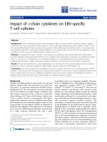
Figure 1 shows the TEM image of hydrothermal synthe-
sized Arg-Eu-HAP, it can be perceived that
unfunctionalized particles appear in short column
shapes and the cross-sections of particles are even,
approximately 50 to 100 nm. The lengthwise size of par-
ticles is in the size range of 50 t o 200 nm (Figure 1a).
After adding in arginine, the particles sizes reduce and
turn to be grain shapes with the sizes of 50 to 80 nm
(Figure 1b). During the process of synthesizing nanopar-
ticles under the hydrothermal equilibrium conditions,
the preferential growth direction of the HA crystal is
[001]. Arginine’s absorption of the seeded out HA crys-
tal face selectively affects particles growth, the positive
electron guanidyl group of arginine is able to have static
effect with the negative electron hydroxyl exposed on
the HA (001) face, resulting in intendancy of arginine to
be absorbed on the (001) face of HA nanoparticles.
Therefore, arginine’s absorption hinders the solution-
synthesized product to be separated out on the H A
(001) face to the greater extent.
Characterization of Arg-Eu-HAP
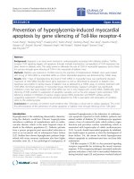
Figure 2 is t he XRD graph of two groups of samples. It
can be seen that all prepared nanoparticles’ XRD graphs
are similar. Their characteristic peaks are sharp and
appar ent, confirming that the resulting europium-doped
HAP had the typical pattern of the pure HAP. All dif-
fraction peaks could be assigned to the standard one
(JCPDs9-432).Thisdemonstratesphenomenonasvar-
ious direction sizes of the Arg-Eu-HAP samples shown
in Figure 2 have concerted tendency and the solid parti-
cles’ characteristics have strengthened.
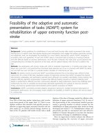
The successful introduction of surfac e functionality
was pr oved by Fourier transform i nfrared (FTIR; Figure
3), showed the infrared spect rometric waveforms of two
sample groups are similar and the main peak positions
of the graph are identical. The stronger peak lines occur
at positions as 565.25, 604.21, 1,035.78, and 3,441.75
cm
-1
, and weaker or broader position peak lines occur
Figure 1 TEM images of Arg-Eu-HAP crystal synthesized by hydrothermal method. (a) Without amino acid; (b) with arginine
Yan-zhong et al. Nanoscale Research Letters 2011, 6:600
/>Page 3 of 8
at positions of 1,106.57, 1,420.30, 1,631.24, and 3,570.12
cm
-1
. The four vibration patterns corresponding peak
positions of phosphate radicals in theory respectively
are: ν
1
peak at around 960 cm
-1
, ν
2
peak at around 470
to 440 cm
-1
region , ν
3
peak at 1,190 to 976 cm
-1
region ,
ν
4
peak at 600 to 560 cm
-1
region. Therefore, the strong
peaks at 565.25, 604.21, and 1035.78 cm
-1
and the weak
peaks of 1,106.57 cm
-1
are generated by the phosphate
radicals of HAP. The water molecule characteristic
peaks in crystal lattice occur at the 3,550 to 3,200 c m
-1
region, thus the peaks of the 3,441.75 and 3,570.12 cm
-1
positions are the reflection of lattice water and hydroxy
group (OH
-
). The characterist ic peak at 1,631.24 cm
-1
is
the vibration peak of H
2
O, indicating the surface of t he
solid samples absorbs a small amount of steam. The
characteristic peak of amino group(-NH
2
) occurs in the
1,400 to 1,420 cm
-1
region and the 1,420.30 cm
-1
peak
is perhaps the reflection of the absorption on HAP of
the ammonium radical (NH
4
+
) and amino acid residue
derived from the raw material ammonium dibasic phos-
phate. For the added arginine sample, the intensity of
this peak is somewhat strengthened, illustrating actual
existence of amino acid residue.
Europium ion (Eu
3+
) could be used as a luminescent
probe in the bimolecular system. And Ca ions on t he
HAP surface could be replaced by the o ther metal
cations with similar ionic radii, especially lanthanide
ions. The formation of Eu-doped HAP could be con-
firmed by the luminescence study. The luminescence
spectrum of Eu-doped HAP is shown in Figure 4. The
emission spectrum with the excitation of 394.4 nm (Fig-
ure 4a) showed the luminescence at the wavelengths of
588.8 and 612.6 nm, which could be ascribed to
5
D
0
-
7
F
1
,and
5
D
0
-
7
F
2
transitions of Eu, respectively. These
emission effects could not be observed in the pure HA
crystallites due to the absence of the featured Eu ele-
ment. Thus, the presence of Eu in the HAP was con-
firmed. In addition, the more efficient emission with a
maximum intensity at 612.6 nm is in the range of the
emission filter chosen for the confocal microscopy. An
excitation at 394.4 nm with the highest intensity is close
to the visible range. However, another excitation peak
was recorded at 464.8 nm, close to the available excita-
tion wavelength in the confocal microscope. Observa-
tions on living cells are possible as this excitation
wavelength is in the visible region.
Zeta potential of Arg-Eu-HAP
Figure 5 shows the Zeta potential of Arg-Eu-HA at the
pH value of 7.5. Results suggested under the weak alka-
lescent condition (pH 7.5), the Zeta potential of Arg-Eu-
HAP is (30.1 ± 6.3 mV) and unmodified HAP is (-10.6
± 4.2 mV). This illustrates arginine surface functionali-
zation of HA nanoparticles, cationic aminated functional
Figure 2 XRD patterns of nanoparticles and Eu-doped nanoparticles.
Yan-zhong et al. Nanoscale Research Letters 2011, 6:600
/>Page 4 of 8
groups increased its zeta p otential value. This change
comes from absorption of amino acids of amino acid
residue on the Arg-Eu-HAP surface. In later researches,
this substance is designed to be extracted from the aqu-
eous solution medium synthesized from Arg-Eu-HAP
and titrated to further discuss the hydrothermal crystal-
line behavior of HAP affected by arginine and the hid-
den mechanism of the surface electronic charge status.
DNA binding of Arg-Eu-HAP
Due to arginine-functionalized on the HA nanoparticles,
this can serve as the foundation for an effective enrich-
men t of negatively charged DNA strands onto the posi-
tively charged nanoparticles surfaces. In this study,
green fluorescence protein plasmid DNA was selected as
a model DNA. Agarose gel electrophoresis demonstrated
that Arg-Eu-HA could bind with DNA to form Arg-Eu-
Figure 3 FTIR spectra of arginine-functionalized nanoparticles: (a) without amino acid; (b) with arginine.
Figure 4 Luminescence excitation (a) and emission (b) spectrum of europium-doped HAP.
Yan-zhong et al. Nanoscale Research Letters 2011, 6:600
/>Page 5 of 8
HA/DNA complexes. As shown in Figure 6 , lane 1,
naked plasmid DNA moved in the electric field, lanes 3
to 5, no uncomplexed pDNA was observed in the lane
when mass ratios of Arg-Eu-HA to pEGFP-N1 plasmid
are 30:1, 50:1, and 70:1, respectively, demonstrating
DNA have fully bound with nanoparticles. The adsorp-
tion ratio is about 1 μgpEGFP-N1pDNAper30μg
HAP. The ultraviolet spectrometer 260-nm light absorp-
tion value measurement also proves the same result
(data not shown).
Cell toxicity of Arg-Eu-HAP
The effect of varying concentrations and exposure time
of Arg-Eu-HAP on cell toxicity was evaluated using
human epithelial lung cancer cell line (A549). The cell
line was chosen as representative models of the various
cellular environments that Arg-Eu-HAP are likely to
interact with in vivo. Results showed that the studied
Arg-Eu-HAP did not affect the cells survival in a con-
centration- and time-dependent manner. The cells
exposed to nanoparticles survived well similar to those
of the controls (Figure 7). Our data indicate that Arg-
Eu-HAP is a potential gene carrier in vitro,andfurther
preclinical and clinical development of this carrier for
cancer gene therapy is warranted.
Cellular uptake studies of Arg-Eu-HAP
Despite the unique advantages of HAP in biomedical
applications , exploration of their interactions with biolo-
gical systems remains at a very early stage. To effectively
develop these systems for application, it is necessary to
systematically delineate its functional properties about
cellular uptake and interactions after arginine functiona-
lized and europium doped. The majority of uptake stu-
dies in vitro have been performed in buffers devoid of
protein. In physiological fluids, howev er, a protein cor-
ona could be formed on a particle surface and affect its
interaction with cells [28,29]. We performed uptake stu-
dies in cell culture medium with free serum. Cellular
uptake of Arg-Eu-HAP was i nvestigated in A549 cell
line.
In order to visualize the luminescence of the euro-
pium-doped nanoparticles and to demonstrate internali-
zation in eucaryotic cells, several microscopic
Figure 5 Zeta potential curve of Arg-Eu-HAP at pH of 7.5.
Figure 6 Agarose gel electrophoresis of Arg-Eu-HAP/DNA complexes (w/w ratio). M, marker; L1, positive control; L2, 101:; L3, 301:; L4, 501:;
L5, 701:; L6, negative control.
Yan-zhong et al. Nanoscale Research Letters 2011, 6:600
/>Page 6 of 8
techniques were utilized. Figure 8a showed the fluores-
cence micrographs of DAPI-stained A549 cells after 2-h
incubation with 30 μg/mL nanoparticles. It can be seen
that most of the A549 cells incubated with Arg-Eu-HAP
(green) were evident in the cytoplasm, nuclei were
counterstained with DAPI dye (blue). These phenomena
indicat ed a higher uptake of nanoparticles in A549 cells.
The Laser scanning confocal microscope studies also
verified the above results and showed that numerous
luminescent nanoparticles were internalized within the
A549 cells after 1 h and were observed in the cytoplasm
of most cells ( Figure 8b). Figure 8b (A magnified and B
magnified) shows an accumulation of luminescent nano-
particles in the perinuclear areas of a cell on sections.
Figure 7 Cell viability assay. Cell viability assay showing the effect of varying concentrations of nanoparticles on growth inhibition of human
lung epithelial (A549) cancer cells cultured in vitro. Results are reported as mean. There is no statistically significant difference between test
groups and control groups (p < 0.05).
Figure 8 Green emission and Laser scanning confocal microscope images. (a) Green emission of the internalized Arg-Eu-HAP into the cells
under fluorescence microscopy. Arg-Eu-HAP (green) were evident in the cytoplasm, nucleus were counterstained with DAPI dye (blue).
Representative images of four different experiments are shown (magnification ×40). (b) Laser scanning confocal microscope images
(magnification ×60, insert magnification ×252). No fluorescent light in the control cells can be detected.
Yan-zhong et al. Nanoscale Research Letters 2011, 6:600
/>Page 7 of 8
No fluorescent light in the contro l cells can be det ected
(Figure 8b, control). Although the nanoparticles were
detected throughout the endoplasm, no evidence of
HAP entering the cell nucleus could be found from
microscopy images in our study.
Conclusions
In conclusion, nontoxic Arg-Eu-HAP have been pre-
pared and characterized in vitro by various physico-
chemical means. As arginine surface functionalization
changes HAP surface electron, its Zeta potential is chan-
ged from the unmodified (-10.6 ± 4 .2 mV) into the
functionalized (30.1 ± 6.3 mV). Meanwhile, arginine-
functionalized and europium-doped hydroxyapatite
nanoparticles with positive zeta potential can effective ly
bind negative plasmid DNA, and can be visualized in
the cytoplasm and perinuclear of A549 cells by fluores-
cence microscope and laser scanning confocal
microscope.
Acknowledgements
This work was partly supported by Project (no. 81071869) supported by the
National Natural Science Foundation of China (NSFC), Scholarship Program
(no. 2009637526) supported by China Scholarship Council and Project (no.
2010QZZD006) supported by the Key Program of Central South University
Advancing Front Foundation.
Author details
1
Medical Experiment Center in the Third Xiangya Hospital, Central South
University, Changsha 410013, China
2
State Key Laboratory of Powder
Metallurgy, Central South University, Changsha 410083, China
3
Research
Center for Medical Material and Instruments, Central South University,
Changsha 410013, China
Authors’ contributions
ZY and HY conceived and designed the study, carried out the experiments,
analyzed the results, and drafted the manuscript. ZJ and LZ assisted in
synthesis and characterization of nanoparticles experiments and assisted in
cell culture; ZS and ZK supervised the research, contributed in interpretation
of data and revision of the manuscript. All the authors have given final
approval of the version to be published.
Competing interests
The authors declare that they have no competing interests.
Received: 13 June 2011 Accepted: 23 November 2011
Published: 23 November 2011
References
1. FDA Places Temporary Halt on Gene Therapy Trials Using Retroviral
Vectors in Blood Stem Cells. US FDA, FDA Talk Paper 2003 [http://www.
fda.gov/bbs/topics/ANSWERS/2003/ANS01190.html].
2. Weiss R: Second boy receiving gene therapy develops cancer. The
Washington Post 2003.
3. Yasuhide N, Takesshi M, Makoto N: High performance gene delivery
polymeric vector: nano-structured cationic star polymers (star vectors).
Curr Drug Deliv 2005, 2:53-57.
4. Stefaan C, DE S, Joeseph D, Wim EH: Cationic polymer based gene
delivery systems. Pharmaceut Res 2000, 17(2):113-126.
5. Jennifer AF, Alexander MK: Highly effective gene transfection in vivo by
alkylated polyethylenimine. J Drug Deliv .
6. Markus E, Senta Ü, Carsten R: Nanocarriers for gene delivery - polymer
structure, targeting ligands and controlled-release devices. Current
Nanoscience 2008, 4:322-353.
7. Ko YT, Kale A, Hartner WC, Torchilin VP: Self-assembling micelle-like
nanoparticles based on phospholipid-polyethyleneimine conjugates for
systemic gene delivery. J Contr Release 2009, 133(2):132.
8. Zhao YZ, Yu ZP, Zhu SH, Huang YY, Zhou KC: Surface modification and
biomedical application of silica nanoparticles. The Chinese Journal of
Nonferrous Metals 2010, 20(7):1412-14917.
9. Legeros RZ: Properties of osteoconductive biomaterials:
calciumphosphates. Clin Orthop Relat Res 2002, 395:81-98.
10. Aoki H, Kutsuno T: An in vivo study on the reaction of hydroxyapatite-sol
injected into blood. J Mater Sci Mater Med 2000, 11:67-72.
11. Jiang W, Cheng J, Dinesh K: Improved mechanical properties of
nanocrystalline hydroxyapatite coating for dental and orthopedic
implants. Mater Res Soc 2009, 1140:1140-HH03-03.
12. Roya M, Amit B, Susmita B: Induction plasma sprayed nano
hydroxyapatite coatings on titanium for orthopaedic and dental
implants. Surf Coat Tech 2011, 205(1):2785-2792.
13. Matsumoyo T, Okazaki M, Inouc M: Hydroxyapatite particles as a
controlled release carrier of protein. Biomateials 2004, 25(17):3807-3812.
14. Boonsonggrit Y, Abe H, Sato K, Naito M, Ichikawa H, Fukumori Y: Controlled
release of bovine serum albumin from hydroxyapatite microspheres for
protein delivery system. Mater Sci Eng B 2008, 148:162-165.
15. Liu ZS, Tang SL, Ai ZL: Effects of hydroxyapatite nanoparticles on
proliferation and apoptosis of human hepatoma BEL-7402 cells. World J
Gastroenterol 2003, 9(9):1968-1971.
16. Zhu SH, Huang BY, Zhou KC, Huang SP, Liu F, Li YM, Xue ZG, Long ZG:
Hydroxyapatite nanoparticles as a novel gene carrier. Journal of
Nanoparticle Research 2004, 6(2):307-311.
17. Tan K, Cheang P, Iaw Ho, Pyp LK: Nanosized bioceramic particles could
function as efficient gene delivery vehicles with target specificity for the
spleen. Gene Therapy 2007, 14:828-835.
18. Sun H, Jiang M, Zhu SH: In vitro and in vivo studies on hydroxyapatite
nanoparticles as a novel vector for inner ear gene therapy. Chinese
Journal of Otorhinolaryngology Head and Neck Surgery 2008, 43(1):51-57.
19. Xie CJ, Yin DG, Li J, Zhang L, Liu BH, Wu MH: Preparation of a novel
amino functionalized fluorescein-doped silica nanoparticle for pH probe.
Nano Biomed Eng 2009, 1(1):27-31.
20. Yin DG, Liu BH, Zhang L, Xie CJ, Zhang L: Synthesis of Ru(bpy)3-doped
silica nanoparticle and its application in fluorescent immunoassay. Nano
Biomed Eng 2010, 2(2):117-120.
21. Zhang HB, Zhou KC, Li ZY, Huang SP, Zhao YZ: Morphologies of
hydroxyapatite nanoparticles adjusted by organic additives in
hydrothermal synthesis. J Cent S Univ Tech 2009, 16:0871-0875.
22. Brooks H, Lebleu B, Vives E: Tat peptide-mediated cellular delivery: back
to basics. Adv Drug Deliv Rev 2005, 57(4):559-577.
23. Umezawa N, Gelman MA, Haigis MC, Raines RT, Gellman SH: Translocation
of a beta-peptide across cell membranes. J Am Chem Soc 2002,
124(3):368-369.
24. Aslan K: Rapid whole blood bioassays using microwave-accelerated
metal-enhanced fluorescence. Nano Biomed Eng 2010, 2(1):1-7.
25. Li YQ, Li ZY, Zhou XP, Yang P: Detection of nano Eu
2
O
3
in cells and study
of its biological effects. Nano Biomed Eng 2010, 2(1):24-30.
26. Yin DG, Zhang L, Xie CJ, Liu BH, Zhang L: Preparation and characterization
of DPPDA-Eu3+ doped silica fluorescent nanoparticles. Nano Biomed Eng
2010, 2(1):40-44.
27. Yin DG, Zhang L, Liu BH, Zhang L, Yan H: Time-resolved fluorescence
immunoassay of mouse IgG using europium(III) chelate-doped silica
nanoparticles. Nano Biomed Eng 2011, 3(1):25-28.
28. Jiang X, Weise S, Hafner M, Rrocker C: Quantitative analysis of the protein
corona on FePt nanoparticles formed by transferrin binding. J R Soc
Interface 2010, 7(Suppl 1):S5-S13.
29. Oleg L, Tatiana S, Cornelia L, Beil J: Differential uptake of functionalized
polystyrene nanoparticles by human macrophages and a monocytic cell
line. American of Chemical Society: Nano 2011, 5(3):1657-1669.
doi:10.1186/1556-276X-6-600
Cite this article as: Yan-zhong et al.: Characteristics of functionalized
nano-hydroxyapatite and internalization by human epithelial cell.
Nanoscale Research Letters 2011 6:600.
Yan-zhong et al. Nanoscale Research Letters 2011, 6:600
/>Page 8 of 8