Báo cáo hóa học: " Visible emission from Ce-doped ZnO nanorods grown by hydrothermal method without post thermal annealing process" ppt
Bạn đang xem bản rút gọn của tài liệu. Xem và tải ngay bản đầy đủ của tài liệu tại đây (508.2 KB, 14 trang )
This Provisional PDF corresponds to the article as it appeared upon acceptance. Fully formatted
PDF and full text (HTML) versions will be made available soon.
Visible emission from Ce-doped ZnO nanorods grown by hydrothermal method
without post thermal annealing process
Nanoscale Research Letters 2012, 7:43 doi:10.1186/1556-276X-7-43
Yong-Il Jung ()
Bum-Young Noh ()
Young-Seok Lee ()
Seong-Ho Baek ()
Jae Hyun Kim ()
Il-Kyu Park ()
ISSN 1556-276X
Article type Nano Express
Submission date 6 September 2011
Acceptance date 5 January 2012
Publication date 5 January 2012
Article URL />This peer-reviewed article was published immediately upon acceptance. It can be downloaded,
printed and distributed freely for any purposes (see copyright notice below).
Articles in Nanoscale Research Letters are listed in PubMed and archived at PubMed Central.
For information about publishing your research in Nanoscale Research Letters go to
/>For information about other SpringerOpen publications go to
Nanoscale Research Letters
© 2012 Jung et al. ; licensee Springer.
This is an open access article distributed under the terms of the Creative Commons Attribution License ( />which permits unrestricted use, distribution, and reproduction in any medium, provided the original work is properly cited.
1
Visible emission from Ce-doped ZnO nanorods grown by hydrothermal
method without a post thermal annealing process
Yong-Il Jung
1,2
, Bum-Young Noh
1,2
, Young-Seok Lee
1
, Seong-Ho Baek
2
, Jae Hyun Kim
*2
,
and Il-Kyu Park
*1
1
LED-IT Fusion Technology Research Center and Department of Electronic Engineering,
Yeungnam University, Gyeongbuk, 712-749, Korea
2
Energy Research Division, Daegu Gyeongbuk Institute of Science & Technology(DGIST),
50-1, Sang-Ri, Hyeonpung-Myeon, Dalseong-gun, Daegu, 711-873, Korea
*Corresponding authors: ;
Email addresses:
YIJ:
BYN:
YSL:
SHB:
JHK:
IKP:
Abstract
Visible light-emitting Ce-doped ZnO nanorods [NRs] without a post thermal annealing
process were grown by hydrothermal method on a Si (100) substrate at a low temperature
of 90°C. The structural investigations of Ce-doped ZnO NRs showed that the Ce
3+
ions
were successfully incorporated into the ZnO lattice sites without forming unwanted Ce-
related compounds or precipitates. The optical investigation by photoluminescence spectra
shows that the doped Ce
3+
ions in the ZnO NRs act as an efficient luminescence center at
540 nm which corresponds to the optical transition of 5d → 4f orbitals in the Ce
3+
ions. The
photoluminescence intensity of the Ce-doped ZnO NRs increased with the increasing
content of the Ce-doping agent because the energy transfer of the excited electrons in ZnO
to the Ce
3+
ions would be enhanced by increased Ce
3+
ions.
Keywords: nanostructures; oxides; crystal growth; optical properties.
2
Introduction
Zinc oxide [ZnO] nanostructures have received much attention due to their remarkable
performance in electronic, piezoelectric, thermoelectric, and optoelectronic applications [1].
High electrical conductivity and transparency in a visible wavelength spectral range of the
ZnO alloys have been regarded as an efficient candidate for transparent conducting
electrodes of thin films and flexible electronics [2]. The absence of a center of symmetry in
its wurtzite structure, along with a large electromechanical coupling, results in strong
piezoelectric and pyroelectric properties which make it an energy recycling material system
[3]. In addition, because ZnO has a large direct bandgap of 3.37 eV and a high exciton
binding energy of 60 meV, it has been much investigated for optoelectronic applications,
such as light-emitting diode [LED] and UV laser diode [4]. Recently, the ZnO
nanostructures doped with rare earth elements such as Ce, Y, and Eu have achieved much
attention for biological tagging as well as optoelectronic applications due to their unique
optical properties [5-7]. Most of all, the Ce element possessing a unique optical
characteristic may be an ideal material for visible light-emitting phosphors in display, high-
power laser, and light-emitting diode [5-9]. However, few have been reported for the
fabrication and optical properties of the Ce-doped ZnO nanostructures especially by
hydrothermal method [7-9] even though this method has many advantages of low
temperature process which makes it favorable to the integration and in-situ fabrication of
various devices. Furthermore, for more compatibility of the Ce-doped ZnO nanorods [NRs]
to be applicable to other devices, visible emission should be possible without post thermal
annealing processes. In this report, we have fabricated visible light-emitting Ce-doped ZnO
NRs by hydrothermal method without a post thermal annealing process.
Experimental details
Growth of undoped and Ce-doped ZnO NRs on Si (100) substrate
The Ce-doped ZnO NRs were grown on a p-type Si (100) substrate. After cleaning the Si
substrate by acetone, methanol, HF, and deionized [DI] water, a seed layer for the ZnO NRs
was formed by dipping the substrate into 40 mM of zinc acetate dihydrate
(Zn(CH
3
COO)
2
·2H
2
O) dissolved in ethanol solution, followed by drying at 100°C for 5 min.
The Ce-doped ZnO NRs were grown by placing the seed layer grown substrate into a mixed
3
solution of 20 mM zinc nitrate hexahydrate (Zn(NO
3
)
2
·6H
2
O), 20 mM
hexamethylenetetramine [HMT] ((CH
2
)
6
N
4
), and cerium nitrate hexahydrate
(Ce(NO
3
)
3
·6H
2
O) in DI water at 90°C for 3 h. The amount of Ce agent was varied from 0.1
to 0.8 mM, which corresponds from 0.5% to 4% in molarity. At a solution temperature
above 70°C, the solution started being cloudy, indicating that a chemical reaction started.
After the reaction, the samples were cleaned in the flowing DI water for 5 min.
Structural characterizations
The structural properties of Ce-doped ZnO NRs were investigated by field emission
scanning electron microscopy [FE-SEM], energy dispersive X-ray spectroscopy [EDS], and
X-ray diffraction [XRD] with an excitation source of Cu Kα radiation. The chemical
composition of the deposited ZnO NRs was observed using EDS attached to a FE-SEM
microscope.
Optical characterizations
The optical properties of Ce-doped ZnO NRs were investigated by photoluminescence [PL]
spectra which were measured by using a 24-mW power 325-nm continuous He-Cd laser at
room temperature. The laser was focused onto the sample surface by an objective lens; the
excitation area was estimated to be about 400 µm in diameter.
Results and discussion
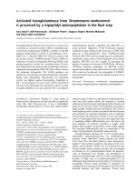
Figures 1a to 1e compare the FE-SEM images of undoped ZnO NRs grown on Si substrate
with an increasing amount of ZnO sources. As the molar content of the ZnO source
increases to 80 mM, the well-faceted six-side surfaces are developed, and the top of the
ZnO NRs shows a hexagonal facet, indicating that the ZnO NRs are single crystals grown
along the [0001] direction [10]. The average length and diameter of the ZnO NRs obtained
from FE-SEM images were plotted versus the ZnO source molar contents in Figure 1f. With
the increasing molar content of the ZnO source from 10 to 80 mM, the diameter increased
linearly from 45 to 123 nm. The length of the ZnO NRs decreased, with the source molar
content up to 20 mM, then increased up to 60 mM, and finally, saturated again. In this way,
we could control the size and morphologies of ZnO NRs by molar contents of sources in
the main solution. This result indicates that the growth mechanism of the ZnO NRs is
governed by the source supplying rate from the solution to the growth front surface of ZnO
4
NRs, not by the surface reaction's limited condition when the source molar contents were
below 60 mM. Therefore, in the source molar content range below 60 mM, the Ce dopant
atoms can be incorporated into the ZnO matrix in a controllable way because the growth is
governed by the source supplying rate.
Figures 2a to 2e show the surface morphologies of the Ce-doped ZnO with different
amounts of the Ce agent, from 0.1 to 0.8 mM, in the ZnO NRs which were grown by 20
mM of ZnO source agent. The diameter of the undoped ZnO NRs is nearly 50 nm. As
shown in Figures 2b to 2e, the morphology of ZnO NRs did not change significantly with
the changing amount of the Ce dopant agent. This indicates that the growth of ZnO NRs is
not influenced by the doping of Ce atoms because the Ce(NO
3
)
3
·6H
2
O agent was used for
supplying Ce
3+
ions to be doped, and the molar contents of the dopant agent, compared to
the ZnO source, was too small to change the morphology. Figure 2f is an EDS pattern of the
0.8-mM Ce-doped ZnO NRs showing that there are Zn, O, and Ce elements in the ZnO
NRs. The other peaks noted Si and Pt to correspond to the substrate and coating material for
SEM measurement. The EDS results show that the amount of Ce in ZnO NRs is 2.6%,
indicating that the Ce dopant is successfully incorporated into the ZnO NRs. The
mechanism for the synthesis of the Ce-doped ZnO NRs using HMT can be summarized in
the following equations:
(
)
( )
( )
2 4 2 2 3
6
3 2 3 2 4
2
3 2 3 2
2
2
2
3
3 2 3 2
3
3
2 3 2
CH N 6H O 6COH 4NH
NH H O NH ·H O NH OH
Zn NO ·6H O Zn 2NO 6H O
Zn 2OH ZnO H O
Ce NO ·6H O Ce 3NO 6H O
4Ce 12OH 2Ce O 6H O
+ −
+ −
+ −
+ −
+ −
+ → +
+ → → +
→ + +
+ → +
→ + +
+ → +
.
HMT plays a very complicated role in the solution during the hydrothermal process [11],
but it supplies OH
-
ions to the Zn
2+
and Ce
3+
ions to form Zn-O and Ce-O bonds here,
respectively. Thereby, Ce
3+
ions substitute the Zn lattice sites during the growth of ZnO
NRs.
Figures 3a and b show the XRD patterns of as-prepared undoped ZnO NRs and 0.8-mM
Ce-doped ZnO NRs, respectively. The diffraction peaks are quite similar to those of bulk
5
ZnO. The diffraction peaks in the pattern can be indexed to the standard hexagonal
wurtzite-structured ZnO (space group: P63mc; a = 0.3249 nm, c = 0.5206 nm), and
diffraction data are in agreement with the JCPDS card for ZnO (JCPDS 36-1451). All the
diffraction peaks were identified as corresponding to (002), (101), (102), (103), (201), and
(004) planes of the hexagonal wurtzite ZnO structure, and no additional peaks
corresponding to Ce-related alloys were observed as shown in Figure 3b. This result shows
that the as-prepared products are single-phase hexagonal ZnO, and the Ce-related
compounds or precipitates are not formed during growth, but the Ce atoms are incorporated
into the lattice sites in the ZnO. Even though the ionic radius of Ce
3+
(1.03 Å) is bigger than
that of Zn
2+
(0.74 Å), there is no change in diffraction peak positions between the undoped
and the 0.8-mM Ce-doped ZnO NRs because the amount of incorporated Ce dopants is
negligibly small to be detectable in the diffraction data.
Figure 4a shows the PL spectrum of as-grown undoped ZnO NRs and 0.8-mM Ce-doped
ZnO NRs measured at room temperature. The PL spectra of both samples exhibit mainly
two emission peaks at UV and visible ranges, which correspond to the band edge emission
of ZnO and deep or Ce-activated atomic levels, respectively. The emission band at a visible
wavelength range of the undoped ZnO NRs arises from a radiative recombination through
point defects in the ZnO lattice, such as oxygen vacancy, zinc vacancy, oxygen interstitial,
zinc interstitial, and anti-site defects, as reported by many other papers [12, 13]. The PL
peak positions of the UV emission for undoped and Ce-doped ZnO NRs are the same at 384
nm. If the Ce dopant incorporated into the ZnO as a donor with the valence state Ce
4+
, the
band edge emission peak position from Ce-doped ZnO NRs should be red-shifted [9]. The
undoped ZnO NR shows a larger UV/visible emission intensity ratio than that of the Ce-
doped ZnO NRs. Therefore, these indicate that the Ce
3+
ion activators are embedded in the
ZnO host matrix and act as a luminescence center in the visible spectral range. The
emission band at a visible range of the Ce-doped ZnO NRs is composed of two elementary
PL peaks at 543 and 590 nm. The PL peak at 590 nm corresponds to the emission by the
mentioned point defects, while the peak at 543 nm is from the emission by electron energy
transition from 5d to 4f orbitals in Ce
3+
ions, as shown in the inset of Figure 4a. The
fluorescence of Ce
3+
-activated compounds usually consists of a broad band with two peaks
because the ground state of the Ce
3+
ion consists of a doublet (
2
F
5/2
and
2
F
7/2
) [14]. To
investigate the effect of the Ce dopant amount on the optical properties, PL spectra were
6
measured precisely in the visible range by enhancing the sensitivity of the detector as
shown in Figure 4b. The PL intensity at 543 nm increases gradually with the increase of the
molar ratio of the Ce dopant agent. The energy of excited electrons in the conduction band
of the ZnO is transferred to the Ce
3+
ion activators to luminescence. Therefore, with the
increase of Ce
3+
ions in the ZnO matrix, the energy transfer rate of excited electrons to Ce
3+
ions can be enhanced, resulting in the increase of the PL intensity at this wavelength. A
visible emission from the ZnO NRs without a post thermal annealing process is a very
important point here because it makes possible for the ZnO NRs to be applicable with the
active layer or wavelength conversion layers of optoelectronic devices, such as visible
wavelengths or white LEDs.
Conclusions
In summary, Ce-doped ZnO NRs were grown on Si substrates by using the hydrothermal
method. The structural properties investigated by FE-SEM, EDS, and XRD showed that the
Ce
3+
ions are successfully incorporated into the ZnO lattice sites; the growth of ZnO NRs
was not influenced by the doping of Ce atoms because the molar content of the dopant was
too small an amount to change the morphology, and the dopant agent provided Ce
3+
ions for
doping. The XRD results showed that the ZnO NRs are single-phase hexagonal ZnO, and
unwanted Ce-related compounds or precipitates were not formed during the growth of Ce-
doped ZnO NRs. The PL results showed that the doped Ce
3+
ions in the ZnO NRs act as a
luminescence center for visible emission at 543 nm even though the ZnO NRs were not
thermally annealed. The PL intensity at a visible range of the Ce-doped ZnO NRs increased
with the increased molarity of the Ce dopant agent because the energy transfer rate of the
excited electrons from the conduction band in the ZnO host to a 5d energy state in the Ce
3+
ion activators could be enhanced by increased Ce
3+
ions. These results demonstrate that Ce
doping in ZnO NRs can be an efficient luminescence center in ZnO NRs without post
thermal annealing processes.
Competing interests
The authors declare that they have no competing interests.
7
Authors' contributions
YIJ and IKP developed the concept and design of the Ce-doped ZnO nanorods. YIJ carried
out the fabrication of the Ce-doped ZnO nanorods. BYN carried out the structural
characterization of the ZnO nanorods. YSL, SHB, and JHK carried out the optical
characterization by photoluminescence measurement. YIJ and IKP analyzed the results and
wrote the manuscript. All authors read and approved the final manuscript.
Acknowledgments
This work was financially supported by the basic research program (11-EN-03) through the
Daegu-Gyeongbuk Institute of Science and Technology (DGIST) funded by the Ministry of
Education, Science and Technology (MEST) and IT/SW Creative Research Program
supervised by the National IT Industry Promotion Agency (NIPA-2011-C1820-1102-0013)
supported by the Ministry of Knowledge Economy (MKE).
References
1. Wang, ZL: Zinc oxide nanostructures: growth, properties and applications. Journal
of Physics: Condensed Matter 2004, 16:R829-R858.
2. Remashan K, Hwang DK, Park SJ, Jang JH: Effect of rapid thermal annealing on the
electrical characteristics of ZnO thin-film transistors. Jpn J App Phys 2008, 47:2848-
2853.
3. Choi MY, Choi D, Jin MJ, Kim I, Kim SH, Choi JY, Lee SY, Kim JM, Kim SW:
Mechanically powered transparent flexible charge-generating nanodevices with
piezoelectric ZnO nanorods. Adv Mater 2009, 21:2185-2189.
4. Lim JH, Kang CK, Kim KK, Park IK, Park SJ: UV electroluminescence emission from
ZnO light-emitting diodes grown by high-temperature radio frequency sputtering.
Adv Mater 2006, 18:2720-2724.
5. Mordkovich VZ, Hayashi H, Haemori M, Fukumura T, Kawasaki M: Discovery and
optimization of new ZnO-based phosphors using a combinatorial method. Adv
Funct Mater 2003, 13:519-524.
6. John JS, Coffer JL: Size control of erbium-doped silicon nanocrystals. Appl Phys Lett
2010, 77:1635-1637.
8
7. Cheng B, Xiao Y, Wu G, Zhang L: Controlled growth and properties of one-
dimensional ZnO nanostructures with Ce as activator/dopant. Adv Func Mater 2004,
14:913-919.
8. Li GR, Lu XH, Zhao WX, Su CY, Tong YX: Controllable electrochemical synthesis of
Ce
4+
-doped ZnO nanostructures from nanotubes to nanorods and nanocages.
Crystal Growth & Design 2008, 8:1276-1281.
9. Lang J, Han Q, Yang J, Li C, Li X, Yang L, Zhang Y, Gao M, Wang D, Cao J:
Fabrication and optical properties of Ce-doped ZnO nanorods. J Appl Phys 2010,
107:074302.
10. Umar A, Karunagaran B, Suh EK, Hahn YB: Structural and optical properties of
single-crystalline ZnO nanorods grown on silicon by thermal evaporation.
Nanotechnology 2006, 17:4072-4077.
11. Yang Y, Lai H, Tao C, Yang H: Correlation of luminescent properties of ZnO and Eu
doped ZnO nanorods . J Mater Sci: Mater Electron 2010, 21:173-178.
12. Ahn CH, Kim YY, Kim DC, Mohanta SK, Cho HK: A comparative analysis of deep
level emission in ZnO layers deposited by various methods. J Appl Phys 2009,
105:013502.
13. Wu C, Qiao X, Luo L, Li L: Synthesis of ZnO flowers and their photoluminescence
properties. Mater Res Bull 2009, 43:1883-1891.
14. Blasse G, Bril A: A new phosphor for flying-spot cathode-ray tubes for color
television: yellow-emitting Y
3
Al
5
O
12
-Ce
3+
. Appl Phys Lett 1967, 11:53-55.
9
Figure 1. FE-SEM images of ZnO NRs grown by various ZnO source molarities. ZnO
source molarities of (a) 10 mM, (b) 20 mM, (c) 40 mM, (d) 60 mM, and (e)
80mM in the main solution.
Figure 2. FE-SEM images and EDS patterns of Ce-doped ZnO NRs. FE-SEM images
of (a) undoped, (b) 0.1-mM (0.5%), (c) 0.2-mM (1%), (d) 0.4-mM (2%), and (e)
0.8-mM (4%) Ce-doped ZnO NRs. (f) EDS patterns of 0.8-mM Ce-doped ZnO
NRs.
Figure 3. Normalized θ-2θ XRD scan spectra. (a) Undoped and (b) 0.8-mM Ce-doped
ZnO NRs in log scale.
Figure 4. Normalized PL sprectra measured at room temperature. (a) Room
temperature PL spectra of as-grown undoped and Ce-doped ZnO NRs. Two
elementary PL spectra shown in dotted lines are obtained by deconvolution of the
PL spectrum of doped ZnO NRs. The inset shows a schematic energy level
diagram of Ce
3+
electronic levels in ZnO bandgap. (b) Normalized PL spectra of
ZnO NRs doped with different amounts of Ce molar contents.
Figure 1
Figure 2Figure 2
Figure 3
Figure 4