Báo cáo hóa học: " Purification and characterization of novel fibrinolytic proteases as potential antithrombotic agents from earthworm Perionyx excavatus" pot
Bạn đang xem bản rút gọn của tài liệu. Xem và tải ngay bản đầy đủ của tài liệu tại đây (3.55 MB, 11 trang )
ORIGINAL Open Access
Purification and characterization of novel
fibrinolytic proteases as potential antithrombotic
agents from earthworm Perionyx excavatus
Tram Thi Bich Phan
1
, Tien Duy Ta
2*
, Dung Thi Xuan Nguyen
3
, Lambertus AM Van Den Broek
4
and
Giang Thi Huong Duong
3
Abstract
Six protease fractions, namely FI, FII, FIII-1, FIII-2, FIII-3 and FIV, were isolated from Perionyx excavatus earthworm
biomass by acetone precipitation, followed by serial chromatography using anion exchange, hydrophobic interaction
and size exclusion chromatography. All fractions exhibited strong hydrolytic activity towards casein. The activity of six
fractions towards fibrin, deter mined by fibrin plate assay, ranged from 44 to 831 plasmin unit.mg
-1
and ranked as FIII-
3 > FIII-2 > FI > FIII-1 > FIV > FII. Casein degradation was optimal at pH 7 and 11, and at 45-60°C. All fractions were
considerably stable at high temperature and wide pH range. They were completely inhibited by
phenylmethylsulfonyl fluoride (PMSF). The molecular weights (MW) and isoelectric points (pI) determined by
2D-electrophoresis were 27.5-34.5 kDa, and 4.3-5.2, respectively. Tandem m ass spectrometry (MS) analysis was used
to deduce the amino acid sequences of some peptides from FIII-1 and FIII-2. The sequences shared 16.9% and 13.2%
similarity, respectively , with the fibrinolytic enzymes from two related earthworm species, Lumbricus rubellus and
Eisenia fetida. The P. excavatus proteases were classified as serine proteases. They could perform rapid hydrolysis on
both coagulated fibrous fibrin and soluble fibrinogen monomers without the presence of activators such as tPA or
urokinase.
Keywords: chromatography, fibrinolysis, Perionyx excavatus, PMSF, serine protease, tandem MS analysis
Introduction
Cardiovascula r di seases have become one of the biggest
concerns all over the world (Grundy et al. 1999). Among
these, thrombosis is the most widespread within the
elderly population. The disease results from severe
blood-clotting, which leads to obstruction of the blood
flow circulation. In the physiological state, fibrin and
platelets are utilized for clotting to prevent blood loss
from injuries in a process called hemostasis (Furie and
Furie 2008). A se rine protease called plasmin acts to
digest blood clots via fibrino lysis to properly terminate
the hemostasis. Plasmin deficiency is one reason that
leads to thrombosis due to insufficient clots degradation.
Fibrin is a fibrous polymer protein that plays an
important role in the final blood coagulation step in
hemostasis. The fibrino gen monomer is a 304 kDa gly-
coprotein containing two sets of three different chains:
Aa,Bb and g (Mosesson 2005). The conversion of fibri-
nogen into fibrin requires the presence of thrombin, a
serine protease that cleaves the N-terminus of Aa and
Bb chain (Mosesson et al. 2001). Fibrinogen level was
reported to be significantly related to the incidence of
cardiovascular disease in both men and women during
the tenth biennial examination of the Framingham
Study (Kannel et al. 1987). Treatment of cerebral venous
thrombosis currently relies on the use of anticoagulants
such as heparin, which is also a medicament for deep
vein t hrombosis (Stam et al. 2003) despite the risk
of consequent occurrence of intracranial hemorrhage
(Einhaup l et al. 1991 and Mehraein et al. 2003). Enzyme
therapy of t hrombosis has been investigated since 1969
by using streptokinase, a fibrinolytic enzyme (Kakkar
et al. 1969), and was reported to be a better treatment
* Correspondence:
2
Faculty of Food Processing Technology, Can Tho University of Technology,
Can Tho, Vietnam
Full list of author information is available at the end of the article
Phan et al. AMB Express 2011, 1:26
/>© 2011 Phan et al; licensee Springer. This is an Open Access article distributed under the terms of th e Cre ative Commons Attribution
License ( which permits unrestricted use, distribution, and reproduction in any medium,
provided the original work is properly cited.
for acute deep vein thrombosis than of heparin (Marder
et al. 1977 and Arnesen et al. 1982).
A no vel fibrinolytic enzyme, namely lumbrokinase, has
been isolated from some earthworm species such as Lum-
bricus rubellus (Cho e t al. 2004; Mih ara et a l. 1991 and
Nakajima et al. 1983) and Eisenia fetida (Yang and Ru
1997), and was thoroughly characterized. They have been
identified as serine protease isozymes, which are highly
thermostable and alkali tolerant. The genes enc oding
strong fibrinolytic enzymes from these earthworms have
been identified (Dong et al. 2004). High -throughput pro-
duction of these enzymes by recombinant DNA technol-
ogy has been conducted in Escheri chia coli (Cho et al.
2004 and Xu et al. 2010) and Pichia pastoris (Ge et al.
2005 and Sugimoto et al. 2005). The recombinant enzymes
expressed strong fibrinolytic activity both in vitro (Sugi-
moto et al. 2005) and in vivo in rats via oral administration
(Cho et al. 2004). Crystallographic data of two compo-
nents of L. rubellus lumbrokinase were obtained, revealing
the structure determinants of their catalytic mechanisms
(Tang et al. 2002 and Wang et al. 2005). The analysis
showed that the structure of component B resembled that
of the trypsyin-like proteases, and was the first reported
glycosylated trypsin-like structure. The study also revealed
the structural basis for high stability and complicated
post-translational modifications of the enzyme.
Application of the fibrinolytic enzymes has also been of
great interest in Vietnam, focusing especially on the use of
local enzyme sources. The preliminary experiments of this
research have discovered the novel and remarkably strong
firbinolytic enzymes from Perionyx exc avatus eart hworm
(family Megascolecidae). This species has been widely cul-
tivated in Southern Vietnam for the production of aquatic
animal feeds and compost. In our study, an extensive puri-
fication of these proteases was carried out together with
the characterization of temperature and pH optimum,
inhibition, substrate specificity, fibrinolytic effect and par-
tial sequencing. We have found the potential of these
enzymes as effective fibrinolytic agents. The study was
thus aimed to understand more deeply the properties of
these enzymes and to initially evaluate their applicability
in thrombosis treatment.
Materials and methods
Materials
Living earthworms (P. excavates; 8-9 weeks old) were pur-
chased from the farms in An Giang province, Vietnam.
Protease substrate s included casein from Merck (USA),
fibrinogen (together with throm bin and human plasmin),
Na -benzoyl-L-arginine p-nitroanilide (BApNA), and
Na-benzoyl-L-tyrosine p-nitroanilide (BTpNA) from
Sigma-Aldrich (USA). The enzyme inhibitors were bought
from Sigma (USA). The 2D-electrophoresis kits were
obtained from Bio-Rad (USA).
Autolysis of P. excavatus
Earthworm biomass was washed and finely homogenized
in distilled water containing 0.1% (w/v) sodium azide.
Autolysis was initially performed at 45°C for 4 hours
and continued at room temperature for 15 days with
stirring. Enzyme activity was measured daily by the
modified method of Anson (Anson 1938) using casein
as substrate. One activity unit (U) was defined as the
amount of enzyme that catalyzed the release of 1 μmol
of tyrosine per minute at 30°C at pH 7.5. Specific activ-
ity (expressed in U.mg
-1
) was defined as the activity per
milligram of t otal protein, which was determined by the
Lowry protein assay (Lowry 1951).
Purification of P. excavatus proteases
The autolysate was centrifuged (11,200 × g) at 4°C for
30 minutes, and the supernatant was precipitated in
pre-chilled acetone at 4°C for 2 hours. The precipitate
was collected by centrifugation (11,200 × g) at 4°C for
30 minutes and was lyophilized. A series of chromato-
graphic techniques were used for purification. Fractions
containing proteases were collected and analyzed by
SDS-PAGE (Hames 1998) after each purification step.
Anion exchange chromatography (AEX)
Precipitated protein was re-dissolved in 20 mM Tris-
HCl buffer pH 8.5 and was loaded on to a 1.5 × 40 cm
Unosphere Q (Bio-Rad, USA) column at a flow-rate of
0.8 ml.min
-1
. Bound proteins were eluted in the same
buffer, with a continuous NaCl gradient from 0 to
0.45 M at a flow-rate of 1 ml.min
-1
.
Hydrophobic interaction chromatography (HIC)
Each active fraction was dialyzed against 20 mM Tris-HCl
buffer pH 8.5 for desalting; subsequently 30% (w/v)
ammonium sulfate (AS) was added, followed by loading
the sample on to a 1.5 × 30 cm Phenyl Sepharose (GE
Healthcare, UK) column at a flow-rate of 1 ml.min
-1
. Elu-
tion was done using a continuous AS gradient from 30%
to 0% at the same flow-rate.
Size exclusion chromatography (SEC)
SEC was carried out for each active HIC fraction on a
Superose 12 column (GE Healthcare, UK) using the Biolo-
gic HR System (Bio-Rad, USA). The running buffer was
20 mM Tris-HCl buffer pH 7.5 containing 15 mM NaCl,
passing through the column at a flow-rate of 0.5 ml.min
-1
.
Eluted fractions were dialyzed against water and freeze-
dried using a VirTis Bench T op M anifold Freeze Dryer
(SP Scientific, USA) and stored at -20°C for later use.
Characterization of P. excavatus protease fractions
Determination of optimal temperature and thermostability
The caseinolytic assay was performed to determine the
optimal temperature for each active SEC fraction in
50 mM sodium phosphate buffer pH 7.5 at different
temperature ranging from 30°C to 80°C in 10 minutes.
Phan et al. AMB Express 2011, 1:26
/>Page 2 of 11
To investigate the thermal stability, each fraction was
incubated at different temperatures between 37 and
70°C for 3 hours, and the remaining activity (expressed
as % of the activity at 37°C) was determined after 30, 60,
120 and 180 minutes.
Determination of optimal pH and pH-tolerance
Each fraction was assayed for caseinolytic activity at
30°C in different pH-buffered solutions f rom 3 to 12,
prepared as follows: 100 mM Glycine-HCl (pH 2),
100 mM c itrate-phosphate (pH 3-5), 100 mM sodium
phosphate (pH 6-8), 100 mM Glycine-NaOH (pH 9-11),
and KCl-NaOH (pH 12-13). The pH tolerance was
determined by incubating each fraction in these buffer
solutions for 16 hours at 4°C; and after neutralization at
30°C the remaining activity was determined (expressed
as % of the activity at pH 7). In addition, the long-term
storage of these enzymes was also investigated using dis-
tilled water and 50 mM sodium phosphate buffer pH 7.5
as preservative solvents. The freeze-dried enzymes were
dissolved in these solvents containing 0.1% (w/v) sodium
azide, and stored in carefully-s ealed glass vials at 4°C for
10 months. The remaining activity, displayed as % of the
initial activity, was determined every month.
Enzyme inhibition assay was done with eight inhibitors
PMSF, N-torsyl-L-phenylalanine c hloromethyl ketone
(TPCK), aprotinin, leupeptin, soybean trypsin inhibitor
(SBTI), ethylene diamine tetraacetic acid (EDTA), chy-
mostatin and peptastatin. The used concentration ranged
from 0.01 to 1 mM. Samples incubated with casein but
without inhibitors w ere used as controls. Each fraction
was incubated separately with each inhibitor in 50 mM
sodium phosphate buffer pH 7.5 containing casein a t
37°C fo r 10 minutes. The measured activity was com-
pared with the controls.
Hydrolytic assay using different substrates
The hydrolytic ability towards different substrates such
as casein, fibrin, BApNA, and BTpNA was examined.
The assays for the last two substrates were performed at
37°C in 50 mM sodium phosphate buffer pH 7.5 based
onthemethodof(Wangetal.2005).Oneactivityunit
(U) on these substrates was defined as the amount of
enzyme that released of 1 μmol of p-nitroaniline under
the given conditions. Fibrinolysis was perfor med on a
fibrin plate as described by (Choi and Sa (2001)). Briefly,
a mixture of 0.6% (w/v) fibrinogen and 2% (w/v) agar
was prepared in 50 mM sodium phosphate buffer pH
7.5, boiled for 2 mi nutes, cooled down to 55°C and sub-
sequently added with thrombin (10 NIH unit.ml
-1
)for
coagulation in a Petri disc. Ten μlofenzymesolution
was pipetted into the small holes created on the plate
and incubated at 37°C for one hour. Fibrinolytic activity,
expressed as plasmin unit (PlasminU), was extrapolated
from the area of the hydrolytic zones based on a human
plasmin standard curve. In vitro cleavage of fibrinogen
was investigated by incubating each protease fraction
(0.2 mg.ml
-1
) with bovine fibrinogen (1 mg.ml
-1
) at 37°C
in 50 mM sodium phosphate buffer pH 7.5 containing
100 mM NaCl. Aliquots of 10 μl were taken out of the
reaction mixture aft er 10, 20, 30, 60, 90, 120, 180, 2 40
and 480 minutes, and were visualized on SDS-PAGE gel.
2D-electrophoresis-coupled MS/MS sequencing
The protease fractions were purified using a 2D-Cleanu p
Kit. Isoelectric focusing (IEF) dimension was run on pH 3-
10 IPG-Strips, and the second dim ension was carried out
on a 12% polyacrylamide gel without stacking layer,
according to the manufacturer’ susermanuals.Images
were captured by GelDocTM XR+ system (Bio-Rad, USA)
and were analyzed using PDQuestTM 2-D Analysis Soft-
ware (BioRad, USA) for MW and pI calculation. Each pro-
tein spot was excised, digested with trypsin, an d purified
using the protocol from the Protein and P roteomics Centre
(NUS, Singapore). Tandem MS analyses for these peptides
were carried out at the Laboratory of Protein and Proteo-
mics Centre (NUS, Singapore) using a MALDI-TOF-TOF
MS: 4800 Proteomics Analyzer instrument (Applied Bio-
systems, Framingham, USA).
Results
Purification of P. excavatus proteases
The hydrolytic activity on casein of the crude extract
before autolysis was determined as 0.02 U.mg
-1
.To
examine the further activati on of these proteases, earth-
worm biomass was subjected to autolysis at 45°C in 4
hours, following by a 15-day autolysis at room tempera-
ture. Consequently, a two-fold increase of caseinolytic
activity was achieved after 10 days.
Precipitation of earthworm proteins from the autoly-
sate by acetone was done after the completion of autoly-
sis. The pre-chilled acetone:lysate 2:1 (v/v) ratio resulted
in the highest reco very yie ld of proteins (31%). Although
the yield was not very high, the total proteolytic activity
was more or less the same as prior to precipitation (76.84
U in comparison to 81.05 U). The specific proteolytic
activity was therefore increased three-fold (0.138 U.mg
-1
in comparison to 0.045 U.mg
-1
).
After desiccation, earthworm proteins were dissolved
in 20 mM Tris-HCl buffer pH 8.5 and subjected to AEX
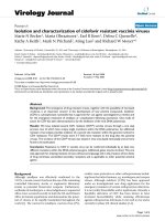
using a Unosphere Q column. Four fractions, namely
from FI to FIV, possessing caseinolytic activity were
collected when applying a NaCl gradient of 0-0.45 M
(Figure 1). FI had the highest activity. SDS-PAGE profile
of all fractions showed impurities (data not shown), thus
requiring further purification processes.
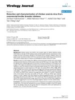
Each fraction was loaded onto a phenyl sepharose col-
umn a fter dialysis, and a gradient of 30-0% AS concen-
tration was used for elution. FI and FII were eluted in
single prominent peaks (Figure 2A and 2B). In contrast,
FIII was further separated into three fractions, namely
Phan et al. AMB Express 2011, 1:26
/>Page 3 of 11
FIII-1, FIII-2 and FIII-3, having caseinolytic activity
(Figure 2C). F IV was fractioned into two partially over-
lapping peaks; however, only the later one had caseino-
lytic activity and thus was referred to as FIV (Figure
2D). After this step, F I and FII showed high purity as
determined by SDS-PAGE (data not shown). The sub-
fractions of FIII and FIV, however, still contained some
low MW contami nants (data not shown). Thus a subse-
quent SEC was necessary to purify these fractions and
to confirm the purity of the others as well.
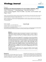
SEC was carried out for a ll ac tive HIC fractions on a
Superose 12 column. Three fractions FI, FII and FIII-1
eluted as a single peak with a MW of 28, 29 and 35 kDa,
respectively. These results were in ac cordance with those
obtained from SD S-PAGE (Figure 3B). The major peak
of FIII-2 appeared to be a single band of 34 kDa on SDS-
PAGE gel and showed protease activity. FIV was fractio-
nated into two peaks but only the large peak of 34 kDa
protein (marked as FIV*) showed proteolytic activity
(Figure 3A). FIII-3 was also separated into two peaks,
namely FIII-3a and FIII-3b, with a MW of 33 and
31 kDa, respectively, and only th e latter one showed pro-
tease activity. Both fractions had a band of 33 kDa on
SDS-PAGE gel. Besides, FIII-3b contained also a 31 kDa
protein which was not present in FIII-3a, as seen in the
SEC chromatogram. M ost fractions were more than 98%
pure as estimated by SDS-PAGE Coomassie Brilliant Blue
staining. On ly FIII-3b contained two proteins with more
or less the same intensity. There were six proteolytic
Figure 1 Anion exchange chromatogram of P. excavatus crude
proteins. Four peaks indicated as FI, FII, FIII, and FIV (arrows) with
caseinolytic activity were obtained when eluting with a continuous
gradient of NaCl concentration (0-0.45 M).
Figure 2 Hydrophobic interaction chrom atograms of four AEX fractions. A continuous gra dient of ammonium sulfate (AS) concentration
(30-0%) was used for eluting. (A) FI and (B) FII were eluted as single prominent peaks. (C) FIII was separated into three sub fractions with
protease activity: FIII-1, FIII-2 and FIII-3. (D) FIV was fractionated into multiple peaks, however, only one fraction showed protease activity. All
active fractions were indicated by arrows.
Phan et al. AMB Express 2011, 1:26
/>Page 4 of 11
fractions in total that were purified. The same number of
fractions was also reported for L. rubellus lumbrokinase
using a similar purification strategy (Mihara et al. 1991).
Effect of temperature and pH on the stability
of P. excavatus proteases
All fractions exhibited maximal proteolytic activity in
the temperature range of 60-65°C. Increasing the tem-
perature to 70°C caused rapid loss of activity, and
between 75-80°C almost complete inactivation was
observed (Figure 4A). The thermostability over time
was examined at different temperatures in a 3-hour
period. These proteases were highly stable at tempera-
ture below 50°C but had rapid activity loss of 10-30%
from 50°C onward. Higher temperatures inactivated
these enzymes very rapidly. FI and FII were more
stable than other fractions, as their activity decrease
only about 10-20% at temperatures from 50 to 60°C
(Figure 4B).
All proteases expressed optimal activity at both pH 7
and 11, except for FIII-1 which showed the highest
activity only at pH 7 (Figure 5A). The proteases of
P. excavatus were stable in a wide pH range from 4 to
12 during 16 hours (Figure 5B). Similar results were
reported for Korean and Japanese L. rubellus (Cho et al.
2004 and Mihara et al. 2004). FIII-1 and FIII-2 were the
two most stable proteases towards a wide range of pH
values in comparison to the others. The long-term pre-
servation of the purified proteases at 4°C was experi-
mentally investigated with two solvents, distilled water
and sodium phosphate buffer pH 7.5. Sodium azide
(0.1% w/v) was used as preservative. After 10 months,
all fractions tended to be more stable in water (with
only 10% activity loss) except for FIII-3, whose activity
reduced t o about 78% of the initial activity. In contrast,
these enzymes were less stable in phosphate buffer since
their activity decreased approximately 25-30% (data not
shown).
Inhibition of P. excavatus proteases
PMSF is known as an effective inhibitor for serine pro-
teases. As shown in Table 1, this compound could
almost completely inhibit all protease fractio ns, causing
92-100% of activity loss. In contrast, EDTA had no inhi-
bitory effect on any of the six fractions, s uggesting that
they were not metalloproteases, because such enzymes
need a metal ion for activity. Only FIII-3 was inhibited
to some extent by TPCK, which is a specific inhibitor
for chymotrypsin-like serine proteases. The other rever-
sible inhibitors such as SBTI, aprotinin, leupeptin and
chymostatin caused different levels of inhibition of these
proteases.
Hydrolytic activity on different substrates
All fractions were assayed for their hydrolytic ability
towards different substrates including casein, fibrin,
BApNA, and BTpNA (Table 2). The last two are syn-
the tic substrates specific for trypsin- and chymotrypsin-
like proteases, respectivel y. None of these fractions
showed hydrolytic activity towards BTpNA, indicating
that they probably do not belong to the chymotrypsin-
like protease group. This result was in accordance with
the inhibitory assay (Table 1) although FIII-3 was to
some extent inhibited by TCPK, a specific inhibitor for
chymotrypsin-like serine protease s. Only three fractions
Figure 3 Profiling of P. excavatus protease fractions:(A)Size
exclusion chromatogram of FIII-3 and FIV presented their
fractionation into two peaks, ones of which marked with an asterisk
showed protease activity. (B) SDS-PAGE profiling of all SEC fractions.
Phan et al. AMB Express 2011, 1:26
/>Page 5 of 11
FIII-1, FIII-2 a nd FIV were able to show hydrolysis of
BApNA.
The caseinolytic activity was more or less the same for
all fractions. Results from the fibrin plate assay, how-
ever, showed that all fractions had different levels of
fib rino lytic activity. Three fractions FIII-3, FIII-2 and FI
expressed the highest activity. Coagulated fibrin seemed
not to be a specif ic substrate for FII since its hydrolysis
towards fibrin was much smaller than those of the
others.
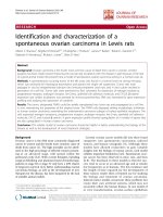
The hydrolytic effect of these proteases towards fibri-
nogen monomers was also investiga ted. As visualized on
the SDS-PAGE gel, all fract ions could completely
degrade the Aa and Bb subunits of fibrinogen within 10
minutes, and FIII-1 and FIII-2 could even cleave off the
g subunit. The hydrolytic ability of fraction FIII-2 was
the highest since almost no protein bands were visible
on the gel after 180 minutes (Figure 6). The hydrolytic
activity was thus ranked as FIII-2 > F III-1 > FI > FIV >
FIII-3 > FII. The activity magnitude was more or less
similar to the hydrolytic effect on intact fibrin in the
fibrin-plate assay (Table 2).
2D-electrophoresis coupled MS/MS sequencing
Each protease fraction was analyzed on 2D-PAGE gel
for determination of their MW and pI. All fractions,
except for FI and FIII-3, appeared as single spots, indi-
cating that they were pure. The pI values of these frac-
tions were d enoted in Table 3, ranging from 4.3 to 5.2.
Interestingly, they shared similar pI’s with the proteases
from E. fet ida (Zhao et al. 2006). The presence of two
protein with similar MW but different pI (5.0 and 5.2),
was observed for FI, thus it might contain two isozymes.
Fraction FIII-3 was hardly visible on the gel, probably
due to the loss during sample preparation for 2D-elec-
trophoresis. Therefore, the pI and MW of this fraction
could not be determined.
Only the MS/MS spe ctra o f FIII-1 and FII I-2 pept ide
fragments were obtained with good signal-to-noise ratios.
The sequence alignments revealed that they shared con-
siderable similarity (16.9% and 13.2%, respectively) with
the segments of fibrinolytic lumbrokinase isozyme C (EC
3.4.21) from L. rubellus and E. fetida (Figure 7). This
enzyme is a serine protease with the length of 242 am ino
acid residues and a MW o f 26 kDa. The two fractions
Figure 4 Effect of temperature on the activity of P. excavatus proteases: (A) Temperature optimum and (B) thermostability of all isolated
fractions. The highest activity of each fraction in (A) and the activity of each fraction at 30°C in (B) were set at 100%.
Phan et al. AMB Express 2011, 1:26
/>Page 6 of 11
FIII-1 and FIII-2 also showed sequence similarity, but at a
lower degree, with the serine proteases from mouse (Mus
musculus). The sp ectra ob tained fr om the other fractions
gave no clear s ignals, so it was not po ssib le t o de termine
any of these peptide sequences.
Discussion
Initial autolysis is necessary for full activation of P.
excavatus proteases
As we could see, the proteolytic activity towards casein
increased and peaked through a 15-day autolysis. The
presence of sodium azide could inhibit the bacterial
growth. The autolytic process could therefore trigger the
release of proteases from the earthworm’stissuesand
exert a subsequent degradation of keratin and lipids,
thus reducing the mixture’ s viscosity. The increase of
proteolytic activity over time suggested that activation of
these enzymes had occurred, probably through self-pro-
teolysis that cleaved parts of the zymogens.
Purification protocol for P. excavatus proteases
The acetone precipitation of the P. excavatus lysate was
more effective than ammonium sulfate (AS) precipita-
tion because of more impurity removal, higher proteoly-
tic activity recovery (data not shown) and less time
consuming since subsequent dialysis is not necessary.
Figure 5 Effect of pH o n the activity of P. excavatus proteases. All fractions except for FIII-1 had dual pH optima. The highest activity for
each fraction at pH 7 was set at 100%, and the activities at other pH were calculated in accordance to this value.
Table 1 Effect of different inhibitors on P. excavatus
proteases.
Inhibitors Conc. (mM) Relative activity (%)
FI FII FIII-1 FIII-2 FIII-3 FIV
Control 100 100 100 100 100 100
PMSF 1 0 0 2 4 0 8
TPCK 0.1 100 100 96 100 76 100
Aprotinin 0.01 29 95 87 95 26 71
Leupeptin 0.1 0 100 98 100 0 73
SBTI 0.01 0 76 81 93 0 0
EDTA 1 100 100 100 100 100 100
Chymostatin 0.1 19 100 76 100 19 90
Pepstatin 1 100 58 60 86 83 75
Relative activity was determined as percentage of the activity of enzyme
without inhibitor (control) (results were rounded up to the integer)
Table 2 Hydrolytic activity of P. excavatus proteases on
different substrates
Specific activity (U.mg
-1
)
Fractions Fibrin Casein BApNA BTpNA
FI 602 1.8 0 0
FII 44 1 0 0
FIII-1 393 1 0.1 0
FIII-2 783 1 0.4 0
FIII-3 831 1.2 0 0
FIV 296 0.9 2.2 0
Phan et al. AMB Express 2011, 1:26
/>Page 7 of 11
The presence of the 33 kDa protein in faction FIII-3
could support the hypothesis of zymogen degradation
mentioned earlier since it could be the zymogen of the
31 kDa peptide, and its presence in the SEC fraction
probably resulted fr om the inco mplete initial autolysis.
Simila r results were reported in the study on L. rubellus
earthworm (Cho et al. 2004) in which a 44 residues
were cleaved off from a 283-r esidue-zymogen to release
the fully active proteolytic enzyme. This hypothesis,
however, requires further validati on through sequencing
of our FIII-3a and FIII-3b proteins.
Generally, the tw o-step chromatography of AEX and
HIC was sufficient for the purification of FIII-1, FIII-2,
and FII, since their SE C and SDS-PAGE profiles repre-
sented pure proteins. Fraction FI actually contained two
isozymes with close MW and pI, which could not be
further separated. For FIII-3 and FIV, SEC was necessary
to achieve the highest purity.
P. excavatus proteases possess dual pH optima
The dual pH optima has not been reported for the
proteases from L. rubellus and E. fetida. However, this
characteristic was found for the intestinal serine
Figure 6 Digestion of fibrinogen subunits (Aa,Bb and g)intimebyP . excavatus proteases. The MW of the three subunits Aa,Bb and g
are 95, 56, and 51.5 kDa (UniProt), respectively. The time of hydrolysis is plotted at the top of the figures.
Table 3 The values of pI and molecular weights (MW) of
all P.excavatus proteases, except for FIII-3, determined by
PDQuest™ 2-D Analysis Software (Bio-Rad, USA).
Fractions pI MW (kDa)
FI (spot 1) 5.0 27.5
FI (spot 2) 5.2 27.5
FII 4.3 29.0
FIII-1 4.5 34.5
FIII-2 4.3 33.5
FIII-3 not determined
FIV 4.5 34.0
Phan et al. AMB Express 2011, 1:26
/>Page 8 of 11
protease of red flour beetle (Tribolium castaneum),
whose optimal pH was determined to be at 4 and 8.5
(Oppert et al. 2003). (Choi et al. (1989)) studied the
proteases from the parasitic protozoa Toxoplasma gon-
dii and discovered that they catalyzed most effectively
at pH 6 and 8.5. Likewise, the dual pH optima charac-
teristic has been observed in various hydrolases such
as b-glucuronidase from human seminal plasma
(Gupta and Singh 1983), Staphylococcus sp. xylanase
(Gupta et al. 2000), reptile lysozyme (Thammasirirak
et al. 2006) and Rhizopus lipase (Upadhyay et al. 1989).
(Gupta et al. (2000)) hypothesized that the xylanase in
their study might contain two distinct active sites that
could perform cat alysis at two distinct pH levels of 7.5
and 9.2, respectively, although no such enzyme has been
reported before. In another study, th e aspartate protease
Plasmepsin I from Plasmodium falciparum was charac-
terized, revealing the existence of two states of this
Figure 7 Sequence alignment of fragments obtained from MS/MS analyses of FIII-1 and FIII-2 from P. excava tus with lumbrokinase
and its precursor from L. rubellus (sp:P83298 and U25647) and E. fetida (gpu:EU167737 and AY438624), with a serine protease from
M. musculus (sp:P69525 and Q8BZ10) and B. antarctica (gpu:DQ507327). Symbols were defined as following: (*) for identical amino acids, (.)
for redundant amino acids with similar 3D-structures, (:) for redundant amino acids with similar physicochemical properties. Significantly similar
sequences were wrapped in dot-lined boxes.
Phan et al. AMB Express 2011, 1:26
/>Page 9 of 11
protease a s monomer and aggregated oligomer (Xiao et
al. 2007). These two co-existing states resulted in the
dual pH optima of the enzyme as determined experi-
mentally. Since all protease fractions from P. excavatus
in our study were completely inhibited by PMSF (Table
1),theywouldnotharboranyactivesitesratherthan
the typical catalytic triad of serine proteases. Addition-
ally, no aggregation was observed by SEC chromatogra-
phy performed at pH 8.5. We therefore hypothesize that
the P. excavatus proteases existed in both monomeric
and aggregated oligomeric form in our assays; and the
aggregation might be triggered at strong alkaline pH, for
instance pH 11.
Serine proteases
The inhibitory effect of PMS F towards all fractions
revealed that P. excavatus proteases are serine proteases,
since PMSF is a specific irreversible inhibitor for this
group of proteases (James 1978). FIII-3 was to some
degree inhibited by TPCK, which is specific for chymo-
trysin-likeprotease(Table1).However,itwasnotable
to hydrolyze the chymotrypsin-like specific BTpNA sub-
strate. Therefore, it was not possible to classify this pro-
tease. Two fractions FI and FII were also unambiguous
since they had no activity towards both BApNA and
BTpNA. In contrast, FIV was more likely a trypsin-like
protease due to its specific hydrolysis of BA pNA and
specific inhibition by SBTI. Two fractions FIII-1 and
FIII-2 displayed much lower hydrolyt ic effect on
BApNA in comparison to FIV but were not inhibited by
SBTI, thus it is still questionable if they were actually
trypsin-like proteases.
On the other hand, t he sequence alignment study
revealed a considerable similarity between FIII-1 and
FIII-2 fragments with the trypsin-like lumbrokinase
fragments from L. rubellus and E. fetida.However,the
sequence homology obtained in our study was
expected to be higher because of the close evolutionary
relationship between these earthworm species. Cho et
al. reported extremely high conservation of the N-
terminal 20-22 residues between L. rubellus protease
fractions (Cho et al. 2004). The sequence alignment
within P. excavatus fractions was not conducted due to
insufficient information from the MS/MS data. There-
fore mass spectrometric analysis for these proteases
should be further elaborated to obtain their full
sequences.
The isozymes expressed strong hydrolytic activity
towards both fibrinogen and fibrin
(Park et al. (2007)) discovered a prote ase from Flammu-
lina velutipes that showed both fibrinolytic and fibrino-
genolytic activity. This enzyme could perform
hydrolyses without the presence of any activators, while
human plasminogen is an inactive precursor and strictly
requires tPA or urokinase for its conversion into fibrino-
lytic plasm in. In our experimen t, all fractions except for
FII displayed remarkable fibrinolysis, which was two to
three times stronger than human plasmin (data not
shown). They rapidly degraded the fibrinogen monomer
as well. Therefore, the P. excavatus proteases would
have a different catalytic mechanism tow ards these two
substrates than human plasminogen. Moreover, each
fraction experimentally displayed a distinct catalytic rate,
thus probably having different kinetic parameters such
as K
M
, V
max
, and K
cat
.
Applicability of P. excavatus proteases
Pure proteins are generally less stable in water due to the
absence of natural intracellular buffering and ionic condi-
tions. There fore, good storage conditions are req uired to
maintain their biological activity. Nakajima et al.found
that the protease fractions from Japanese L. rubellus
could maintain approximately 80% of their activity after
five ye ars in 100 mM Tris-HCl buffer at pH 8 (Nakajima
et al. 2000). Interestingly, our results revealed that water
is more appropriate than phosphate buffer as a storage
medium for P. exca vatus proteases. In addition, the pre-
sence of plasminogen activators was declared to be unne-
cessary for the enzymes that could be able to hydrolyze
both fibrinogen an d fibrin (Park et al. 2007). These prop -
erties are favourable for convenient and cost-effective
formulation of these enzymes. (Cho et al. (2004))
reported that all six protease fractions from L. rubellus
had similar cas einolytic activity, only one of which exhib-
ited remarkable fibrinolysis. This fraction w as the first
earthworm pr otease to be investigated for thrombosis
therapy in Korea. Therefore, the proteases from P. exca-
vatus characterised in the present study seem promising
candidates for that purpose in Vietnam. Fraction FIII-2 is
the most interesting fraction because of its strong fibri-
nolysis activity and high stability over long-term storage.
Acknowledgements
The authors are greatly thankful to Professor Les Copeland (Faculty of
Agriculture, Food and Natural Resources - The University of Sydney, New
South Wales, Australia) and Professor Kaeko Kamei (Department of
Biomolecular Engineering, Kyoto Institute of Technology Matsugasaki, Sakyo-
ku, Kyoto, Japan), who have kindly given the proof reading to the
manuscript of this paper. The research was financially supported by the the
Vietnamese Ministerial Research Project (B2007-16-56).
Author details
1
College of Agriculture and Applied Biology, Can Tho University, Can Tho,
Vietnam
2
Faculty of Food Processing Technology, Can Tho University of
Technology, Can Tho, Vietnam
3
Biotechnology Research and Development
Institute, Can Tho University, Can Tho, Vietnam
4
Wageningen UR Food &
Bio-based Research, 6708 WG, Wageningen, The Netherlands
Competing interests
The authors declare that they have no competing interests.
Phan et al. AMB Express 2011, 1:26
/>Page 10 of 11
Received: 13 September 2011 Accepted: 30 September 2011
Published: 30 September 2011
References
Anson ML (1938) The Estimation of Pepsin, Trypsin, Papain, and Cathepsin with
Hemoglobin. J Gen Physiol 22(1):79–89. doi:10.1085/jgp.22.1.79.
Arnesen H, Hoiseth A, Ly B (1982) Streptokinase of heparin in the treatment of
deep vein thrombosis. Follow-up results of a prospective study. Acta Med
Scand 211(1-2):65–8.
Cho IH, Choi ES, Lim HG, Lee HH (2004) Purification and characterization of six
fibrinolytic serine-proteases from earthworm Lumbricus rubellus. J Biochem
Mol Biol 37(2):199–205. doi:10.5483/BMBRep.2004.37.2.199.
Cho IH, Choi ES, Lee HH (2004) Molecular cloning, sequencing, and expression of
a fibrinolytic serine-protease gene from the earthworm Lumbricus rubellus.J
Biochem Mol Biol 37(5):574–81. doi:10.5483/BMBRep.2004.37.5.574.
Choi WY, Nam HW, Youn JH (1989) Characterization of proteases of Toxoplasma
gondii. Kisaengchunghak Chapchi 27(3):161–70
Choi HS, Sa YS (2001) Fibrinolytic and antithrombotic protease from Spirodela
polyrhiza. Biosci Biotechnol Biochem 65(4):781–6. doi:10.1271/bbb.65.781.
Dong GQ, Yuan XL, Shan YJ, Zhao ZH, Chen JP, Cong YW (2004) Molecular
cloning and characterization of cDNA encoding fibrinolytic enzyme-3 from
earthworm Eisenia foetida. Acta Biochim Biophys Sin (Shanghai) 36(4):303–8.
doi:10.1093/abbs/36.4.303.
Einhaupl KM, Villringer A, Meister W, Mehraein S, Garner C, Pellkofer M, Haberl RL,
Pfister HW, Schmiedek P (1991) Heparin treatment in sinus venous
thrombosis. Lancet 338(8767):597–600. doi:10.1016/0140-6736(91)90607-Q.
Furie B, Furie BC (2008) Mechanisms of thrombus formation. N Engl J Med
359(9):938–49. doi:10.1056/NEJMra0801082.
Ge T, Sun ZJ, Fu SH, Liang GD (2005) Cloning of thrombolytic enzyme
(lumbrokinase) from earthworm and its expression in the yeast Pichia
pastoris. Protein Expr Purif 42(1):20–8. doi:10.1016/j.pep.2005.04.005.
Grundy SM, Pasternak R, Greenland P, Smith S, Fuster V (1999) Assessment of
Cardiovascular Risk by Use of Multiple-Risk-Factor Assessment Equations.
Circulation 100:1481–1492
Gupta S, Bhushan B, Hoondal GS (2000) Isolation, purification and
characterization of xylanasefrom Staphylococcus sp. SG-13 and its application
in biobleaching of kraft pulp. J Appl Microbiol 88(2):325–34.
Gupta GS, Singh GP (1983) Isolation and characterization of the major form of
beta-glucuronidase from human seminal plasma. Biochim Biophys Acta
748(3):398–404. doi:10.1016/0167-4838(83)90185-1.
Hames BD (1998) Gel electrophoresis of proteins. A practical approach, 3rd edn.
Oxford University.
James GT (1978) Inactivation of the protease inhibitor phenylmethylsulfonyl
fluoride in buffers. Anal Biochem 86(2):574–9. doi:10.1016/0003-2697(78)
90784-4.
Kakkar VV, Flanc C, Howe CT, O’Shea M, Flute PT (1969) Treatment of deep vein
thrombosis. A trial of heparin, streptokinase, and arvin. Br Med J , 5647:
806–10.
Kannel WB, Wolf PA, Castelli WP, D’Agostino RB (1987) Fibrinogen and risk of
cardiovascular disease. The Framingham Study. JAMA 258(9):1183–1186.
doi:10.1001/jama.258.9.1183.
Lowry OH, Rosenberg WJ, Farr AL, Randall RJ (1951) Quantization of protein
using Folin-Ciocalteau reagent. J Biol Chem 193:265–275.
Marder VJ, Soulen RL, Atichartakarn V, Budzynski AZ, Parulekar S, Kim JR,
Edward N, Zahavi J, Algazy KM (1977) Quantitative venographic assessment
of deep vein thrombosis in the evaluation of streptokinase and heparin
therapy. J Lab Clin Med 89(5):1018–29.
Mehraein S, Schmidtke K, Villringer A, Valdueza JM, Masuhr F (2003) Heparin
treatment in cerebral sinus and venous thrombosis: patients at risk of fatal
outcome. Cerebrovasc Dis 15(1-2):17–21. doi:10.1159/000067117.
Mihara H, Sumi H, Yoneta T, Mizumoto H, Ikeda R, Seiki M, Maruyama M (1991) A
novel fibrinolytic enzyme extracted from the earthworm, Lumbricus rubellus.
Jpn J Physiol 41(3):461–72. doi:10.2170/jjphysiol.41.461.
Mosesson MW (2005) Fibrinogen and fibrin structure and functions. J Thromb
Haemost 3(8):1894–904. doi:10.1111/j.1538-7836.2005.01365.x.
Mosesson MW, Siebenlist KR, Meh DA (2001) The structure and biological
features of fibrinogen and fibrin. Ann N Y Acad Sci 936:11–30.
Nakajima N, Mihara H, Sumi H (1993) Characterization of potent fibrinolytic
enzymes in earthworm, Lumbricus rubellus. Biosci Biotechnol Biochem
57(10):1726–30. doi:10.1271/bbb.57.1726.
Nakajima N, Sugimoto M, Ishihara K (2000) Stable earthworm serine proteases:
application of the protease function and usefulness of the earthworm
autolysate. J Biosci Bioeng 90(2):174–9.
Oppert B, Morgan TD, Hartzer K, Lenarcic B, Galesa K, Brzin J, Turk V, Yoza K,
Ohtsubo K, Kramer KJ (2003) Effects of proteinase inhibitors on digestive
proteinases and growth of the red flour beetle, Tribolium castaneum (Herbst)
(Coleoptera: Tenebrionidae). Comp Biochem Physiol C Toxicol Pharmacol
134(4):481–90. doi:10.1016/S1532-0456(03)00042-5.
Park SE, Li MH, Kim JS, Sapkota K, Kim JE, Choi BS, Yoon YH, Lee JC, Lee HH,
Kim CS, others (2007) Purification and characterization of a fibrinolytic
protease from a culture supernatant of Flammulina velutipes mycelia. Biosci
Biotechnol Biochem 71(9):2214–22. doi:10.1271/bbb.70193.
Stam J, de Bruijn S, deVeber G (2003) Anticoagulation for cerebral sinus
thrombosis. Stroke 34(4):1054–5. doi:10.1161/01.STR.0000062344.87396.72.
Sugimoto M, Nakajima N (2001) Molecular cloning, sequencing, and expression
of cDNA encoding serine protease with fibrinolytic activity from earthworm.
Biosci Biotechnol Biochem 65(7):1575–80. doi:10.1271/bbb.65.1575.
Tang Y, Liang D, Jiang T, Zhang J, Gui L, Chang W (2002) Crystal structure of
earthworm fibrinolytic enzyme component a: revealing the structural
determinants of its dual fibrinolytic activity. J Mol Biol 321(1):57–68.
doi:10.1016/S0022-2836(02)00559-4.
Thammasirirak S, Ponkham P, Preecharram S, Khanchanuan R, Phonyothee P,
Daduang S, Srisomsap C, Araki T, Svasti J (2006) Purification, characterization
and comparison of reptile lysozymes. Comp Biochem Physiol C Toxicol
Pharmacol 143(2):209–17. doi:10.1016/j.cbpc.2006.02.004.
Upadhyay CM, Nehete PN, Rothari RM (1989) A lipase preparation with dual pH
optima, wide temperature optima and broad substrate specificity for
multiple applications. Biotechnology Letters 11(11):793–796. doi:10.1007/
BF01026099.
Wang F, Wang C, Li M, Zhang JP, Gui LL, An XM, Chang WR (2005) Crystal
structure of earthworm fibrinolytic enzyme component B: a novel,
glycosylated two-chained trypsin. J Mol Biol 348(3):671–85. doi:10.1016/j.
jmb.2005.02.055.
Xiao H, Tanaka T, Ogawa M, Yada RY (2007) Expression and enzymatic
characterization of the soluble recombinant plasmepsin I from Plasmodium
falciparum. Protein Eng Des Sel 20(12):625–33. doi:10.1093/protein/gzm066.
Xu ZR, Yang YM, Gui QF, Zhang LN, Hu L (2010) Expression, purification, and
characterization of recombinant lumbrokinase PI239 in Escherichia coli.
Protein Expr Purif 69(2):198–203. doi:10.1016/j.pep.2009.08.013.
Yang JS, Ru BG (1997) Purification and characterization of an SDS-activated
fibrinolytic enzyme from Eisenia fetida. Comp Biochem Physiol B Biochem
Mol Biol 118(3):623–31. doi:10.1016/S0305-0491(97)00223-X.
Zhao XY, Liu ZM, Jing TY, Wu JX, Zhao ZY (2006) A component of earthworm
fibrinolytic enzyme having higher thrombolytic activity than total
components in vivo. Yao Xue Xue Bao 41(11):1068–73.
doi:10.1186/2191-0855-1-26
Cite this article as: Phan et al.: Purification and characterization of novel
fibrinolytic proteases as potential antithrombotic agents from
earthworm Perionyx excavatus. AMB Express 2011 1:26.
Submit your manuscript to a
journal and benefi t from:
7 Convenient online submission
7 Rigorous peer review
7 Immediate publication on acceptance
7 Open access: articles freely available online
7 High visibility within the fi eld
7 Retaining the copyright to your article
Submit your next manuscript at 7 springeropen.com
Phan et al. AMB Express 2011, 1:26
/>Page 11 of 11