báo cáo hóa học: " Can we make the basilic vein larger? maneuvers to facilitate ultrasound guided peripheral intravenous access: a prospective cross-sectional study" ppt
Bạn đang xem bản rút gọn của tài liệu. Xem và tải ngay bản đầy đủ của tài liệu tại đây (285.56 KB, 5 trang )
ORIGINAL RESEARCH Open Access
Can we make the basilic vein larger? maneuvers to
facilitate ultrasound guided peripheral intravenous
access: a prospective cross-sectional study
Simon A Mahler
1*
, Greta Massey
2
, Liliana Meskill
3
, Hao Wang
4
and Thomas C Arnold
5
Abstract
Background: Studies have shown that vein size is an important predictor of successful ultrasound-guided vascular
access. The objective of this study is to evaluate maneuvers designed to increase basilic vein size, which could be
used to facilitate ultrasound-guided peripheral intravenous access (USGPIV) in the Emergency Department (ED)
setting.
Methods: This was a prospective non-randomized trial. Healthy volunteers aged 18-65 were enrolled. Basilic veins
were identified and the cross-sectional area measured sonographically. Following baseline measurement, the
following maneuvers were performed: application of a tourniquet, inflati on of a blood pressure (BP) cuff,
application of a tourniquet with the arm lowered, and BP cuff inflation with the arm lowered. Following each
maneuver there was 30 s of recovery time, and a baseline measurement was repeated to ensure that the vein had
returned to baseline. Change in basilic vein size was modeled using mixed model analysis with a Tukey correction
for multiple comparisons to determine if significant differences existed between different maneuvers.
Results: Over the 5-month study period, 96 basilic veins were assessed from 52 volunteers. All of the maneuvers
resulted in a statistically significant increase in basilic vein size from baseline (p < 0.001). BP cuff inflation had the
greatest increase in vein size from baseline 17%, 0.87 mm 95% CI (0.70-1.04). BP cuff inflation statistically
significantly increased vein size compared to tourniquet placement by 3%, 0.16 mm 95% CI (0.02-0.30).
Conclusions: The largest increase in basilic vein size was due to blood pressure cuff inflation. BP cuff inflation
resulted in a statistically significant increase in vein size compared to tourniquet application, but this difference
may not be clinically significant.
Background
Intravenous (IV) access is often required in Emergency
Department (ED) pa tients. Landmark techniques for
obtaining peripheral IV access are usually succes sful, but
patients with prior IV drug abuse, obesity, and chronic
medical conditions are more likely to have failed attempts
[1,2]. Several studies have demonstrated that ultrasound
can be used to successfully place peripheral IVs in patients
who have failed landmark techniques [1,3-6]. Prior to
ultrasound-guided peripheral intravenous access (USG-
PIV), patients with failed landmark techniques often
required central venous cannulation, a procedure with a
higher complication rate and demanding more staff
resources than peripheral access [2,7].
Studies have shown that vein size is an important pre-
dictor of successful ultra sound-guided vascular ac cess
[8,9]. While several studies have investigated maneuvers
to increase femoral and jugular vein size to faci litate
ultrasound-guided central line placemen t [10-14], few
have evaluated maneuvers to increase basilic vein size.
Studies evaluating basilic vein size have mainly focused
on the creation of an AV fistula for dialysis rather than
facilitating USGPIV [15-20]. The objective of this study is
to evaluate maneuvers practical for ED use that could be
utilized to improve t he success of USGPIV by increasing
basilic vein size.
* Correspondence:
1
Department of Epidemiology and Prevention, Department of Emergency
Medicine, Wake Forest University School of Medicine, Winston-Salem, NC,
USA
Full list of author information is available at the end of the article
Mahler et al. International Journal of Emergency Medicine 2011, 4:53
/>© 2011 Mahler et al; licensee Springer. This is an Open Access article distribute d under the terms of the Creative Commons Attribution
License (http://creativeco mmons.or g/licenses/by/2.0), which permits unrestrict ed use, distribution, and reproduction in any medium,
provided the orig inal work is properly cited.
Methods
This was a prospective non-randomized trial, which was
approved by the Institutional Review Board of the spon-
soring organization. Healthy volunteers aged 18-65 were
enrolled over a 5-month period (January to May 2010)
at Louisiana State University Health Sciences Center-
Shreveport (LSUHSC-S). LSUHSC-S is a tertiary care
facility, level one trauma center, and academic center
home to a 3-year EM residency program training seven
residents per year. Written informed consent was
obtained from all volunteers. Volunteers were excluded
from the study if they had any acute medical illness or
were pregnant.
Volunteers were given a questionnaire to determine if
they had undergone venopuncture or vascular access
within the previous week, history of upper extremity
thrombosis, history of humerus fracture, upper extremity
deformity , or upper extremi ty surgery. If the subjects had
any of the above in both arms they were excluded from
the study. If the items in the questionnaire were present
in only one arm, the volunteer was allowed to participate,
but could only use the unaffected arm for the study
measurements.
The basilic veins of each subject were identified using a
high-frequency linear probe (8-12 MHz, L25 probe on a
Sonosite M-Turbo or S-series, Sonosite, Inc., Bothell, WA,
USA). After the ba silic vei n had been identified, two skin
marks were made overlying the vein at a point of optimal
vein visualization approximately 2-4 cm above the medial
epicondyle. If a branching point off the basilic vein was
identified within the 2-4 cm area, it was also used as a
landmark. The skin marking and branch points were used
to ensure that measurements of the vein during differ ent
maneuvers occurred at the same location.
Basilic vein measurements for each maneuver were
obtained using the following procedures: First, the vein
was identified at the location of skin markings o n the
short axis. Then, the zoom function was used to obtain an
enlarged view of the vein, and electronic calipers measured
the vein diameter in two dimensions: anterior-posterior
and m edial-lateral. Using the measurements obtained
above, an average vein diameter was calculated. Sono-
graphic measurements were completed by GH and LM,
4th year medical students who had received 1.5 h of didac-
tic and proctored hands-on training in vascular ultrasound
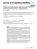
prior to the start of this study (see Figure 1).
The first measurement obtained was a baseline mea-
surem ent in which subjects had their arms supported at
the level of the heart. Following baseline measurement,
the following maneuvers were performed: application of
a tourniquet, inflation of a blood pressure cuff (above
diastolic pressure), holding the arm below the level of
the heart for more than 30 s prior to the application of
a tourniquet, and holding the arm below the level of the
Figure 1 Ultrasound of basilic vein with measurement of anterior to posterior and medial to lateral diameters.
Mahler et al. International Journal of Emergency Medicine 2011, 4:53
/>Page 2 of 5
heart for more than 30 s prior to inflation of a blood
pressure cuff. Measurements of basilic vein size were
made before and after each maneuver. Following each
maneuver the subject was allow ed at least 30 s of recov-
ery time, and a ba seline measurement was repeated
to ensure that the vein had returned to its normal size
(± 0.1 mm).
The effect of each maneuver on vein size was modeled
using mixed model analysis. Tukey post hoc analysis
was performed to determine if significant differences
existed between different maneuvers and adjust for mul-
tipl e comparisons. Covariance structure was determined
by minimizing the AIC (Akaike information criterion),
resulting in unstructured covariance. Statistical analysis
was preformed with SAS 9.2 (Cary, NC) for Windows.
Results
Over the 5-month study period from January to May
2010, 96 basilic veins were assessed from 52 volunteers.
Of the 52 healthy volunteers, 44 had basilic veins mea-
sured in both arms, and 8 subjects had one basilic vein
measured. The mean age of the volunteer s was 25 (± SD
4 years), 14 (27%) were male, and 38 (73%) were female.
Themeanbaselinediameterofthebasilicveinswas
5.1 mm (± SD 1.3 mm ). Application of a tourniq uet
with the arm supported at the level of the heart
increased size by 14%, a difference of 0.71 mm 95% CI
(0.55, 0.88), p < 0.001. Inflati on of a blood pressure cuff
above diastolic pressure, w ith the arm supported at the
level of the heart, increased basilic vein diameter by
17%, 0.87 mm 95% CI (0.70-1.04), p < 0.001. BP cuff
inflation statistically significantly increased vein size
compared to tourniquet placement by 3%, 0.16 mm 95%
CI (0.02-0.30), p = 0.018 (see Figure 2). All post hoc
pairwise comparisons are summarized in Table 1.
Discussion
All of the maneuvers tested in our study resulted in a sta-
tistically significant increase in basilic vein size. Basilic
vein size was increased the most by inflation of a blood
pressure cuff above diastolic pressure with the arm sup-
ported at the level of the heart. The blood pressure cuff
inflated with the arm resting below the heart resulted in
the second largest increase in vein size. Blood pressure
cuff inflation increased vein size more than a tourniquet
or tourni quet applied with the arm below the level of the
heart. However, the difference in vein size between BP
cuff inflation and tourniquet application was small (3%,
0.16 mm). This difference would result in a change in
cross-sectional area of only 5.5% (1.46 mm
2
), which may
not be a clinically significan t difference for clinicians
attempting USGPIV.
Application of a tourniquet with the arm below the
level of the heart was the least effective maneuver to
increase vein size. On post hoc analysis this maneuver
was statistically significantly inferior to all of the other
maneuvers. In theory, lowering the arm below the level
of the heart for 30 s should have resulted in venous
pooling. Therefore, it was expected that the application
of a tourniquet in this position would increase basilic
vein size compared to a heart level arm postition. It was
also expected that lowering the arm before inflation of a
blood pressure cuff would increase basilic vein size com-
pared to a blood pressure cuff used at the level of the
heart, but this also did not occur. Another study enrol-
ling healthy volunteers and dialysis patients also failed
to show a significant differe nce in cephalic vein size fol-
lowing lowering of the arm and a combination of lower-
ing the arm and warm water emersion [16].
It is unclear why lowering the arm seemed to have a
negative impact on basilic vein size compared to the
maneuvers performed at heart level. It is possible t hat
despite ensuring that the vein returned to within ± 0.1
mm of its baseline diameter between different maneu-
vers, recovery time may have been inadequate. Further-
more, volunteers underwent each maneuver in an
ordered fashion with maneuvers placing the arm at heart
level pe rformed before maneuvers placing the arm below
the heart. It is possible that w ith each maneuver there
was some attenuation in the ability of the vein to distend.
Our study differs from prior studies that have exam-
ined maneuvers to increase upper extremity vein size,
which have mostly evaluated commercial devices or
were designed to facilitate vein mapping for dialysis
access rather than USGPIV [16,18-20]. Nee et al. investi-
gated antecubital fossa vein size for I V access with the
application of a tourniquet versus a tourniquet used in
combination with one of two commercially available
devices, an Esmarch bandage and a Rhys-Da vies exsan-
guinator. They determined that the combination of
Figure 2 Difference from baseline basilic vein size (mm) for
each manuever.
Mahler et al. International Journal of Emergency Medicine 2011, 4:53
/>Page 3 of 5
either device with a tourniquet was superior to a tourni-
quet alone [17]. Another study evaluated a vacuum
device used with a tourniquet to significantly increase
vein size [15]. Other studies have failed to demonstrate
significant difference in vein sizes comparing different
vein-dilating maneuvers including BP cuff inflation and
tourniquets [16,18]. In a study by Planken et al. on
patients requiring dialysis access, no significant differ-
ence in vein size (cephalic) was found between a tourni-
quet and a graduated pressure cuff [18]. It is unclear
why our results differ from those of Planket et al., but it
could be related to differences in the ability to distend
veins in dialysis patients compared to healthy volunteers.
Several studies have also investigated maneuvers to
increase femoral and jugular vein size to facilitate ultra-
sound-guided central line placement [10-14]. However,
we are not aware of any prior studies investigating man-
euvers with the aim of fac ilitating USGPIV. While USG-
PIV has a high success rate amo ng patients who have
failed landmark techniques, several studies have shown
that vein size is an important predictor of successful
ultrasound-guided vascular access [1,3-6]. Therefore,
maneuvers that can be practically implemented in the
ED to increase basilic vein size may improve the success
rate of USGPIV [1,8,9].
Limitations
This study was performed on healthy volunteers, mostly
young and female, rather than on patients requiring diffi-
cult IV access. Therefore, the results of this study may not
be generalizable to patients requiring USGPIV. In addi-
tion, sonographic measurements were completed by two
relatively inexperienced sonographers, and inter-observer
reliability was not assessed. However, prior studies have
shown that vein size measurements do not differ signifi-
cantly between sonographers [18,19].
Temperature changes are known to affect vein size,
with warmer temperatures increasing vein size. Warm
water emersion has been used as a t echnique to
increased vein size [16,20]. However, our study did not
evaluate warm water emersion, because it did not seem
practical in the ED setting. While temperature was not
directly accounted for in this study, we do not believe
that it functioned as a confounder since all of the sub-
jects served as their own controls. Volunteers were pre-
sent in the same climate-controlled environment
throughout their exposure to the different maneuvers.
As previously mentioned, the decreased effectiveness of
maneuvers completed with the arm resting below the level
of the heart may have been the result of bias. Although
procedures were utilized to ensure that the basilic vein
returned to baseline size between different maneuvers, it is
possible that our results were biased by inadequate recov-
ery time. Also, sequence bias may have occurred, as the
ability of veins to dilate may have been attenuated over
time or with repetitive maneuvers. Future studies should
have longer recovery periods and vary the sequence of the
maneuvers studied.
In addition, some of the differences between maneuvers,
while statistically significant, were small and may not be
clinically s ignificant. Fu rthermore, although prior studies
have demonstrated that larger vein size improves USGPIV
success, the subjects in this study were volunteers and did
not have USGPIV performed. Therefore, further study is
required to determine if s pecific maneuvers used for
venous distention increase USGPIV success relative to
other maneuvers.
Conclusions
All of the maneuvers tested resulted in a statistically sig-
nificant increase in basilic vein size. Inflation of a blood
pressure above diastoli c pressure with the arm supported
at the level of the heart produced the largest increase in
basilic vein size. BP cuff inflation resulted in a statistically
significant increase in vein size compared to tourniquet
application, but th is difference may not be clinically
Table 1 All pairwise comparisons of maneuvers used to dilate the basilic vein
Maneuver A Maneuver B (A-B) Difference in
vein diameter (mm)
Adjusted 95% confidence interval of difference (mm)* Adjusted p*
Baseline BP cuff 0.87 0.70-1.04 < 0.0001
Baseline Tourniquet 0.71 0.55-0.88 < 0.0001
Baseline BP cuff below heart 0.73 0.52-0.94 < 0.0001
Baseline Tourniquet below heart 0.45 0.25-0.65 < 0.0001
Tourniquet below heart BP cuff 0.43 0.24-0.62 < 0.0001
Tourniquet below heart BP cuff below heart 0.29 0.10-0.48 0.0006
Tourniquet below heart Tourniquet 0.27 0.09-0.44 0.0005
Tourniquet BP cuff 0.16 0.02-0.30 0.0176
Tourniquet BP cuff below heart 0.02 -0.20-0.16 0.9983
BP cuff below heart BP cuff 0.14 -0.01-0.29 0.0843
Differences measured in mm.
*p values and 95% confidence intervals have been adjusted for multiple comparisons using the Tukey method.
Mahler et al. International Journal of Emergency Medicine 2011, 4:53
/>Page 4 of 5
significant. The least effecti ve maneuver was the applica-
tion of a tourniquet with the arm resting below the level
of the heart. Future investigat ion of these maneuvers
desi gned to facilitate USGPIV should study patients with
failed landmark IV techniques, have long recovery peri-
ods betwe en maneuvers, and vary the sequence of the
maneuvers studied.
Patient Consent
Written informed consent was obtained from all study
volunteers.
Author details
1
Department of Epidemiology and Prevention, Department of Emergency
Medicine, Wake Forest University School of Medicine, Winston-Salem, NC,
USA
2
Department of Emergency Medicine, West Virginia University School of
Medicine, Morgantown, WV, USA
3
Department of Anesthesiology, The
University of Texas School of Medicine San Antonio, San Antonio, TX, USA
4
Department of Emergency Medicine, John Peter Smith Health Network, Fort
Worth, TX, USA
5
Department of Emergency Medicine, Louisiana State
University Health Sciences Center-Shreveport, Shreveport, LA, USA
Authors’ contributions
SM was involved in the study design, statistical analysis, and manuscript
preparation. GM and LM were involved in the study design and carrying out
study measurements. HW provided statistical support and was involved in
the study design. TA was involved in manuscript preparation. All authors
read and approved the final manuscript.
Competing interests
The authors declare that they have no competing interests.
Received: 28 June 2011 Accepted: 25 August 2011
Published: 25 August 2011
References
1. Keyes LE, Frazee BW, Snoey ER, Simon BC, Christy D: Ultrasound-guided
brachial and basilic vein cannulation in emergency department patients
with difficult intravenous access. Ann Emerg Med 1999, 34(6):711-714.
2. Chinnock B, Thornton S, Hendey GW: Predictors of success in nurse-
performed ultrasound-guided cannulation. J Emerg Med 2007,
33(4):401-405.
3. Mahler SA, Wang H, Lester C, Conrad SA: Ultrasound-guided peripheral
intravenous access in the emergency department using a modified
seldinger technique. J Emerg Med 2010, 39(3):325-9.
4. Costantino TG, Kirtz JF, Satz WA: Ultrasound-guided peripheral venous
access vs. the external jugular vein as the initial approach to the patient
with difficult vascular access. J Emerg Med .
5. Brannam L, Blaivas M, Lyon M, Flake M: Emergency nurses’ utilization of
ultrasound guidance for placement of peripheral intravenous lines in
difficult-access patients. Acad Emerg Med 2004, 11(12):1361-1363.
6. Costantino TG, Parikh AK, Satz WA, Fojtik JP: Ultrasonography-guided
peripheral intravenous access versus traditional approaches in patients
with difficult intravenous access. Ann Emerg Med 2005, 46(5):456-461.
7. Steele R, Irvin CB: Central line mechanical complication rate in
emergency medicine patients. Acad Emerg Med 2001, 8(2):204-207.
8. Panebianco NL, Fredette JM, Szyld D, Sagalyn EB, Pines JM, Dean AJ: What
you see (sonographically) is what you get: vein and patient
characteristics associated with successful ultrasound-guided peripheral
intravenous placement in patients with difficult access. Acad Emerg Med
2009, 16(12):1298-1303.
9. Witting MD, Schenkel SM, Lawner BJ, Euerle BD: Effects of Vein Width and
Depth on Ultrasound-guided Peripheral IV Success Rates. J Emerg Med
2010, 39(1):70-5.
10. Fronek A, Criqui MH, Denenberg J, Langer RD: Common femoral vein
dimensions and hemodynamics including Valsalva response as a
function of sex, age, and ethnicity in a population study. J Vasc Surg
2001, 33(5):1050-1056.
11. Mallory DL, Shawker T, Evans RG, McGee WT, Brenner M, Parker M,
Morrison G, Mohler P, Veremakis C, Parrillo JE: Effects of clinical maneuvers
on sonographically determined internal jugular vein size during venous
cannulation. Crit Care Med 1990, 18(11):1269-1273.
12. Rippey JC, Pascu O, Jacobs I: Abdominal compression effectively increases
the size of the common femoral vein, as measured by ultrasonography.
Ann Emerg Med 2008, 52(4):446-452.
13. Stone MB, Price DD, Anderson BS: Ultrasonographic investigation of the
effect of reverse Trendelenburg on the cross-sectional area of the
femoral vein. J Emerg Med 2006, 30(2):211-213.
14. Verghese ST, Nath A, Zenger D Patel RI, Kaplan RF, Patel KM: The effects of
the simulated Valsalva maneuver, liver compression, and/or
Trendelenburg position on the cross-sectional area of the internal
jugular vein in infants and young children. Anesth Analg 2002,
94(2)
:250-4.
15. Hedges JR, Weinshenker E, Dirksing R: Evaluation of venous distension
device: potential aid for intravenous cannulation. Ann Emerg Med 1986,
15(5):540-543.
16. Korten E, Spronk S, Hoedt MT, et al: Distensibility of forearm veins in
haemodialysis patients on duplex ultrasound testing using three
provocation methods. Eur J Vasc Endovasc Surg 2009, 38(3):375-380.
17. Nee PA, Picton AJ, Ralston DR, Perks AG: Facilitation of peripheral
intravenous access: an evaluation of two methods to augment venous
filling. Ann Emerg Med 1994, 24(5):944-946.
18. Planken RN, Keuter XH, Hoeks AP, Kooman JP, van der Sande FM,
Kessels AG, Leiner T, Tordoir JH: Diameter measurements of the forearm
cephalic vein prior to vascular access creation in end-stage renal disease
patients: graduated pressure cuff versus tourniquet vessel dilatation.
Nephrol Dial Transplant 2006, 21(3):802-806.
19. Planken RN, Keuter XH, Kessels AG, Hoeks AP, Leiner T, Tordoir JH: Forearm
cephalic vein cross-sectional area changes at incremental congestion
pressures: towards a standardized and reproducible vein mapping
protocol. J Vasc Surg 2006, 44(2):353-358.
20. Van Bemmelen PS, Kelly P, Blebea J: Improvement in the visualization of
superficial arm veins being evaluated for access and bypass. J Vasc Surg
2005, 42(5):957-962.
doi:10.1186/1865-1380-4-53
Cite this article as: Mahler et al.: Can we make the basilic vein larger?
maneuvers to facilitate ultrasound guided peripheral intravenous access: a
prospective cross-sectional study. International Journal of Emergency
Medicine 2011 4:53.
Submit your manuscript to a
journal and benefi t from:
7 Convenient online submission
7 Rigorous peer review
7 Immediate publication on acceptance
7 Open access: articles freely available online
7 High visibility within the fi eld
7 Retaining the copyright to your article
Submit your next manuscript at 7 springeropen.com
Mahler et al. International Journal of Emergency Medicine 2011, 4:53
/>Page 5 of 5