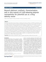
báo cáo hóa học: " Beyond platinum: synthesis, characterization, and in vitro toxicity of Cu(II)-releasing polymer nanoparticles for potential use as a drug delivery vector" ppt
Bạn đang xem bản rút gọn của tài liệu. Xem và tải ngay bản đầy đủ của tài liệu tại đây (1.28 MB, 10 trang )
NANO EXPRESS Open Access
Beyond platinum: synthesis, characterization,
and in vitro toxicity of Cu(II)-releasing polymer
nanoparticles for potential use as a drug
delivery vector
Alesha N Harris, Barbara R Hinojosa, Montaleé D Chavious and Robby A Petros
*
Abstract
The field of drug delivery focuses primarily on delivering small organic molecules or DNA/RNA as therapeutics and
has largely ignored the potential for delivering catalytically active transition metal ions and complexes. The delivery
of a variety of transition metals has potential for inducing apoptosis in targeted cells. The chief aims of this work
were the development of a suitable delivery vector for a prototypical transition metal, Cu
2+
, and demonstration of
the ability to impact cancer cell viability via exposure to such a Cu-loaded vector. Carboxylate-functionalized
nanoparticles were synthesized by free radical polymerization and were subsequently loaded with Cu
2+
via binding
to particle-bound carboxylat e functional groups. Cu loading and release were characterized via ICP MS, EDX, XPS,
and elemental analysis. Results demonstrated that Cu could be loaded in high weight percent (up to 16 wt.%) and
that Cu was released from the particles in a pH-dependent manner. Metal release was a function of both pH and
the presence of competing ligands. The toxicity of the particles was measured in HeLa cells where reductions in
cell viability greater than 95% were observed at high Cu loading. The combined pH sensitivity and significant
toxicity make this copper delivery vector an excellent candidate for the targeted killing of dis ease cells when
combined with an effective cellular targeting strategy.
Keywords: copper, polymer nanoparticles, copper ion release, drug delivery, oxidative stress, HeLa cells
Introduction
The field of drug delivery focuses primarily on deliver-
ing small organic molecules or DNA/RNA as therapeu-
tics and has largely ignored the potential for delivering
catalytically active transition metal ions and complexes
[1-3]. Some success has been realized in the case of
cisplatin [4-7]; however, vectors designed to deliver
other metal species are rare [8-11]. Thus, a significant
opportunity exists for examining the impact of selec-
tively delivering a variety of metal ions and complexes
to cells. Rational design of a vector capable of sequester-
ing and re leasing metals is therefore needed. Nanoparti-
cles based on nanoscale metal/organic frameworks and
infinite coordination polymers are being pursued
actively as drug delivery vectors; however, the metal is
used as a structural component of the particle, and in
general is not the therapeutically active moiety [12,13].
We have developed a prototypical approach that allows
us to accomplish reversible metal binding to polymeric
nanoparticles that a re stable in aqueous solutions and
that are capable of releasing bound metal in a pH-
dependent manner. We also postulate that release could
be triggered by a change in reduction potential. Sensitiv-
ity to pH allows one to capitalize on the drop in pH
known to occur along the endosomal/lysosomal pathway
for endocytosis to facilitate release, while sensiti vity to a
reducing environment could stimulate release in
response to the reducing nature of cytosol [1].
If targeted delivery can be achieved, transition metal
species would be expected to display a range of activities
inside the cell ranging from redox catalysis to the tar-
geted binding of biomolecules [14-17]. Recent findings
[18-26] indicate that many types of nanoparticles are
* Correspondence:
Department of Chemistry, University of North Texas, 1155 Union Circle,
CB#305070, Denton, TX, 76203-5017, USA
Harris et al. Nanoscale Research Letters 2011, 6:445
/>© 2011 Harris et al; licensee Springer. This is an Open Access article distributed under the terms of the Creative Commons Attribution
License ( which perm its unrestricted use, distr ibution, and reproduction in a ny medium,
provided the original work is properly cited.
capable of inducing oxidative stress, which i s of great
concern in terms of the nanotoxicology of particles
being pursued for a variety of consumer products.
Furthermore, some colloidal metal particles have been
shown to be particularly effective at generating reactive
oxygen species (ROS) presuma bly through the slow
leaching of metal ions from the particle core [19-21,25].
Increased ROS production is capable of inducing biolo-
gical damage and has been linked to a variety of disease
states including cancer, cardiovascular disease, arthritis,
diabetes, Alzheimer’s disease, and Parkinson’s disease
[27]. Cancer cells use ROS to suppress apoptosis, accel-
erate proliferation, induce metastasis and angiogenesis,
and promote genetic instability through DNA damage
[27-32]. However, the inherent toxicity of increased
ROS production represents an opportunity if it can be
harnessed by selectiv ely targeting ROS-generating parti-
cles to diseased cells [28,30]. In this case, it would be
desirable to release large amounts of metal ions in a
short period of time, which is opposite to what is
observed for the slow leaching of metal ions from colloi-
dal metal particles. Increased ROS production has the
pot ential to induce cell death by altering the expression
of apoptosis-related genes, such as Fas, c-fos, c-ju n, p53,
and Bcl-2 [22,24,33,34]. It is important to note that
most chemotherapeutics display high levels of toxicity,
and that their maximum tolerated dose is often dictated
by the maximum tolerable off-target toxicity. Transition
metal complexes also routinely e xhibit high levels of
toxicity; however, such toxicity does not limit their
potential for treating disease [17]. For example, a series
of Cu
2+
-containing compounds that exhibit high levels
of cytotoxicity and genotoxicity are being actively pur-
sued as cancer chemotherapeutics [35,36].
We have therefore designed a carboxylate-functiona-
lized, polymer-based nanoparticle capable of sequester-
ing a prototypical metal, Cu
2+
, for the ultimate goal of
delivering Cu
2+
to cancer cells to facilitate apoptosis.
The particles described here represent a single example
of a multitude containing other metal/ligand combina-
tions that can be envisaged [37]. Here, we report the
synthesis, characterization, and metal binding properties
of our Cu-binding particles, as well as preliminary
in vitro toxicity in cancer cells
Results
Synthesis and characterization of Cu-loaded polymeric
nanoparticles
Carboxylate-function alized, acrylate-based nanoparti cles
were synthesized via standard microwave-assisted, free
radical polymerization techniques [38]. Nanoparticles
used for all experiments described in this work were
prepared from an aqueous pre-polymer solution
containing 50 wt.% of an acrylic acid monomer. Nano-
particles were synthesized in aqueous solution and
remained well dispersed over the course of several
weeks. Excess unreacted monomer was removed via dia-
lysis and nanoparticle concentration was determined by
lyophilizing a sample of purified particles and weighing
the resultant solid.
Cu
2+
loading to form Cu-loaded carboxylate-functio-
nalized nanoparticles (CuCNPs) was accomplished by
first adjusting the pH of the particle-containing solu-
tion to 7.0 using 1.0 M NaOH, which deprotonated
the carboxylic acid groups, followed by the ad dition of
CuSO
4
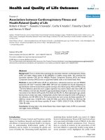
in a 1:1 molar ratio to NaOH (Figure 1). The
representation shown in Figure 1 for Cu binding to
CuCNPs represents a mononuclear complex; however,
a dinuclear complex like that observed for molecular
copper acetate (also shown in Figure 1) is equally likel y
(note: the schematic in Figure 1 shows carboxylic acid
groups only on the surface of the particle; however,
the particle is a p orous hydrogel, which allows copper
to freely diffuse throughout the polymer network and
bind to carboxylate groups on the interior of the parti-
cle as well). The particle solution was then dialyzed to
remove unbound copper. Particle size of approximately
215 nm in solution was determined via dynamic light
scattering (see Additional file 1) and a scanning elec-
tron microscope (SEM) image of dried CuCNPs is
showninFigure2A.
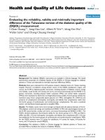
The amount of Cu bound to CuCNPs was investigated
using several analytical techniques including: inductively
coupled plasma mass spectrometry (ICP MS), X-ray
photoelectron spectroscopy (XPS), and energy-dispersive
X-ray analysis (EDX). For ICP MS studies, the amount
of unbound copper released during purification by dialy-
sis was monitored for 48 h from a sample containing
a known mass of particles (see Additional file 2).
Figure 1 Cu-loading chemistry for CuCNPs (left) and the structure of dinuclear Cu
2
(OAc)
4
(H
2
O)
2
(right).
Harris et al. Nanoscale Research Letters 2011, 6:445
/>Page 2 of 10
The difference between the amount released at 48 h and
that contained in the original loading solution deter-
mined Cu loading, resulting in values ranging between
12 and 16 wt.% based on these reactions conditions.
XPS was used to further confirm Cu loading and to
probe Cu coordination sphere (Figure 2C). Cu weight
percent measured by XPS was 15 wt.%; one of the peaks
in the spectrum (933.9 eV) was consistent with that of a
copper acetate complex.
Only peaks for C, O, and Cu were observed in EDX
spectra obtained for CuCNPs (Figure 2B), and the mea-
sured weight percents were consistent with both ICP
MS and XPS. EDX was also performed on samples
immediately before and after the addition of CuSO
4
.
Before CuSO
4
treatment,onlypeaksforC,O,andNa
were observed; after treatment, the Na peak disappeared
and a Cu peak appeared. The amount of Cu
2+
loaded
in CuCNPs could be varied trivially by adding a sub-
stoichiometric amount of CuSO
4
. CuCNPs with 16, 12,
5, and 3 wt.% Cu were synthesized in this manner, and
loading quantified via ICP MS.
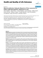
Cu release from CuCNPs
The applicability of CuCNPs for triggered release [1] has
been studied by examining the rate of Cu release in
response to changes in p H. Three identical samples of
purified CuCNPs were dialyzed in either ultrapure
water, 100 mM TRIS buffer at pH 7, or 100 mM citrate
buffer at pH 5 and the release of Cu was monitored for
48 h by ICP MS (Fig ure 3A). Virtually no release was
BA
C
Figure 2 Characterization of CuCNPs.(A) SEM image of CuCNPs, (B) EDX spectra of CNPs before (left) and after (right) addition of Cu, (C) XPS
spectra for CuCNPs (right) and control particles containing no Cu (left).
Harris et al. Nanoscale Research Letters 2011, 6:445
/>Page 3 of 10
observed in ultrapure water with approximately 95% of
the loaded Cu remaining bound to the particles over the
course of the experiment. Cu release was observed at
pH 7; however, release was much slower at this pH
(particles at pH 7 had released approximately 55% of
theirCuat12h)comparedtopH5.AtpH5,CuCNPs
had released over 93% of their loaded Cu at 12 h, and at
48 h, complete release was observed. Cu weight percents
determined by ICP MS at the end of this set of experi-
ments were 12.1, 1.7, 0.0 wt.% Cu for CuCNPs dialyzed
in ultrapure water, pH 7 buffer, and pH 5 buffer,
respectively.
Qualitatively, a color change was observed in CuCNPs
upon release of Cu where the particle color gradually
turned from blue to white. CuCNPs dialyzed at pH 5
turned white within 12 h; wherea s, those dialyzed at pH
7 remained faintly blue even at the end of 48 h. Particles
were then collected from the dialysis tubing and
analyzed further for Cu content by EDX. Cu weight per-
cents were 12.7, 3.3, and 0.7 for particles in ultrapure
water, pH 7, and pH 5, respectively, consistent with ICP
MS data. Elemental analysis by an outside vendor of
CuCNPs dialyzed in ultrapure water (Cu wt.% = 10.92)
and at pH 5 (Cu wt.% = 0.04) further confirmed our
0
20
40
60
80
100
0 5 10 15 20 2
5
10 mM acetate + 1 M NaCl
pH 4
pH 5
pH 6
% Cu released
Time
(
h
)
0
20
40
60
80
100
0 5 10 15 20 25
100 mM citrate, pH 5
100 mM Tris, pH 7
ultrapure water
% Cu released
Time (h)
0
20
40
60
80
100
0 5 10 15 20 25
100 mM citrate
100 mM acetate
10 mM acetate
% Cu released
Time (h)
AB
CD
0
20
40
60
80
100
0 5 10 15 20 25
10 mM acetate + 1 M NaCl
10 mM acetate + 1 M NaBr
10 mM acetate
% Cu released
Time
(
h
)
Figure 3 Release of Cu under various reaction conditions.(A) Initial release data simulating endosome/lysosome pH conditions, (B) release
as a function of buffering species, (C) release as a function of added competing ligand, (D) release as a function of pH in the absence of
competing ligand effects.
Harris et al. Nanoscale Research Letters 2011, 6:445
/>Page 4 of 10
experimental findings. T able 1 contains a summary of
Cu weight percents determined for each sample by the
various the experimental methods employed.
The lack of release in ultrapure water compared to
buffered solutions implied that competing ligands other
than water must be present in order to facilitate Cu
release, which led us to further investigate Cu release as
a function of competing ligand. Cu release experiments
conducted in 100 mM citrate buffer at pH 4, 5, and 6
revealed faster metal release at pH 6 than at pH 4 (see
Additional file 3), which was surprising and further illu-
strated that metal release is affected by more than just
pH. Here, the differences in rates can be attributed to
difference s in concentrations of the various species pre-
sent in the buffer as the pH is lowered (see Discussio n).
Figure 3B shows that Cu release was accelerated in 100
mM citrate buffer compared to 100 mM acetate buffer
at pH 5 also implying that the conjugate base of the
buffering species plays an important role. Buffer
strength also influenced Cu release rates as can also be
seen in Figure 3B (10 and 100 mM acetate buffer at pH
5). Cu release rate in 10 mM acetate buffer at pH 5
incre ased upon the introduction of an appropriate com-
peting ligand, such as chloride (Figure 3C). The identity
of the competing ligand that was added also influenced
the rate of Cu release as was observed on substituting
bromide for chloride (Figure 3C). In the absence of
competing ligand effects (see Discussion), Cu release
displayed pH-dependent behavior with faster release
being observed as the pH was lowered from 6 to 4
(Figure 3D). These combined experiments illustrate both
pH- and competing ligand-dependent effects on the rate
of Cu release (see Discussion).
In vitro toxicity of CuCNPs in HeLa cells
The in vitro toxicity of CuCNPs in HeLa cells (a cervi-
cal adenocarcinoma) was investigated via an assay
based on the MTT reagent (3-(4,5-dimethylthiazol-2-
yl)-2, 5-diphenyl-tetra-zolium bromide). Particles were
added to wells with cells at the desired particle con-
centrations; the plates were incubated for 48 h, fol-
lowed by an assessment of cell survival via the MTT
reagent. Control particles (particles without added Cu)
showed no toxicity up to the highest dosing. In con-
trast, Cu-loaded particles displayed significant toxicity
with an IC
50
of approximately 100 μg/mL (Figure 4A).
The toxicity of free copper acetate was measured to
allow for direct comparison with the amount of Cu
contained in CuCNPs (see Additional file 4). We
found that Cu contained in CuCNPs was significantly
more toxic than an equivalent amount of free Cu
dose, implying that the observed Cu toxicity was parti-
cle mediated. Finally, the amount of Cu loaded in
CuCNPs was varied and its effect on toxicity investi-
gated (Figure 4B). CuCNPs became significantly less
toxic as the Cu loading was reduced, with little or no
toxicity being observed for CuCNPs containing 3, or 5
wt.% Cu.
Discussion
Synthesis and characterization of Cu-loaded polymeric
nanoparticles
A prototypical approach for sequestering and releasing
metal ions from a delivery vector has been demon-
strated. In the current example, Cu
2+
was loaded to
acrylate-based nanoparticles with Cu loadings as high
as 16 wt.%. This strategy relied on functionalizing the
nanoparticle with carboxylate ligands to bind Cu
2+
;
however, other metal/ligand/polymer combinations
could be synthesized including t hose employing other
polymers routinely used in targeted drug delivery, such
as PLGA, chitosan, or dextran. Thus, the metal/ligand
chemistry is readily adaptable to, and independent
from, the desired polymeric material used as the deliv-
ery vector. The rational design of carriers to deliver
other metal species should be possible using this
approach.
Cu release from CuCNPs
The loading and stimuli-responsive release of transition
metals and any drug molecule in general from a delivery
are major factors that ultimately determine the success
or failure of that vector whe n applied t o targeted drug
delivery. One o f the goals of this work was to demon-
strate that CuCNPs were capable of responding to
changes in pH to facilitate Cu release. A general
schematic for the expected in vitro behavior is shown in
Figure 5 (targeting ligands were not used in the experi-
ments described here, but will be incorporated in the
future). Initial Cu release experiment s were conducted
at pH 5 and 7 to mimic conditions that would be pre-
sent during endocytosis of the nanoparticle along an
endosomal pathway. Those experiments (Figure 3A)
were promising and showed release to be much faster at
pH 5 vs. pH 7, which would trigger Cu release upon
particle internalization. It was postulated that protona-
tion of the carboxylate groups on the nanoparticle
would reduce the binding affinity of the ligand for Cu
thereby facilitating release. Somewhat surprising ly, how-
ever, CuCNPs rele ased virtually no Cu in ultrapure
Table 1 Cu content (in weight percent) for CuCNPs used
in Cu release experiments
ICP MS EDX Elemental analysis
Ultrapure water 12.1 12.7 10.92
pH 7 buffer 1.7 3.3 Not measured
pH 5 buffer 0.0 0.7 0.04
Harris et al. Nanoscale Research Letters 2011, 6:445
/>Page 5 of 10
water, which has a neutral to slightly acidic pH. While
this feature is promising in terms of the stability of solu-
tions of CuCNPs over long periods of time, Cu release
cannot simply be a function of pH but must also depend
on the presence of ligands that can co mpete with the
particle-bound carboxylate groups in Cu binding. This
feature led to a series of additional experiments to eluci-
date the effect of competing ligands on Cu release with
the idea that the underlying principles governing release
could be used in the design/optimizati on of this class of
delivery vectors.
Release of Cu from CuCNPs in citrate buffer at p H 4,
5, and 6 (see Additional file 3) illustrated the effect of
competing ligand concentration on the rate of Cu
release, which was actually faster at pH 6 compared to
pH 4. This can be explained b y looking at the various
protonation states of citrate as a function of pH to
determine the competing ligands present in solution.
Equations 1-4 were used to determine the relative con-
centrations of L
3-
,LH
2-
,LH
2
-
,andLH
3
(L = citrate)
using pK
a
values of 3.13, 4.76, and 6.40 for citric acid
( K
1
=7.40×10
-4
, K
2
=1.70×10
-5
, K
3
=4.00×10
-7
).
The relative concentrations of L
3-
,LH
2-
,LH
2
-
,andLH
3
are 27%, 69%, 4%, and 0% at pH 6 while at pH 4 the
relative concentrations for the same species are 0%,
13%, 77%, and 10%. At low pH, the predominate species
is LH
2
-
whereas at high pH the predominate species is
LH
2-
with a substantial amount of L
3-
being present as
well. One would expect the affinity of these ligands for
Cu to increase with increasing negative charge, and that
is clearly what is observed. So, even though the particle-
bound carboxylate is protonated to a lesser extent at pH
6, the presence of the di- and tri-anion form of citrate
effectively compete out Cu.
%LH
3
=
[H
+
]
3
[H
+
]
3
+ K
1
[H
+
]
2
+ K
1
K
2
[H
+
]+K
1
K
2
K
3
× 100
(1)
%LH
2
−
=
K
1
[H
+
]
2
[H
+
]
3
+ K
1
[H
+
]
2
+ K
1
K
2
[H
+
]+K
1
K
2
K
3
× 100
(2)
%LH
2
−
=
K
1
K
2
[H
+
]
[H
+
]
3
+ K
1
[H
+
]
2
+ K
1
K
2
[H
+
]+K
1
K
2
K
3
× 100
(3)
%L
3
−
=
K
1
K
2
K
3
[H
+
]
3
+ K
1
[H
+
]
2
+ K
1
K
2
[H
+
]+K
1
K
2
K
3
× 100
(4)
In an effort to decouple competing ligand effects due
to the presence of changing buffering species with actual
pH-dependent Cu release, we sought to reduce the
AB
Figure 4 In vitro toxicity of CuCNPs.(A) H eLa cell viability as measured via an assay based on MTT at 48 h showing toxicity only after the
addition of Cu to the nanoparticles, (B) a similar experiment showing the reduced toxicity of CuCNPs upon reduction of Cu content.
Figure 5 Proposed intracellular release mechanism based
on pH.
Harris et al. Nanoscale Research Letters 2011, 6:445
/>Page 6 of 10
buffer effect while concomitant ly introducing competing
ligands that were unaffected by solution pH. The use of
citrate buffer was less than ideal in this case due to the
multiple acidic protons capable of generating four possi-
ble species in solution. The use of acetate buffer in
place of citrate was expected to reduce this complexity.
Acetate buffer g enerates only two species with rel ative
concentrations of A
-
and HA being 95% and 5% at pH 6
and 15 and 85% at pH 4, respectively (pK
a
=4.75).
Furthermore, even though the concentrat ion of the
anion changes in this case as well, and is higher at pH
6, this species is identical to the particle-bound carboxy-
late groups making it a less effective competitor com-
pared to the species present in citrate buffer at the same
pH. A direct comparison of Cu release at pH 5 in acet-
ate buffer and citrate buffer (Figure 3B), both at 100
mM, confirmed this assumption. Cu release was much
slower in acetate buffer most likely a result of the
reduced competitive nature of A
-
compared to LH
2-
.Cu
release in acetate buffer could be further reduced by
lowering the buffer strength to 10 mM (Figure 3B).
With buffer effects reduced, a competing ligand was
then introduced. Chloride was chosen as an appropriate
competing ligand because its concentration would not
be effected by the pH of the solutions being investi-
gated. Indeed, the introduction of chloride increased the
rate of Cu release in acetate buffer (Figure 3C). Again,
Cu release was highly dependent on the identity of com-
peting ligand as illustrated by comparing the effect of
added chloride vs. bromide (Figure 3C). Next, Cu release
was monitored in 10 mM acetate buffer at pH 4, 5,
and 6 containing a large excess of chloride (1 M NaCl,
Figure 3D). With changes in competing ligand concen-
trations effectively minimized, pH-dependent Cu release
was clearly demons trated and release was accelerated as
the pH was reduced.
In vitro toxicity of CuCNPs in HeLa cells
The toxicity of CuCNPs to cancer cells was investigated
and results demonstrated that the delivery vector itself,
particles containing no Cu, was not toxic and that
CuCNPs displayed significant toxicity depending on dos-
ing (Figure 4A). Furthermore, the delivery of Cu con-
tained in CuCNPs was more toxic than an equivalent
amount of free Cu dose implying that the phenomenon
was particle mediated (see Additional file 4). This effect
is likely attributable to the differences in modes of inter-
nalization for the two forms of Cu. Free copper would
be taken up by the cell via normal metal trafficking
pathways that utilize metal-binding proteins located on
the cell surf ace. As the cell begins to experience metal
overload, those receptors would be internalized and
degraded to prevent further metal accumulation.
CuCNPswouldbeexpectedtobeinternalizedbyan
entirely different pathway that is no t subject to the nor-
mal cellular mechanisms for controlling metal homeos-
tasis. Thus, the cell’ s normal metal overload defenses
were likely bypassed leading to unregulated Cu uptake.
As the Cu-loading in CuCNPs was reduced, cell survival
improved with little or no toxicity being observed for
CuCNPs containing 3 or 5 wt.% Cu (Figure 4B). The
most likely source of toxicity was induced oxidative
stress (see Introduction) and future experiments will
probe the mechanism of cell death to determine the
validity of this hypothesis.
Conclusions
In summary, we have synthesized Cu-loaded polymeric
nanoparticles that release boun d Cu in a pH- dependent
manner. Cu loading and release were characterized by
several analytical techniques where we demonstrated the
ability to load up to 16 wt.% Cu. The release of bound
Cu from CuCNPs was found to be both pH and com-
peting ligand dependent. Decoupling these effects was
non-trivial, but was accomplished through careful selec-
tion of reaction conditions. Based on the behavior
observed, we conclude that simple protonation of the
particle-bound carboxylate, while rate accelerating was
not sufficient to promote release rather the presence of
a ligand capable of displacing the carboxylate was
required. The complexities described here will undoubt-
edly increase dramatically when CuCNPs are introduced
to biologically relevant media containing a plethora of
potential ligands. Our coupling strategy allows us to
capitalize on the pH gradient observed along the endo-
some/lysosome pathway for particle internalization for
targeted delivery of Cu [1]. CuCNPs were capable of
inducing toxicity in cancer cells where reductions of
>95% viability were observed at high Cu loadings. The
stimuli- responsive release and toxicity of Cu in CuCNPs
meets the requirements for application in targeted drug
delivery.
Methods
General considerations
Methyl methacrylate, acrylic acid, and poly(ethylene gly-
col) (n) diacrylate (n = 200 = MW of PEG block) were
purchased from Polysciences, Inc. (Warrington, PA,
USA) and used as received. Potassium persulfate, copper
sulfate, copper acetate, nitric acid (trace metal grade)
were from Fisher Scientific (Pennsylvania , PA, USA). All
materials were used as received unless o therwise noted.
Microwave reactions were conducted in a Synthos 3000
from Anton Paar (Ashland, VA, USA). ICP MS experi-
ments were conducted on a Varian 820-MS (Varian
Inc., L ake Forest, CA, USA) usin g the followi ng
parameters: plasma flow 17.5 L/min, auxiliary flow 1.65
L/min, sheath gas 0.13 L/min, and neb ulizer flow 0.89
Harris et al. Nanoscale Research Letters 2011, 6:445
/>Page 7 of 10
L/min. The torch alignment had a sampling depth of 5
mm. The RF power was set at 1.3 kW. The pump rate
was 3 rpm, and the stabilization delay was 30 s. The ion
optics parameters were: first extraction lens -1 V, sec-
ond extraction lens -191 V, third extraction lens -206 V,
corner lens -236 V, mirror lens left 56 V, mirror lens
right49V,mirrorlensbottom16V,entrancelens0V,
fringe bias -2.5 V, entrance plate -31 V, and pole bias 0
V. CRI parameters were skimmer gas off, sampler gas
off, skimmer flow 0 mL/min, and sampler flow 0 mL/
min. ICP MS tubing was rinsed in between samples to
avoid sample contamination.
Synthesis of nanoparticles
An aqueous soluti on (58.8 mL ) containing acrylic acid
(0.57 g), methyl methacrylate (0.575 g), PEG diacrylate
(0.053 g), and potassium persulfate (0.164 g) was pre-
pared in a PTFE vessel for a Synthos 3000 16MF100
rotor in a freshly regenerated inert atmosphere glove-
box. The vessel was sealed, removed from the glovebox,
and placed in the 16MF100 rotor along with seven
other vessels containing 60 mL of water each. The rotor
was placed in the microwave and then heated to 90°C
for 60 min with a maximum microwave power of 1400
W (see Additional file 5). The internal temperature and
pressure of the vessel containing the monomer solution
were monitored via a p/T sensor accessory (Anton
Paar). The resulting nanoparticle solution was dialyzed
in 4 L of ultrapure water for 48 h with a change in the
water after the first 24 h. The particle concentration
after purification was determined by lyophi lizing a
known volume and then weighing the resulting solid,
which resulted in a final particle concentration of 12.8
mg/mL. Based on this number, a total of 0.896 g of par-
ticles was synthesized with approximately 75% conver-
sion of monomer to particles.
Copper loading
A 3-mL aliquot of the nanoparticle solution was
adjusted to a pH of 7 using NaOH followed by the addi-
tion of copper sulfate in a 1:1 molar ratio with amount
of NaOH added. The particle solution was then dialyzed
in 1.5 L of ultrapure water for 48 h to remove unbound
copper. Particle size of approximately 215 nm was deter-
mined via dynamic light scattering (DLS).
ICP MS Cu-loading studies
For Cu-loading studies, the Cu-loading solution contain-
ing CuSO
4
and nanoparticles was dialyzed in 1.5 L of
ultrapure water. Samples (1 mL each) w ere removed at
1, 2, 3, 4, 5, 6, 12, 24, and 48 h, diluted in 1% nitric acid
and then analyzed for
63
Cu content via ICP MS. Cu con-
tent was determined by comparison with a calibration
curve generated using known samples (see Additional
file 6).
ICP MS Cu release studies
Purified CuCNP-containing solutions (3 mL) were dia-
lyzed in 1.5 L of the desired buffering solution. Samples
(1mLeach)wereremovedat0.08,1,2,3,4,5,6,12,
24, and 48 h, diluted in 1% nitric acid and then analyzed
for
63
Cu content via ICP MS. Cu content was deter-
mined by comparison with a calibration curve generated
using known samples.
X-ray photoelectron spectroscopy
XPS spectra were acquired with a PHI 5000 VersaP-
robe™ Scanning XPS Microprobe (Physical Electronics
Inc., Chanhassen, MN, USA). Samples were prepared by
spotting 5 μL of the desired particle-containing solution
onto a glass slide and then drying under vacuum.
Scanning electron microscopy and energy-dispersive X-
ray analysis
SEM images and EDX spectra were obtained with a
Quanta ESEM microscope (FEI, Hillsboro, OR, USA)
equipped with a Sapphire Si(Li) detecting unit for EDX
(EDAX Inc., Mahwah, NJ, USA). Samples were prepared
by spotting 5 μL of the desired particle-containing solu-
tion onto a glass slide, drying under vacuum, and then
repeating the spot/dry three times to produce samples
with enough thickness to prevent interference from the
glassslideduringEDXanalysis.Sampleswerethen
coated with Au (2-5 nm thickness) using a Cressington
108 Manual Sputter Coater (Ted Pella, Redding, CA,
USA). Images w ere obtained with an acceleration vol-
tage of 5-15 kV and EDX spectra were obtained with an
acceleration voltage of 5 kV.
Elemental analysis
Microanalysis was performed by Columbia Analytics
(formerly Desert Analytics) in Tuc son, AZ. Samples
(100 mg) for elemental analysis were prepared by lyo-
philizing the desired nanoparticle-containing solution,
which were further dried for 4 h at 25°C under vacuum
prior to analysis.
Cell viability measurements
HeLa cells were purchased from ATCC (cat. # CCL-2),
and maintained in Eagle’s Minimum Essential Medium
(ATCC, cat. # 30-2003) with 10% FBS (Thermo Scienti-
fic HyClone, South Logan, UT, USA). Five thousand
cells per well seeded on 96-well plates and incubated
overnightat37°C(5%CO
2
). The desired particle
amounts were added to the wells and the plates were
incubated for an additional 48 h at 37°C (5% CO
2
).
Harris et al. Nanoscale Research Letters 2011, 6:445
/>Page 8 of 10
After the incubation, cell viability was evaluated with the
MTT reagent. Media was removed each well and
replaced with fresh media containing 1 mg/mL MTT.
The cells were incubated for 4 h at 37°C (5% CO
2
)after
which time the media was removed and replaced with
DMSO. Light absorption was measured on a Synergy 2
multi-mode microplate reader (BioTek, Winooski, VT,
USA). The viability of the cells exposed to particles was
expressed as a percentage of the viability of c ells grown
in the absence of particles on the same plate.
Additional material
Additional file 1: DLS results for purified CuCNPs. graph showing
particle size as determined by Dynamic Light Scattering.
Additional file 2: Release of unbound Cu over time during
purification of CuCNPs as monitored by ICP MS. graph showing all
copper that is not bound to the particle is removed by dialysis for 48 h.
Additional file 3: Release of Cu from purified CuCNPs over time in
100 mM citrate buffer at pH 4, 5, and 6. graph showing that Cu
release is actually slower as the pH is lowered due to competing ligand
effects.
Additional file 4: In vitro toxicity for comparison of Cu in CuCNPs
versus similar dosing of free Cu(OAc)
2
. graph showing copper
contained in nanoparticles was more toxic than an equivalent amount of
copper dosed as a free complex.
Additional file 5: Graph of reaction time vs. temperature, pressure,
and microwave power during nanoparticle synthesis. graphs
showing microwave conditions used for nanoparticle synthesis.
Additional file 6: Typical calibration curve used for determining the
Cu concentration in unknown samples. calibration curve generated
from samples containing a known amount of copper.
Acknowledgements
We thank Guido Verbeck and William Hoffmann for their help with ICP MS
studies, Nancy Bunce, David Diercks, and David Garrett for aid with EDX,
XPS, and SEM analysis. Portions of this work were conducted at the UNT
Laboratory of Imaging and Mass Spectrometry and the UNT Center for
Advanced Research and Technology. MC was an NSF-REU scholar (grant
CHE-1004878) at the University of North Texas.
Authors’ contributions
AH carried out Cu loading and release studies via ICP MS, participated in the
design and coordination of ICP MS studies, and helped draft the manuscript.
BH optimized microwave conditions for the free radical polymerization
reaction used in the synthesis of polymeric nanoparticles. MC carried out
particle synthesis, and metal loading. RP conceived of the study, and
participated in its design and coordination, carried out in vitro toxicity
studies, SEM and EDX analysis and drafted the manuscript.
Competing interests
The authors declare that they have no competing interests.
Received: 7 January 2011 Accepted: 11 July 2011
Published: 11 July 2011
References
1. Petros RA, DeSimone JM: Strategies in the design of nanoparticles for
therapeutic applications. Nat Rev Drug Discov 2010, 9:615-627.
2. Davis ME, Chen Z, Shin DM: Nanoparticle therapeutics: an emerging
treatment modality for cancer. Nat Rev Drug Discov 2008, 7:771-782.
3. Zhang L, Gu FX, Chan JM, Wang AZ, Langer RS, Farokhzad OC:
Nanoparticles in medicine: Therapeutic applications and developments.
Clin Pharmacol Ther 2008, 83:761-769.
4. Dhar S, Daniel WL, Giljohann DA, Mirkin CA, Lippard SJ: Polyvalent
Oligonucleotide Gold Nanoparticle Conjugates as Delivery Vehicles for
Platinum(IV) Warheads. J Am Chem Soc 2009, 131:14652-14653.
5. Dhar S, Gu FX, Langer R, Farokhzad OC, Lippard SJ: Targeted delivery of
cisplatin to prostate cancer cells by aptamer functionalized Pt(IV)
prodrug-PLGA-PEG nanoparticles. Proc Natl Acad Sci USA 2008,
105:17356-17361.
6. Rieter WJ, Pott KM, Taylor KML, Lin WB: Nanoscale coordination polymers
for platinum-based anticancer drug delivery. J Am Chem Soc 2008,
130:11584-11585.
7. Haxton KJ, Burt HM: Polymeric Drug Delivery of Platinum-Based
Anticancer Agents. J Pharm Sci 2009, 98:2299-2316.
8. Treiber C, Quadir MA, Voigt P, Radowski M, Xu SJ, Munter LM, Bayer TA,
Schaefer M, Haag R, Multhaup G: Cellular Copper Import by Nanocarrier
Systems, Intracellular Availability, and Effects on Amyloid beta Peptide
Secretion. Biochemistry 2009, 48:4273-4284.
9. Withey ABJ, Chen G, Nguyen TLU, Stenzel MH: Macromolecular Cobalt
Carbonyl Complexes Encapsulated in a Click-Cross-Linked Micelle
Structure as a Nanoparticle To Deliver Cobalt Pharmaceuticals.
Biomacromolecules 2009, 10:3215-3226.
10. Chen H, Ahn R, Van den Bossche J, Thompson DH, O’Halloran TV: Folate-
mediated intracellular drug delivery increases the anticancer efficacy of
nanoparticulate formulation of arsenic trioxide. Mol Cancer Ther 2009,
8:1955-1963.
11. Neuse E: Macromolecular Ferrocene Compounds as Cancer Drug Models.
J Inorg Organomet Polym Mater 2005, 15:3-31.
12. Spokoyny AM, Kim D, Sumrein A, Mirkin CA: Infinite coordination polymer
nano- and microparticle structures. Chem Soc Rev 2009, 38:1218-1227.
13. Della Rocca J, Lin WB: Nanoscale Metal-Organic Frameworks: Magnetic
Resonance Imaging Contrast Agents and Beyond. Eur J Inorg Chem 2010,
3725-3734.
14. Gianferrara T, Bratsos I, Alessio E: A categorization of metal anticancer
compounds based on their mode of action. J Chem Soc, Dalton Trans
2009, 7588-7598.
15. Jung YW, Lippard SJ: Direct cellular responses to platinum-induced DNA
damage. Chem Rev
2007, 107:1387-1407.
16.
Messori L, Casini A, Gabbiani C, Sorace L, Muniz-Miranda M, Zatta P:
Unravelling the chemical nature of copper cuprizone. J Chem Soc Dalton
Trans 2007, 2112-2114.
17. Gasser G, Ott I, Metzler-Nolte N: Organometallic Anticancer Compounds. J
Med Chem 2011, 54:3-25.
18. Thubagere A, Reinhard BM: Nanoparticle-Induced Apoptosis Propagates
through Hydrogen-Peroxide-Mediated Bystander Killing: Insights from a
Human Intestinal Epithelium In Vitro Model. ACS Nano 2010, 4:3611-3622.
19. George S, Pokhrel S, Xia T, Gilbert B, Ji Z, Schowalter M, Rosenauer A,
Damoiseaux R, Bradley KA, Mädler L, Nel AE: Use of a Rapid Cytotoxicity
Screening Approach To Engineer a Safer Zinc Oxide Nanoparticle
through Iron Doping. ACS Nano 2010, 4:15-29.
20. AshaRani PV, Low Kah Mun G, Hande MP, Valiyaveettil S: Cytotoxicity and
Genotoxicity of Silver Nanoparticles in Human Cells. ACS Nano 2009,
3:279-290.
21. Xia T, Kovochich M, Liong M, Mädler L, Gilbert B, Shi H, Yeh JI, Zink JI,
Nel AE: Comparison of the Mechanism of Toxicity of Zinc Oxide and
Cerium Oxide Nanoparticles Based on Dissolution and Oxidative Stress
Properties. ACS Nano 2008, 2:2121-2134.
22. Ahamed M, Akhtar MJ, Siddiqui MA, Ahmad J, Musarrat J, Al-Khedhairy AA,
AlSalhi MS, Alrokayan SA: Oxidative stress mediated apoptosis induced by
nickel ferrite nanoparticles in cultured A549 cells. Toxicology 2011,
283:101-108.
23. Park E-J, Yi J, Chung K-H, Ryu D-Y, Choi J, Park K: Oxidative stress and
apoptosis induced by titanium dioxide nanoparticles in cultured BEAS-
2B cells. Toxicol Lett 2008, 180:222-229.
24. Lunov O, Syrovets T, Büchele B, Jiang X, Röcker C, Tron K, Nienhaus GU,
Walther P, Mailänder V, Landfester K, Simmet T: The effect of
carboxydextran-coated superparamagnetic iron oxide nanoparticles on
c-Jun N-terminal kinase-mediated apoptosis in human macrophages.
Biomaterials 2010, 31:5063-5071.
Harris et al. Nanoscale Research Letters 2011, 6:445
/>Page 9 of 10
25. Ahamed M: Toxic response of nickel nanoparticles in human lung
epithelial A549 cells. Toxicol in Vitro 2011, 25:930-936.
26. Ahamed M, Siddiqui MA, Akhtar MJ, Ahmad I, Pant AB, Alhadlaq HA:
Genotoxic potential of copper oxide nanoparticles in human lung
epithelial cells. Biochem Bioph Res Co 2010, 396:578-583.
27. Valko M, Leibfritz D, Moncol J, Cronin MTD, Mazur M, Telser J: Free radicals
and antioxidants in normal physiological functions and human disease.
Int J Biochem Cell Biol 2007, 39:44-84.
28. Wondrak GT: Redox-Directed Cancer Therapeutics: Molecular
Mechanisms and Opportunities. Antioxid Redox Sign 2009, 11:3013-3069.
29. Federico A, Morgillo F, Tuccillo C, Ciardiello F, Loguercio C: Chronic
inflammation and oxidative stress in human carcinogenesis. Int J Cancer
2007, 121:2381-2386.
30. Pelicano H, Carney D, Huang P: ROS stress in cancer cells and therapeutic
implications. Drug Resist Update 2004, 7:97-110.
31. Halliwell B: Oxidative stress and cancer: have we moved forward?
Biochem J 2007, 401:1-11.
32. Erez A, Shchelochkov Oleg A, Plon Sharon E, Scaglia F, Lee B: Insights into
the Pathogenesis and Treatment of Cancer from Inborn Errors of
Metabolism. Am J Hum Genet 2011, 88:402-421.
33. Mao XW, Green LM, Mekonnen T, Lindsey N, Gridley DS: Gene Expression
Analysis of Oxidative Stress and Apoptosis in Proton-irradiated Rat
Retina. In Vivo 2010, 24:425-430.
34. Kannan K, Jain SK: Oxidative stress and apoptosis. Pathophysiology 2000,
7:153-163.
35. Alemón-Medina R, Bravo-Gómez ME, Gracia-Mora MI, Ruiz-Azuara L:
Comparison between the antiproliferative effect and intracellular
glutathione depletion induced by Casiopeína IIgly and cisplatin in
murine melanoma B16 cells. Toxicol in Vitro 2011, 25:868-873.
36. Alemón-Medina R, Breña-Valle M, Muñoz-Sánchez J, Gracia-Mora M, Ruiz-
Azuara L: Induction of oxidative damage by copper-based antineoplastic
drugs (Casiopeínas). Cancer Chemoth Pharm 2007, 60:219-228.
37. Petros RA: Synthesis and Use of Metal Ion-Containing Polymeric Particles.
US Patent Appl 61/370,682 2010.
38. An Z, Tang W, Hawker CJ, Stucky GD: One-Step Microwave Preparation of
Well-Defined and Functionalized Polymeric Nanoparticles. J Am Chem
Soc 2006, 128:15054-15055.
doi:10.1186/1556-276X-6-445
Cite this article as: Harris et al.: Beyond platinum: synthesis,
characterization, and in vitro toxicity of Cu(II)-releasing polymer
nanoparticles for potential use as a drug delivery vector. Nanoscale
Research Letters 2011 6:445.
Submit your manuscript to a
journal and benefi t from:
7 Convenient online submission
7 Rigorous peer review
7 Immediate publication on acceptance
7 Open access: articles freely available online
7 High visibility within the fi eld
7 Retaining the copyright to your article
Submit your next manuscript at 7 springeropen.com
Harris et al. Nanoscale Research Letters 2011, 6:445
/>Page 10 of 10