báo cáo hóa học: " Imaging the effect of receptor for advanced glycation endproducts on angiogenic response to hindlimb ischemia in diabetes" docx
Bạn đang xem bản rút gọn của tài liệu. Xem và tải ngay bản đầy đủ của tài liệu tại đây (2.5 MB, 9 trang )
PRELIMINARY RESEARCH Open Access
Imaging the effect of receptor for advanced
glycation endproducts on angiogenic response to
hindlimb ischemia in diabetes
Yared Tekabe
1
, Xiaoping Shen
2,3
, Joane Luma
1
, Drew Weisenberger
4
, Shi Fang Yan
2,3
, Roland Haubner
5
,
Ann Marie Schmidt
2,3
and Lynne Johnson
1*
Abstract
Background: Receptor for advanced glycation endproducts (RAGE) expression contributes to the impaired
angiogenic response to limb ischemia in diabetes. The aim of this study was to detect the effect of increased
expression of RAGE on the angiogenic response to limb ischemia in diabetes by targeting a
v
b
3
integrin with
99m
Tc-labeled Arg-Gly-Asp (RGD).
Methods: Male wild-type (WT) C57BL/6 mice were either made diabetic or left as control for 2 months when they
underwent femoral artery ligation. Four groups were studied at days 3 to 7 after ligation: WT without diabetes
(NDM) (n = 14), WT with diabetes (DM) (n = 14), RAGE
-/-
NDM (n = 16), and RAGE
-/-
DM (n = 14). Mice were
injected with
99m
Tc-HYNIC-RGD and imaged. Count ratios for ischemic/non-ischemic limbs were measured. Muscle
was stained for RAGE, a
v
b
3
, and lectins.
Results: There was no difference in count ratio betwe en RAGE
-/-
and WT NDM groups. Mean count ratio was
lower for WT DM (1.38 ± 0.26) vs. WT NDM (1.91 ± 0.34) (P<0.001). Mean count ratio was lower for the RAGE
-/-
DM
group than for RAGE
-/-
NDM group (1.75 ± 0.22 vs. 2.02 ± 0.29) (P<0.001) and higher than for the WT DM group
(P<0.001). Immunohistopathology supported the scan findings.
Conclusions: In vivo imaging of a
v
b
3
integrin can detect the effect of RAGE on the angiogenic response to limb
ischemia in diabetes.
Background
The prevalence of peripheral artery disease in the gen-
eral population is 12% to 14%, a ffecting 20% of those
>70 years and contributes to significant morbidity. Limb
ischemia in diabetics take s a particularly malignant
course leading to impaired wound healing, gangrene,
amputati ons, and even death [1,2]. A major and distinct
adaptive process that contributes to restoring nutrient
blood flow to ischemic limbs is angiogenesis/arterio gen-
esis. Angiogenesis refers to the process of endothelial
sprouting. Arteriogenesis is the f ormation of larger
“arteriol e” like vessels. Both processes are essential for
the development of subsequent collateral growth [3].
Tissue hypoxia activates genes that code for angiogenic
growth factors and cytokines. Investigational studies
have documented the involvement of receptor for
advanced glycation endproducts (RAGE) in the impaired
angiogenic response to limb ischemia in diabetes [4-7].
The expression of a
v
b
3
integrin, a cell adhesion recep-
tor that plays a crucial role in the angiogenesis process,
can be targ eted with rad iolabeled peptides for in vivo
imaging [8]. Comparing in vivo imaging in animals with
genetic alteration of pathways implicated in angiogenesis
allows exploration of downstream effects in live animals.
In this study, we investigated the value of imaging the
effectsofRAGEexpressionontheangiogenicresponse
to limb ischemia in live animals. We used
99m
Tc-labeled
Arg-Gly-Asp (RGD) peptide that targets a
v
b
3
integrin
expression occurring during capillary sprouting. Our
hypothesis was that using genetically altered mice,
* Correspondence:
1
Department of Medicine, Columbia University Medical Center, New York, NY
10032, USA
Full list of author information is available at the end of the article
Tekabe et al . EJNMMI Research 2011, 1:3
/>© 2011 Tekabe et al; licensee S pringer . This is an Open Ac cess article distributed under the terms of the Creative Commons Attribution
License
99m
Tc-labeled RGD imaging can detect in vivo the effect
of RAGE expression on angiogenic response to limb
ischemia in diabetes.
Methods
Experimental protocol
Allanimalexperimentswereperformed in accordance
with the approval of the Institutional Animal Care and
Use Committee of Columbia University. Homozygous
male RAGE null (RAGE
-/-
) mic e (backcrossed >10 gen-
erations into C57BL/6) wer e generated as described pre-
viously[9].Malewild-type(WT)C57BL/6micewere
obtained (Jackson Laboratories). At age 6 weeks, half of
the WT and half of the RAGE
-/-
mice were treated with
streptozotocin (STZ; Sigma). Two months later, all mice
underwent femoral artery (FA) ligation.
Induction of diabetes
Mice were tr eated with five consecutive daily doses of
STZ dissolved in citrate buffer ( 55 mg/kg, pH 4.5) via
the intraperitoneal route. One week after the first
dose, glucose levels were assessed by glucometer. The
criteria of two consecutive glucose levels >250 mg/dL
was used to indicate diabetes. If glucose levels were
<250 mg/dL, then the mice received two additional
doses of STZ (55 mg/kg).
Femoral artery ligation
Under isoflurane anesthesia, the hair on the abdominal
wall and pelvis and both upper legs was shaved and the
skin prepped with iodine and alcohol. An incision was
made on the upp er thigh of both the left and right legs
of each mouse. The inguinal ligament and the upper
half of the femoral artery were exposed. On the left side,
the vascular bundle was iso lated from bel ow the ingu-
inal ligament proximally to just above the bifurcation
into the superficial and deep femoral arteries distally.
The femoral artery was dissected free, and two ligatures
were placed around it with 8/0 non-absorbable sutures
and tied. Both skin incisions were closed with sterile 5/0
nylon suture.
Preparation of radiotracer
Aliquots of 5 μg of HYNIC-RGD were incubated with
0.5 m l of tricine solution (70 mg/ml in distilled w ater)
and approximately 0.5 ml of
99m
TcO
4
-
solution (50 mCi
=1,850MBq)and20μl of tin(II) solution (10 mg of
SnCl
2
·2H
2
Oin10mlofnitrogen-purged0.1NHClfor
20 min) at room temperature. To test the s pecificity of
the HYNIC-RGD, cyclo [Arg-Ala-Asp-D -Phe-Lys
(HYNIC)] (Peptides International, Lo uisvill e, KY, USA)
was s imilarly radiolabeled and used as control peptide.
Radiochemical purity was >94% by Tec-co ntrol chroma-
tography (Biodex, Shirley, NY, USA).
Injection of radiotracer and imaging
Under isoflurane anesthesia (1.5% isoflurane at a flow
rate of 0.5% L/min oxygen per mouse), a cutdown was
made over the jugular vein and a specially designed vas-
cular catheter was placed (Braintree Scientific, Braintree,
MA, USA). Mice in each of four groups were injected
with
99m
Tc-HYNIC-RGD and imaged 3 or 7 days after
FA ligation: WT without diabetes (n =14),WTwith
diabetes (n = 14), RAGE
-/-
without diabetes (n = 16),
RAGE
-/-
with diabetes (n = 14), and five WT without
diabetes were injected with control peptide. All mice
were injected through the jugular vein catheter with 1 ±
0.2 mCi of
99m
Tc-HYNIC-RGD in 0.05 to 0.1 ml (corre-
sponding to 1 μg of peptide ) or control peptide. Blood
pool clearance was measured in five mice injected
with
99m
Tc-HYNIC-RGD. By 60 to 75 min after injec-
tion, residual blood pool activity was below 10% of peak.
Whole-body planar gamma images in the anteroposter-
ior view were acquired on a high-resolution high-sensi-
tivity dedicated small animal camera with parallel hole
collimator (provided by Jefferson Lab, Newport News,
VA, USA). The camer a is based on a 5-in. Hamamatsu
position sensitive photomultiplier type R3292 with an
active field of view of about 95 mm diameter. The scin-
tillator sensor is 1.6-mm-step 6-mm-thi ck pixelated NaI
(Tl) scintillator array. The photo peak was set at 140
keV with a 15% energy window.
Ex vivo tissue counting
At completion of the imaging session, each animal was
euthanized by an intraperitoneal injection of pentobarbi-
tal (100 mg/kg). The anterior tibialis muscles were dis-
sected, wei ghed, and counted in a gamma co unter
(Wallac Wizard 1470, PerkinElmer, Waltham, MA,
USA) for determination of the percent injected dose of
radiotracer per gram (%ID/g) tissue. The radiotrac er
activity in the samples was corrected for background,
decay time, and tissue weight. Limb counting was per-
formed in 28 animals. The remaining animals wer e used
for immunohistochemistry.
Histopathology
For immunohistochemical analyses, tibialis a nterior
muscles were harvested and fixed in 10% f ormalin for
48 h. Specimens were embedded in paraffin, and tissue
slices (5 μm in thickness) were prepared. Serial sections
were stained with hematoxylin and eosin (H&E) for
morphology. Immunostaining was performed for capil-
lary sprouting using biotinylated Griffonia Bandeiraea
Simplicifolia Isolectin I (V ector Laboratories, Burlin-
game, CA, USA) for b
3
(1:50;Abcam,Cambridge,MA,
USA.) and for a
ν
(1:100; Millipore, Temecula, CA,
USA). Serial sections were also stained for RAGE using
a monoclonal antibody against RAGE (50 μg/ml).
Tekabe et al . EJNMMI Research 2011, 1:3
/>Page 2 of 9
Secondary stains were performed using avidin -biotin
visualization systems (Vectastain ABC Kit, Vector
Laboratories). All brown staining capillaries were
counted for each of 5 to 6 sections for both the left and
right anterior tibialis muscles for each experiment and
then were averaged. The average number of capillaries
for the left anterior tibialis muscle was divided by the
average n umber for the right (control) anterior tibialis
muscle. RAGE staining was quantified as area staining
positive for the brown chromagen per 100× field.
Immunofluorescence
Dual immunofluorescent studies were undertaken to
determine the cell types expressing a
ν
integrin. Serial
sections (5 μm in thickness) obtained from the ischemic
hindlimb were d eparaffinized in xylene and incubated
with a
ν
(rat anti-mouse integrin a
ν
, 1:100) and co-
stained with endothelial cell marker (FVIII, 1:200) or
macrophage marker (Mac-3, 1 :50). Secondary fluores-
cent antibodies were Texas Red anti-rabbit and FITC
anti-mouse. The images were captured and processed
using confocal fluorescence microscope (Nikon, Tokyo,
Japan) and SPOT imaging software (Diagnostic Instru-
ments, Inc., Sterling Heights, MI, USA).
Image analysis
Radiotracer counts in the ischemic hindlimb were deter-
mined from the in vivo scans by using the region of
interest (ROI) method in the mini gamma camera image
using public domain Image J software (NIH, Bethesda,
MD, USA). A region was drawn around the focal
uptake, and the mean counts were determined. Radioac-
tivity in the contralateral control limb was similarly
determined using a comparable R OI (same anatomic
location and the number of pixels). The counts from
each of these areas were used to determine the ischemic
to non-ischemic ratios.
Statistical analysis
Continuous variables were expressed as mean ± stan-
dard deviation. Normality was assessed using the Sha-
piro-Wilk test. Comparisons between two groups were
made using the Student’s t test. Correlation was assessed
using the Pearson product-moment correlation coeffi-
cient. All statistical tests were two-tailed, with P <0.05
denoting significance. All statistical analyses were per-
formed using STATA 10.1 (StataCor p, College Station,
TX, USA).
Results
Scan analysis
Mean uptake ratios of counts betwe en the left and right
limbs were not different between days 3 and 7 for any
of the four groups: WT non-diabetic (P = 0.52), WT
diabetic (P = 0.39), RAGE
-/-
non-diabetic (P = 0.41), and
RAGE
-/-
diabetic (P = 0.39). The refore, days 3 and 7
data were combined as the early time period.
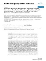
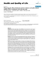
Representative scans from the four groups and the
control p eptide are shown in Figure 1. All scans in the
non-diabetic WT group were positive visually, while
three of the left limbs in the diabetic group were nega-
tive, one was equivocal, and one weakly positive. Scans
of the WT mice in jected with control peptide showed
no tracer uptake in either limb.
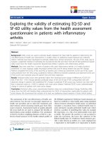
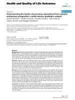
Data from scans and ex vivo well counting for both
hindlimbs are shown in Figure 2. For the WT non-dia-
betic group, the mean scan count ratio for L/R hin-
dlimbs was 1.91 ± 0.34 (range, 1.46 to 2. 79), and for the
WT diabetic group, it was 1.38 ± 0.26 (range, 1.05 to
1.74) (P < 0.001) (Figure 2A). The mean value for the
RAGE
-/-
non-diabetic group was 2.02 ± 0.29 (range, 1.54
to 2.62) not statistically significantly different from the
WT non-diabetic group. The mean value for the
RAGE
-/-
diabetic group was 1.75 ± 0.22 (range, 1.53 to
2.35) which was significantly lower than the RAGE
-/-
non-diabetic group (P < 0.001) and was significantly
higher than the WT diabetic group (P < 0.001).
Figure2Bshowsvaluesas%ID/gforthefourgroups
for the left and right hindlimbs. The counts in the left
(ischemic) hindlimbs showed the same patt ern of dif-
ferences among the four groups as shown f or the scan
ratios except for values for the WT diabetic and
RAGE
-/-
diabetic (1.42 and 1.43). However, the ratios
of L/R hindlimb %ID/g for RAGE
-/-
diabetic was
higher than for WT diabetic (2.85 ± 0.40 vs. 2.13 ±
0.67, P = 0.03) (Figure 2C). This difference is due to
lower mean %ID/g in the right limb for RAGE
-/-
dia-
betic group. For the remaining limb ratios for %ID/g
values, WT non-diabetic was significantly higher than
WT diabet ic (3.04 ± 0.95 vs. 2.13 ± 0.67, P =0.03)
and RAGE
-/-
non-diabetic was higher t han RAGE
-/-
diabetic (4.08 ± 1.00 vs. 2.85 ± 0.40, P = 0.02). All of
these significance levels for intergroup differences
were lower than for the scan data (Figure 2A) possibly
due to the technical challenge to cleanly dissect the
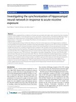
anterior tibialis muscles in the mouse. This limitation
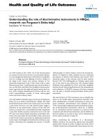
may have weakened the correlation for the plot o f the
ratios of L/R limbs against %ID/g (R = 0.059),
although the correlation is highly significant (P =
0.001) (Figure 3).
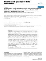
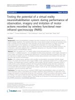
Histopathology
Examples of tissue sections stained for H&E, a
ν
, b
3
,and
lectin are shown in Figure 4A. Quanti tative lectin stain-
ing for capillaries from anterior tibialis muscle sections
(n = 20 per group) for both the left (ischemic) and right
(sham operated) hindlimbs of WT non-diabetic, WT
diabetic, RAGE
-/-
non-diabetic, and RAGE
-/-
diabetic are
Tekabe et al . EJNMMI Research 2011, 1:3
/>Page 3 of 9
shown in Figure 4B. Th e average capillary staining for
the WT non-diabetic left limbs was significantly lower
than the RAGE
-/-
non-diabetic left limbs (P =0.05)and
significantly higher than the WT diabetic left limbs (P <
0.001). The capillary staining for the WT diabetic left
limbs was borderline significantly lower than for the
RAGE
-/-
diabetic left limbs (P = 0.06). These histological
results support the scan findings. Co-staining of sections
for both endothelial cells and macrophage s showed
colocalization with a
ν
(Figure 5).
RAGE staining also supported the scan findings. There
was posit ive staining for RAGE in the ischemic sections
of hindlimbs from both diabetic and non-diabetic mice,
and no staining in the contralateral control limbs (Fig-
ure6).TheRAGE
-/-
mice both non-diabetic and dia-
betic showed no RAGE staining.
Discussion
In this study, we used radiolabeled RGD targeting integ-
rin expression and in vivo gamma imaging to look at
the effects of both diabetes and RAGE expression on
the a ngiogenic response to hindlimb isch emia in mice.
By measuring the ratio of tracer uptake in the ischemic
limb to the contralateral control limb, we were able to
show in live animals that in the absence of RAGE, the
angiogenic response to ischemia is ameliorated both in
diabetic and non-diabetic mice.
Diabetics have an attenuated angiogenic response to
tissue hypoxia which contributes to long-term complica-
tions including poor collateral formation in the heart
and in the lower extremiti es which is further aggravated
by poor wound healing and ulcers. Several factors have
been identified that contribute to this impaired angio-
genic response in diabetics which include maladaptive
regulation of vascular endothelial growth factor (VEGF)
ligand signaling [10-12], impaired release of endothelial
progenitor cells from the bone marrow [13], and defec-
tive function of the released cells [13,14]. Shoji and co-
workers using a matrigel patch model showed that the
RAGE system is involved in impaired angiogenesis in
diabetes [4].
Under hypoxic conditions, the expression of hypoxia
inducible factor (HIF-1) is increased which turns on
several genes incl uding genes that code for VEGF that
promote angiogenesis to restore perfusion and nor-
moxia in normal subjects. However, exogenous VEGF
has no effect to restore blood flow to diabetic mice
with limb ischemia and there is reduced downstr eam
VEGF signaling in diabetic animals [10-12]. Tamarat
and co-investigators proposed a mechanism involving
Figure 1 Representa tive scans from the four groups and the control peptide. Images from each of the four groups of mice injected with
99m
Tc cyclo-RGD and imaged on days 3 to 7 after left femoral artery ligation with mean values for ratios for L/R hindlimb below each image.
Image in the right shows a representative scan from an animal injected with control peptide. The yellow arrows point to the tracer uptake. The
color table shows the highest counts in red through purple to blue and green is background. The bladder is labeled.
Tekabe et al . EJNMMI Research 2011, 1:3
/>Page 4 of 9
inhibition of the matrix metalloproteinases (MMPs)
proteolytic enzymes that degrade the extracellular
matrix, a process that is necessary for the sprouting
capillaries as the neovascular mass grows [5]. Af ter 3
days of limb ischemia following femoral artery ligation,
MMP-2, MMP-3, and MMP-13 were increased in dia-
betic mice compared to controls, but collagenolysis
was decreased, indicating a suppression of the
response. Treatment with aminoguanidine, a thiamine
derivative known to inhibit three of the major bio-
chemical pathways in the pathways of angiogenesis
including AGE formation, restored the collagenolysis
process [5]. Tchaikovski and co-workers investigated
mechanisms whereby AGEs and RAGE expression
inhibit the response of circulating macrophages and
progenitor cells to promote angiogenesis in limb ische-
mia in diabetes and found activation of VEGFR-1-
related signal transduction pathways in monocytes
making them resistant to stimulation by VEGF-A [6].
Shen and co-workers showed that both diabetic and
Figure 2 Data from scans and ex vivo well counting for both hindlimbs.(A) Bars represent mean ± standard deviation values for the ratios
of left/right (L/R) hindlimbs from the scan count data. (B) Bars represent mean ± standard deviation values for %ID/g for both the left and right
legs. (C) Bars in graph represent mean ± standard deviation values for the ratios for %ID/g for L/R hindlimbs. WT, wild-type; DM, diabetes
mellitus; NDM, non-diabetes mellitus.
Figure 3 Correlation of ratio of counts in L/R hindlimb.From
ROIs drawn on the scans correlated with counts from ex vivo
gamma well counting of the muscles from the left and right
hindlimbs and corrected for decay and expressed as %ID/g of
tissue.
Tekabe et al . EJNMMI Research 2011, 1:3
/>Page 5 of 9
non-diabetic mice that received marrow transplanta-
tion from RAGE
-/-
donors had improved limb blood
flow at 28 days following ligation compared to mice
receiving marrow from RAGE
+/+
donors [8].
Integrins are cell adhesion receptors expressed on
endothelial cells, and a
v
b
3
integrin is responsible for
cell-cell interaction and the interaction between cells
and the extracellular matrix, processes that are neces-
sary for angiogenesis [15-18]. U pon activation of the
complex t ertiary structure, integrins unfold, revealing a
recognition site for the Arg-Gly-Asp (RGD) s equence
to bind extracellular matrix (ECM) proteins such as
vitronectin, fibrinogen, and fibronectin [19]. This
unique peptide binding site was used to develop linear
and cyclic peptides with RGD sequence to target a
v
b
3
integrin for imaging [19,20]. Because a
v
b
3
integrin is
expressed on both endothelial cells and monocyte/
macrophages and the inflammatory response to i sche-
mia is increased in diabet es, angiogenesis based on
uptake of
99m
Tc-HYNIC-RGD in the ischemic hin-
dlimbs may have been overestimated in the diabetic
mice. Nevertheless, the diabetic mice had significantly
lower uptake of
99m
Tc-HYNIC-RGD in the ischemic
limb compared to the non-diabetic mice, suggesting
that the binding to endothelial cells in this model had
the d ominant effect.
Using an RGD mimetic peptide (
99m
Tc-NC100692),
Hua and co lleagues imaged a
v
b
3
expression in a murine
limb ischemia model [21]. Integrin expression has also
been targeted for in vivo nuclear imaging in myocardial
infarction and remodeling and in response to VEGF
therapy in chronic low flow dysfunctional myocardium
[22,23]. Our study extends these reports to document
the value of this imaging approach to molecular path-
ways involved in diabetes.
Conclusions
We confirmed in live animals the role of RAGE expres-
sion to inhibit the angiogenic response to limb ischemia
in diabetes. B oth the diabetic and non-diabetic RAGE
-/-
mice showed improved angiogenesis compared to the
RAGE
+/+
mice (WT diabetic and non-diabetic) based on
Figure 4 Tissue sections stained for H&E, a
ν
, b
3
, and lectin.(A) An example of histological and immunohistochemi cal staining for anterior
tibialis muscle sections for a wild-type non-diabetic mouse. (B) The bar graph for quantitative lectin staining. Each bar represents average ± SD
of lectin-stained capillaries from sections of left anterior tibialis (ischemic limb) (light gray bars) and right anterior tibialis muscle (sham surgery)
(dark gray bars) for animals from each of the four groups.
Tekabe et al . EJNMMI Research 2011, 1:3
/>Page 6 of 9
Figure 5 Dual immunofluorescent staining for cells expressing a
v
in ischemic limb sections.Sitesofa
v
expression were shown to be
mainly endothelial cells based on colocalization of a
v
(Texas Red) with FVIII (green, fluorescein isothiocyanate) in the merged image.
Colocalization of a
v
with macrophages (Mac-3, fluorescein isothiocyanate) was also seen in the merged image. Areas in yellow represent
colocalization. EC, endothelial cells. (Magnification ×200).
Figure 6 Repre sentative section s of anterior tibialis muscles stained for RAGE (brown chromagen) and displayed at 20×.Theleftsetof
images shows sections from a left (L) ischemic hindlimb (top) and control right (R) limb (bottom) from a WT non-diabetic (NDM) mouse 7 days (D)
after femoral artery ligation. The center set of images shows sections from a left ischemic hindlimb (top) and control right limb (bottom) from a WT
diabetic (DM) mouse 7 days after femoral artery ligation. The right set of images shows sections from a left ischemic hindlimb from a RAGE
-/-
non-
diabetic mouse at day 7 after femoral artery ligation (top) and from a RAGE
-/-
diabetic mouse at day 7 after femoral artery ligation (bottom).
Tekabe et al . EJNMMI Research 2011, 1:3
/>Page 7 of 9
greater uptake of radiolabeled RGD targeting a
v
b
3
expression, a biomarker of tissue changes accompanying
early angiogenesis. While femoral artery occlusion in a
mouse is a simple model for limb ischemia compared to
theslowlyprogressivedisease in humans, the results of
this study support the value of radiolabeled RGD as a
non-invasive tool to follow the angiogenic response to
modifications in factors affecting t he angiogenic
response to tissue hypoxia.
Limitations
Planar imaging was used. As reported in a similar model,
planar imaging tends to underestimate relative uptake
within lower limbs as compared with SPECT and gamma
well counting [21]. Since all experiments were performed
the same way, differences among groups are probably not
affected; however, such an underestimation would
explain the slope of the regression line for the plot of %
ID from the scans vs. %ID/g from the tissue.
Abbreviations
AGEs: advanced glycation endproducts; DM: diabetes mellitus; FA: femoral
artery; MMPs: matrix metalloproteinases; NDM: non-diabetes mellitus; %ID/g:
percent injected dose per gram; ROI: region of interest; RAGE: receptor for
advanced glycation endproducts; VEGF: vascular endothelial growth factor;
WT: wild-type.
Acknowledgements
We thank Stan Majewski, Ph.D. from Jefferson Laboratories for loaning us the
dedicated small animal gamma camera and Geping Zhang for her assistance
in histology.
Author details
1
Department of Medicine, Columbia University Medical Center, New York, NY
10032, USA
2
Department of Surgery, Columbia University Medical Center,
New York, NY 10032, USA
3
Department of Medicine, New York University
Medical Center, New York, NY 10032, USA
4
Thomas Jefferson National
Accelerator Facility, Newport News, VA 23606, USA
5
Department of Nuclear
Medicine, Medical University of Innsbruck, Innsbruck, Austria
Authors’ contributions
YT prepared the tracers, performed the experiments, and revised the
manuscript. JL helped in the acquisition of data. DW provided the high-
resolution gamma imaging device. AMS and SFY developed the RAGE
-/-
animal model. RH provided us the RGD peptide. LJ has been involved in
designing the experiments, analysis and interpretation of data, and in
drafting and revising the manuscript.
Competing interests
The authors declare that they have no competing interests.
Received: 15 February 2011 Accepted: 7 June 2011
Published: 7 June 2011
References
1. Ouriel K: Peripheral arterial disease. Lancet 2001, 358:1257-1264.
2. Currie CJ, Morgan CL, Peters JR: The epidemiology and cost of inpatient
care for peripheral vascular disease, infection, neuropathy, and
ulceration in diabetes. Diabetes Care 1998, 21:42-48.
3. Scholz D, Ziebeglhoeff er T, Helisch A, Wagner S, Friedrich C, Podzuweit T,
Schaper W: Contribution of arteriogenesis and angiogenesis to post
occlusive hindlimb perfusion in mice. J Mol Cell Cardiol 2002,
34:775-787.
4. Shoji T, Koyama H, Morioka T, Tanaka S, Kizu A, Motoyama K, Mori K,
Fukumoto S, Shioi A, Shimogaito N, Takeuchi M, Yamamoto Y, Yonekura H,
Yamamoto H, Nishizawa Y: Receptor for advanced glygation endproducts
is involved in imapaired angiogenic response in diabetes. Diabetes 2006,
55:2245-2255.
5. Tamarat R, Silvestre J-S, Huijberts M, Benessiano J, Ebrahimian TG, Duriez M,
Wautier MP, Wautier JL, Lévy BI: Blockade of advanced glycation
endproduct formation restores ischemia-induced angiogenesis in
diabetic mice. Proc Natl Acad Sci 2003, 100:8555-8560.
6. Tchaikovski V, Olieslagers S, Bohmer FD, Waltenberger J: Diabetes mellitus
activates signal transduction pathways resulting in vascular endothelial
growth factor resistance of human monocytes. Circulation 2009,
120:150-159.
7. Shen X, Song F, Rosario R, Morales A, Zou YS, Schmidt AM, Yan SF: Abstract
5757: RAGE in bone marrow derived cells blunts restoration of blood
flow in mice subjected to femoral artery ligation. Circulation 2009, 120:
S1142.
8. Haubner R: α
v
β
3
-integrin imaging: a new approach to characterize
angiogenesis? Eur J Nucl Med Mol Imaging 2006, 33:S54-S63.
9. Liliensiek B, Weigand MA, Bierhaus A, Nicklas W, Kasper M, Hofer S,
Plachky J, Gröne HJ, Kurschus FC, Schmidt AM, Yan SD, Martin E,
Schleicher E, Stern DM, Hämmerling GG, Nawroth PP, Arnold B: Receptor
for advanced glycation endproducts (RAGE) regulates sepsis but not the
adaptive immune response. J Clin Invest 2004, 113:1641-1650.
10. Roguin A, Nitecki S, Rubinstein I, Nevo E, Avivi A, Levy NS, Abassi ZA,
Sabo E, Lache O, Frank M, Hoffman A, Levy AP: Vascular endothelial
growth factor (VEGF) fails to improve blood flow and to promote
collateralization in a diabetic mouse ischemic hindlimb model.
Cardiovascular Diabetology 2003, 2:1-6.
11. Hazarika S, Dokun AO, Li Y, Popel AS, Kontos CD, Annex BH: Impaired
angiogenesis after hindlimb ischemia in type 2 diabetes mellitus. Circ
Res 2007, 101:948-956.
12. Loomans CJ, de Koning EJ, Staal FJ, Rookmaaker MB, Verseyden C, de
Boer HC, Verhaar MC, Braam B, Rabelink TJ, van Zonneveld AJ: Endothelial
progenitor cell dysfunction: a novel concept in the pathogenesis of
vascular complications of type 1 diabetes. Diabetes 2004, 53:195-199.
13. Tepper OM, Galiano RD, Capla JM, Kalka C, Gagne PJ, Jacobowitz GR,
Levine JP, Gurtner GC: Human endothelial progenitor cells from type II
diabetics exhibit impaired proliferation, adhesion, and incorporation into
vascular structures. Circulation 2002, 106:2781-2786.
14. Sneider EB, Nowicki PT, Messina LM: Regenerative medicine in the
treatment of peripheral arterial disease. J Cell Biochem 2009, 108:753-761.
15. Tsou R, Isik F: Integrin activation is required for VEGF and FGF receptor
protein presence on human microvascular endothelial cells. Mol Cell
Biochem 2001, 224:81-89.
16. Heil M, Clauss M, Suzuki K, Buschmann IR, Willuweit A, Fischer S, Schaper W:
Vascular endothelial growth factor (VEGF) stimulates monocyte
migration through endothelial monolayers via increased integrin
expression. Eur J Cell Biol 2000, 79:850-857.
17. Senger DR, Ledbetter SR, Claffey KP, Papadopoulos-Sergiou A, Perruzzi CA,
Detmar M: Stimulation of endothelial cell migration by vascular
permeability factor/vascular endothelial growth factor through
cooperative mechanisms involving the α
v
β
3
integrin, osteopontin, and
thrombin. Am J Pathol 1996, 149:1.
18. Bayless KJ, Salazar R, Davis GE: RGD-dependent vacuolation and lumen
formation observed during endothelial cell morphogenesis in three-
dimensional fibrin matrices involves the α
v
β
3
and α
v
β
1
integrins. Am J
Pathol 2000, 156:1673-1683.
19. Haubner R, Wester HJ, Burkhart F, Senekowitsch-Schmidtke R, Weber W,
Goodman SL, Kessler H, Schwaiger M: Glycosylate RGD-containing
peptides: tracer for tumor targeting and angiogenesis imaging with
improved biokinetics. J Nucl Med 2001, 42:326-336.
20. Decristoforo C, Faintuch-Linkowski B, Rey A, von Guggenber g E,
Ruppricha M, Hernandez-Gonzales I, Rodrigo T, Haubner R: [
99m
Tc]
HYNIC-RGD for imaging integrin α
v
β
3
expression. Nucl Med Biol 2006,
33:945-952.
21. Hua J, Dobrucki LW, Sadeghi MM, Zhang J, Bourke BN, Cavaliere P, Song J,
Chow C, Jahanshad N, van Royen N, Buschmann I, Madri JA, Mendizabal M,
Sinusas AJ: Noninvasive imaging of angiogenesis with a
99m
Tc-labeled
peptide targeted at α
v
β
3
integrin after murine hindlimb ischemia.
Circulation 2005, 111:3255-3260.
Tekabe et al . EJNMMI Research 2011, 1:3
/>Page 8 of 9
22. Meoli DF, Sadeghi MM, Krassilnikova S, Bourke BN, Giordano FJ, Dione DP,
Su H, Edwards DS, Liu S, Harris TD, Madri JA, Zaret BL, Sinusas AJ:
Noninvasive imaging of myocardial angiogenesis following experimental
myocardial infarction. J Clin Invest 2004, 113:1684-1691.
23. Johnson LL, Schofield L, Donahay T, Bouchard M, Poppas A, Haubner R:
Radiolabeled arginine-glycine-aspartic acid peptides to image
angiogenesis in swine model of hibernating myocardium. J Am Coll
Cardiol Img 2008, 1:500-510.
doi:10.1186/2191-219X-1-3
Cite this article as: Tekabe et al.: Imaging the effect of receptor for
advanced glycation endproducts on angiogenic response to hindlimb
ischemia in diabetes. EJNMMI Research 2011 1:3.
Submit your manuscript to a
journal and benefi t from:
7 Convenient online submission
7 Rigorous peer review
7 Immediate publication on acceptance
7 Open access: articles freely available online
7 High visibility within the fi eld
7 Retaining the copyright to your article
Submit your next manuscript at 7 springeropen.com
Tekabe et al . EJNMMI Research 2011, 1:3
/>Page 9 of 9