Báo cáo hóa học: " New potential antitumoral fluorescent tetracyclic thieno[3,2-b]pyridine derivatives: interaction with DNA and nanosized liposomes" pptx
Bạn đang xem bản rút gọn của tài liệu. Xem và tải ngay bản đầy đủ của tài liệu tại đây (1.17 MB, 8 trang )
NANO EXPRESS Open Access
New potential antitumoral fluorescent tetracyclic
thieno[3,2-b]pyridine derivatives: interaction with
DNA and nanosized liposomes
Elisabete MS Castanheira
1*
, Maria Solange D Carvalho
1,2
, Ana Rita O Rodrigues
1
, Ricardo C Calhelha
2
and
Maria-João RP Queiroz
2
Abstract
Fluorescence properties of two new potential antitumoral tetracyclic thieno[3,2-b]pyridine derivatives were studied
in solution and in liposomes of DPPC (dipalmitoyl phosphatidylcholine), egg lecithin (phosphatidylcholine from
egg yolk; Egg-PC) and DODAB (dioctadecyldimethylammonium bromide). Compound 1, pyrido[2’,3’:3,2]thieno[4,5-d]
pyrido[1,2-a]pyrimidin-6-one, exhibits reasonably high fluorescence quantum yields in all solvents studied (0.20 ≤
F
F
≤ 0.30), while for compound 2, 3-[(p-methoxyphenyl)ethynyl]pyrido[2’ ,3’ :3,2]thieno[4,5-d]pyrido[1,2-a ]pyrimidin-6-
one, the values are much lower (0.01 ≤ F
F
≤ 0.05). The interaction of these compounds with salmon sperm DNA
was studied using spectroscopic methods, allowing the determination of intrinsic binding constants, K
i
= (8.7 ± 0.9)
×10
3
M
-1
for compound 1 and K
i
= (5.9 ± 0.6) × 10
3
M
-1
for 2, and binding site sizes of n = 11 ± 3 and n =7±2
base pairs, respectively. Compound 2 is the most intercalative compound in salmon sperm DNA (35%), while for
compound 1 only 11% of the molecules are intercalated. Studies of incorporation of both compounds in
liposomes of DPPC, Egg-PC and DODAB revealed that compound 2 is mainly located in the hydrophobic region of
the lipid bilayer, while compound 1 prefers a hydrated and fluid environment.
Introduction
Liposomes are among technological delivery develop-
ments for chemotherapeutic dr ugs in the treatment o f
cancer. This technique can potentially overcome many
common pharmacologic problems, such as those invol-
ving solubility, pharmacokinetics, in vivo stability and
toxicity [1-3]. Liposomes are closed spherical vesicles
consisting of a lipid bilayer that encapsulates an aqueous
phase in which hydrophilic drugs can be stored, while
water insoluble co mpounds can be incorporated i n the
hydrophobic region of the lipid bilayer [4].
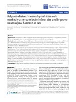
In this work, two new potential antitumoral fluorescent
planar tetracyclic thieno[3,2-b]pyridine derivatives 1 and 2
(Figure 1), previously synthesized by some of us [5], were
encapsulated in liposomes of DPPC (dipalmitoyl phospha-
tidylcholine), egg lecithin (phosphatidylcholine from egg
yolk) and DODAB (dioctadecyldimethylammonium
bromide). DPPC and egg lecithin [egg yolk phosphatidyl-
choline (Egg-PC)] are neutral components of biological
membranes, while cationic liposomes based on the syn-
thetic lipid DODAB have been used as vehicles for DNA
transfection and drug delivery [6]. These studies are
important keeping in mind future drug delivery applica-
tions using these compounds as anticancer drugs.
Due to the antitumoral potential of the two com-
pounds 1 and 2, related with their possible intercalation
between the DNA base pairs, interactions with natural
double-stranded salmon sperm DNA were studied.
These interactions can be assessed using spectroscopic
measurements, which are import ant tools for monitor-
ing DNA-binding processes. The investigation based on
DNA interactions has a key importan ce in order to
understand the mechanisms of action of antitumor and
antiviral drugs and to design new DNA-targeted drugs
[7,8]. Small molecules are stabilized on groove binding
and interca lation with DNA through a series of associa-
tive interactions such as π-stacking, hydrogen bonding,
attractive van der Waals and hydrophobic interactions
* Correspondence:
1
Centre of Physics (CFUM), University of Minho, Campus de Gualtar, Braga,
4710-057, Portugal
Full list of author information is available at the end of the article
Castanheira et al. Nanoscale Research Letters 2011, 6:379
/>© 2011 Castanheira et al; licensee Springer. This is an Open Access article distributed under the terms of the Creative Commons
Attribution License (http://creative commons.org/licenses/by/2.0), which permits unrestri cted use, distribution, and reproduction in
any medium, provided the original work is properly cited.
[8]. The occurrence of intercalation seems to be an
essential (but not sufficient) step for antitumoral act ivity
[7]. Fluorescence quenching experiments using external
quenchers are also very useful to distinguish between
DNA binding modes [9] since intercalated molecules are
less accessible to anionic quenchers due to electrostatic
repulsion with negatively charged DNA [10].
Experimental
Salmon sperm DNA from Invitrogen (Carlsbad, CA,
USA) and compounds stock solutions were prepared in
10 mM Tris-HCl buffer (pH = 7.4), with 1 mM EDTA.
The DNA concentration in number of b ases was deter-
mined from the molar absorption coefficient, ε =6600
M
-1
cm
-1
at 260 nm [11]. Fluorescence spectra of several
solutions with different [DNA]/[compound] ratios and
constant compound concentration (5 × 10
-6
M) were
recorded. The solutions were left several hours to
stabilize.
Dipalmitoyl phosphatidylcholine (DPPC), egg yolk
phosphatidylcholine (Egg-PC), from Sigma-Aldrich (St.
Louis, Missouri, USA), and dioctadecyldimethylammo-
nium bromide (DODAB), from Tokyo Kasei (Tokyo,
Japan), were used as received. Liposomes were prepared
by the ethanolic injection method, previously used for
the preparation of Egg-PC and DPPC liposomes [12-15]
and DODAB vesicles [16,17]. An ethanolic solutio n of a
lipid/compound mixture was injected in an aqueous
buffer solution under vigorous st irring, above the melt-
ing transition temperature of the lipid (approx. 41°C for
DPPC [18] and 45°C for DODAB [19]). T he final lipid
concentration was 1 mM, with a compound/lipid molar
ratio of 1:500. One millilitre solutions of liposome dis-
persions were placed in 3 mL disposable polystyrene
cuvettes for dynamic light scattering (DLS) measure-
ments in a M alvern ZetaSizer Nano ZS particle analyzer
(Worcestershire, UK). Five independent measurements
were performed for each sample. Malvern Dispersion
Technology Software (DTS) (Worcestershire, UK) was
used with multiple narrow mode (high resolution) data
processing, and mean size (nm) and error values were
considered.
Abso rption spectra were recorded in a Shimad zu UV-
3101PC UV-Vis-NIR spectrophotometer (Kyoto, Japan)
and fluorescence measurements were obtained in a
Fluorolog 3 spectrofluorimeter (HORIBA Scientific,
Kyoto, Japan) equipped wit h Glan-Thompson polarizers.
Fluorescence spectra were corrected for the instrumen-
tal response of the system. The fluorescence quantum
yields were determined by the standard method [20,21],
using 9,10-diphenylanthracene in ethanol as reference,
F
r
= 0.95 [22]. The solutions were previously bubbled
for 20 min with ultrapure nitrogen.
Results and discussion
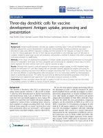
The size and size distribution of the liposomes prepared
was obtained by DLS. All the liposomes have a mean
hydrodynamic radius lower than 150 nm and generally
low p olydispersity. For Egg-PC and DODAB liposomes,
the size distributions are bimodals and broader than for
DPPC liposomes, the Egg-PC being the more polydi s-
perse (Figure 2). The ethanolic injection method was
described to produce phospholipid small unilamellar
vesicles (SV) [12-15]. Accordingly, DPPC and Egg-PC
liposomes obtained here are in this category, with a
mean diameter of around 90 nm for DPPC and 50 nm
for Egg-PC. DODAB liposomes exhibit a significantly
larger mean diameter (around 270 nm) than the phos-
pholipid ones. The size of DODAB vesicles strongly
depends on the preparation method, sonication and
ethanolic injection giving small DODAB vesicles
[17,23,24], while injection using chloroform yielded
large DODAB vesicles [16]. Besides, spontaneously pre-
pared DODAB liposomes have a much larger size
N
N
S
N
O
N
N
S
N
O
OCH
3
1
2
Figure 1 Structure of the compounds 1 and 2.
Figure 2 Size distributions obtained by dynamic light
scattering (DLS) for DPPC, Egg-PC and DODAB liposomes
prepared by the ethanolic injection method.
Castanheira et al. Nanoscale Research Letters 2011, 6:379
/>Page 2 of 8
(hydrodynamic radius around 337 nm [25]), being con-
sidered giant unilamellar vesicles (GUV). The DODAB
liposomes mean diameter obtained here (ca. 270 nm)
compares well with the reported value of 249 nm for
DODAB SV [16]. In all samples, no experimental evi-
dence of the presen ce of open bil ayer fragments (dia-
meter lower than 10 nm [17]) was obtained (Figure 2).
The absorption and fluorescence properties of com-
pounds 1 and 2 were studied in several solvents (Table
1). The normalized fluorescence spectra of compounds
1 and 2 are shown in Figures 3 and 4. The fluores-
cence emission maximum of both compounds displays
a loss of vibrational structure in polar solvents
together with a small red shift (Figures 3 and 4), i ndi-
cating some charge transfer character of the excited
state [26]. The red shifts are more significant for com-
pound 2 (Table 1), which may be due to a higher cap-
ability of this compound to establish hydrogen bonds
with protic solvents (especially with water), due to the
presence of the OCH
3
group. Compound 1 has signifi-
cantly higher fluorescence quantum yields (betw een 20
and30%)thancompound2 (F
F
between 1 and 5%),
showing that the functionalization of the pyridine ring
with a triple bond linked to a p-methoxyphenyl group
causes a significant enhance of the non-radiative deac-
tivation pathways. The fluorescence quantum yields of
compound 1 are also higher than the ones of a benzo
[b]thiophene derivative of the same type, a benzot hie-
nopyridopyrimidone [27], in which the benzene ring
linked to the thiophene is substituted in compound 1
by a pyridine ring. The intrinsic fluorescence of
compounds 1 and 2 can be used to monitor interac-
tions with DNA and compounds behaviour when
encapsulated in liposomes.
Both compounds 1 and 2 were tested for their interac-
tion with natural salmon sperm DNA using spectro-
scopic methods. For co mpound 1, fluorescence intensity
decreases with increasing DNA concentration, while the
opposite happens for compound 2 (Figures 5 and 6).
This behaviour, also previo usly observed for differently
substituted tetracyclic lactams [28], may indicate a dif-
ferent type of interaction of both compounds with the
DNA molecule. For the two compounds, full saturation
(corresponding to spectral invariance with increasing
DNA concentration) is attained at [DNA]/[compound]
= 200, meaning that total binding is achieved at this
ratio. The high [DNA]/[compound] ratio needed for
total binding, together with the negligible changes
observed in absorption spectra (not shown), point to a
weak interaction of these molecules with the nucleic
acid.
The intrinsic binding c onstants (K
i
) and binding site
sizes (n) were determined (Table 2) through the
McGhee and von Hippel modification of Scatchard plot
(Equation 1) [29],
r
c
f
= K
i
(
1 − nr
)
(
1 − nr
)
(
1 −
(
n − 1
)
r
)
n−
1
(1)
where K
i
is the intrinsic binding constant, n the bind-
ing site size, r the ratio c
b
/[DNA] and c
b
and c
f
the con-
centrations of bound and free compound, respectively,
calculated by
Table 1 Maximum absorption (l
abs
) and emission (l
em
) wavelengths, molar absorption coefficients (ε) and
fluorescence quantum yields of compounds 1 and 2 in several solvents
Solvent l
abs
(nm) (ε/10
4
M
-1
cm
-1
) l
em
(nm) F
F
121212
Cyclohexane 398 (0.84); 377 (1.24); 360 (1.27); 305
(0.95); 258 (3.93)
411 sh (0.33); 354 (2.19); 347 (2.37); 308 (1.25); 291
(1.12); 270 (1.40)
402; 426;
452 sh
417;
441
0.20 0.047
Dioxane 398 (0.76); 377 (1.18); 359 (1.20); 305
(1.17); 258 (3.60)
411 sh (0.66); 356 (5.36); 346 (5.40); 309 (3.23); 291
(2.98); 272 (3.33)
407; 428;
455 sh
425;
449
0.29 0.054
Dichloromethane 397 (0.58); 377 (0.91); 360 (0.93); 305
(0.97); 259 (2.70)
410 sh (0.55); 357 (4.37); 311 (2.28); 290 (2.29); 273
(2.78)
408; 429 427;
448
0.26 0.022
Acetonitrile 395 (0.68); 376 (1.06); 358 (1.06); 304
(1.09); 256 (3.32)
409 sh (0.66); 355 (5.76); 308 (3.41); 289 (3.20); 271
(3.67)
408; 428 450 0.21 0.036
N,N-
Dimethylformamide
a
397 (0.78); 377 (1.19); 360 (1.16); 305
(1.19)
411 sh (0.69); 356 (5.52); 311 (3.11); 290 (2.86) 411; 430 453 0.30 0.047
Dimethylsulfoxide
a
397 (0.77); 378 (1.17); 361 (1.14); 305
(1.17)
412 sh (0.61); 357 (4.70); 313 (2.52) 413; 432 455 0.28 0.048
Ethanol 396 (0.69); 375 (1.13); 358 (1.17); 304
(1.40); 256 (3.59)
408 sh (0.72); 355 (5.50); 311 (2.95); 272 (3.69) 412; 431 452 0.27 0.041
Methanol 395 (0.67); 374 (1.08); 358 (1.10); 304
(1.34); 256 (3.43)
408 sh (0.62); 354 (5.00); 311 (2.80); 272 (3.41) 413; 433 453 0.26 0.040
Water 394 (0.41); 374 (0.57); 361 (0.58); 303
(0.93); 256 (2.07)
420 sh (0.26); 358 (0.87); 314 (0.94); 278 (0.97) 413 sh; 433 505 0.22 0.012
a
Solvent cut-offs: N,N-Dimethylformamide: 275 nm; Dimethylsulfoxide: 280 nm; sh: shoulder.
Castanheira et al. Nanoscale Research Letters 2011, 6:379
/>Page 3 of 8
c
b
=
I
F,0
−
I
F
I
F
,
0
− I
F
,
b
× c
total
; c
total
= c
f
+ c
b
(2)
being I
F,0
the fluorescence intensity of the free com-
pound and I
F,b
the fluorescence intensity of the bound
compound at total binding. The binding constants
(Table 2) are moderately low, with a large number of
base pairs between consecutive intercalated compound
molecules (n).
Anionic quenchers can be useful in distinguishing
between DNA binding modes [9,10]. Compounds that
Figure 3 Normalized fluorescence s pectra (l
exc
= 360 nm) of compound 1 (4 × 10
-6
M) in several solvents; the inset shows the
absorption spectrum of 1 in dichloromethane (1 × 10
-4
M) as an example.
Figure 4 Normalized fluorescence s pectra (l
exc
= 360 nm) of compound 2 (4 × 10
-6
M) in several solvents; the inset shows the
absorption spectrum of 2 in dichloromethane (2 × 10
-5
M) as an example.
Castanheira et al. Nanoscale Research Letters 2011, 6:379
/>Page 4 of 8
are bound at the DNA surface (groove binding or elec-
trostatic binding) are more accessible and emission from
these molecules can be quenched more efficiently.
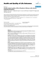
Fluorescence quenching measurements using iodide ion
showed that the usual Stern-Volmer plots (plots of the
fluorescence intensity ratio in the absence, I
0
,and
presence, I, of q uencher vs. quencher concentration) are
not linear and exhibit a downward curvature (Figure
7A). This indicates that some compound molecules are
not accessible to the anionic quencher, being interca-
lated between DNA base pairs. The modified Stern-Vol-
mer plot [30] ( Equation 3) allows the determination of
Figure 5 Fluorescence spectra of compound 1 (5 × 10
-6
M) in 0.01 M Tris-HCl buffer (pH = 7.2), with increasing DNA content.
Figure 6 Fluorescence spectra of compound 2 (5 × 10
-6
M) in 0.01 M Tris-HCl buffer (pH = 7.2), with increasing DNA content.
Castanheira et al. Nanoscale Research Letters 2011, 6:379
/>Page 5 of 8
the fraction of compound molecules accessible to
quencher,
I
0
I
=
1
f
a
+
1
f
a
K
SV
[Q]
(3)
where I
0
is the fluorescence intensity in the absence of
quencher, ΔI = I
0
- I, K
SV
the Stern-Volmer constant,
[Q] the quencher concentration and f
a
the fraction of
molecules accessible to quencher.
The representations of the modified Stern-Volmer plot
are reasonably linear (Figure 7B) and the f
a
values are in
Table 2. Both compounds exhibit some intercalation in
DNA, compound 2 being the more intercalative one,
with a lower fraction (65%) of molecules accessible to
anionic quencher. The higher hydrophobic character of
compound 2, promoted by the functionalization of the
pyridine with a triple bond linked to a p-methoxyphenyl
group, may justify this behaviour. As both compounds 1
and 2 are neutral molecules (and electrostatic interac-
tion with the negatively charged DNA molecule is not
expected), the h igh f
a
values indicate that the main type
of interaction with the nucleic acid must be the binding
to DNA grooves [28].
Fluorescence experiments of both compounds e ncap-
sulated in liposomes of DPPC, DODAB and Egg-PC
were carried out (Figure 8), in both gel (below T
m
)and
liquid-crystal line (above T
m
) phases. The melting transi-
tion temperature of Egg-PC is very low [31] and this
lipid is in the fluid liquid-crystalline phase at room tem-
perature. Fluorescence spectra of compound 1 incorpo-
rated in liposomes (Figure 8, Table 3) are roughly
similar to the one obtained in pure water , regarding the
band shape and maximum emission wavelength. Com-
pound 2 in liposomes presents emission spectra similar
to those in polar solvents, significantly blue-shifted rela-
tive to water. In Egg-PC, a band enlargement is
obse rved in the blue region, which can ind icate two di f-
ferent locations of compound 2 in these liposomes, one
deep in the hydrophobic region and another more close
to the lipid polar heads.
Fluorescence anisotropy (r) measurements (Table 3)
can give relev ant information about the location of the
compounds in liposomes, as r increases with the rota-
tional correlation time of the fluorescent molecule
(and, thus, with the viscosity of the fluorophore envir-
onment) [26]. Anisotropy values in a viscous solvent
(glycerol) were also determined, for comparison. Ani-
sotropy results (Table 3) allow to conclude that com-
pound 2 is mainly located in the inner region of the
lipid bilayer, feeling the penetration of some water
molecules. The transition from the rigid gel phase to
Table 2 Values of the intrinsic binding constants (K
i
) and
binding site sizes (n) and fraction of compound
molecules accessible to external quenchers (f
a
) for
interaction with salmon sperm DNA
Compound K
i
(M
-1
) nf
a
1 (8.7 ± 0.9) × 10
3
11 ± 3 0.89
2 (5.9 ± 0.6) × 10
3
7 ± 2 0.65
Figure 7 Stern-Volmer plots for quenching with iodide ion of compounds 1 and 2 for [DNA]/[compound] = 200 (A) and corresponding
modified Stern-Volmer plots (B).
Castanheira et al. Nanoscale Research Letters 2011, 6:379
/>Page 6 of 8
the liquid-crystalline phase is clearly detected by a sig-
nificant decrease in anisotropy at 55°C observed in
DPPC and DODAB liposomes. Compound 1 exhibits a
different behaviour and anisotropy is very low in all
types of liposomes (and much lower than in glycerol,
Table 3). Overall, the results indicate that compound 1
prefers a hydrated and fluid environment and the tran-
sition from the g el phase to the liquid-crystalline phase
is not detected. To further clarify the location of com-
pound 1, the solutions of liposomes with incorporated
compound were passed through filters of 0.05 μmdia-
meter. The fluorescence emission of the filtered solu-
tions was negligible, indicating that compound 1 is
mainly in the liposome aqueous interior or located at
the interfaces, with a very hydrated environment. This
behaviour is similar to the observed previously for a
benzothienopyridopyrimidone in lipid vesicles [27].
The encapsulation assays performed here may be
important for future drug delivery applications of these
potential antitumoral compounds using liposomes as
drug carriers.
Conclusions
The interaction with DNA of two new p otential antitu-
moral fluorescent pla nar thieno[3,2-b]pyridine deriva-
tives was studied using spectroscopic methods.
Compound 2 was shown to be the most intercalative
compound in salmon sperm DNA (35%). The binding to
DNA grooves seems to be the main type of interaction
with the nucleic acid. Studies of incorporation of both
compounds in liposomes of DPPC, Egg-PC and DODAB
revealed that compound 2 is mainly located in the
hydrophobic region of the lipid bilayer, while compound
1 prefers a hydrated and fluid environment. Our data
thus suggest that both potential antitumoral compounds
may be transported in lipo somes for drug delivery
applications.
Abbreviations
DLS: dynamic light scattering; DODAB: dioctadecyldimethylammonium
bromide; DPPC: dipalmitoyl phosphatidylcholine; DTS: Dispersion Technology
Software; Egg-PC: egg yolk phosphatidylcholine; GUV: giant unilamellar
vesicles; SV: small unilamellar vesicles.
Acknowledgements
This work was funded by FCT-Portugal through CFUM, CQ-UM, Project
PTDC/QUI/81238/2006 (cofinanced by program FEDER/COMPETE, ref.
Figure 8 Normalized fluorescence emission spectra of compounds 1 and 2 incorporated in liposomes of DPPC, Egg-PC and DODAB.
Table 3 Steady-state fluorescence anisotropy (r) values
and maximum emission wavelengths (l
em
) of compounds
1 and 2 incorporated in liposomes
Compound 1 Compound 2
l
em
/nm r l
em
/nm r
DPPC (25°C) 433 0.009 453 0.111
DPPC (55°C) 434 0.008 454 0.032
Egg-PC (25°C) 432 0.008 453 0.095
DODAB (25°C) 433 0.011 454 0.112
DODAB (55°C) 432 0.007 455 0.051
Glycerol (25°C) 437 0.166 472 0.202
Values in glycerol are also shown for comparison.
Castanheira et al. Nanoscale Research Letters 2011, 6:379
/>Page 7 of 8
FCOMP-01-0124-FEDER-007467) and PhD grants of M.S.D. Carvalho (SFRH/
BD/47052/2008) and R.C. Calhelha (SFRH/BD/29274/2006).
Author details
1
Centre of Physics (CFUM), University of Minho, Campus de Gualtar, Braga,
4710-057, Portugal
2
Centre of Chemistry (CQ-UM), University of Minho,
Campus de Gualtar, Braga, 4710-057, Portugal
Authors’ contributions
EMSC conceived the study, was responsible for its coordination and for the
interpretation of results, and drafted the manuscript. MSDC carried out the
liposome preparation and the fluorescence studies in liposomes. AROR
carried out the experimental studies of the compounds interaction with
DNA. RCC carried out the synthesis, purification and characterization of the
new compounds. MJRPQ supervised the organic synthesis and participated
in the draft of the manuscript. All authors read and approved the final
manuscript.
Competing interests
The authors declare that they have no competing interests.
Received: 28 September 2010 Accepted: 12 May 2011
Published: 12 May 2011
References
1. Andresen TL, Jensen SS, Jorgensen K: Advanced strategies in liposomal
cancer therapy: Problems and prospects of active and tumor specific
drug release. Prog Lipid Res 2005, 44:68.
2. Ochekpe NA, Olorunfemi PO, Ngwuluka NC: Nanotechnology and drug
delivery. Part 1: Background and applications. Tropical J Pharm Res 2009,
8:265.
3. Ochekpe NA, Olorunfemi PO, Ngwuluka NC: Nanotechnology and drug
delivery. Part 2: Nanostructures for Drug Delivery. Tropical J Pharm Res
2009, 8:275.
4. Malam Y, Loizidou M, Seifalian AM: Liposomes and nanoparticles:
nanosized vehicles for drug delivery in cancer. Trends Pharmacol Sci 2009,
30:592.
5. Calhelha RC, Queiroz M-JRP: Synthesis of new thieno[3,2-b]pyridine
derivatives by palladium-catalyzed couplings and intramolecular
cyclizations. Tetrahedron Lett 2010, 51:281.
6. Pedroso de Lima MC, Simões S, Pires P, Faneca H, Düzgünes N: Cationic
lipid-DNA complexes in gene delivery: from biophysics to biological
applications. Adv Drug Deliv Rev 2001, 47:277.
7. Lyne PD: Structure-based virtual screening: an overview. Drug Discovery
Today 2002, 7:1047.
8. Mahadevan S, Palaniandavar M: Spectroscopic and voltammetric studies
of copper(II) complexes of bis(pyrid-2-yl)-di/trithia ligands bound to calf
thymus DNA. Inorg Chim Acta 1997, 254:291.
9. Kumar CV, Asuncion EH: DNA-binding studies and site-selective
fluorescence sensitization of an anthryl probe. J Am Chem Soc 1993,
115:8547.
10. Kumar CV, Punzalan EHA, Tan WB: Adenine-thymine base pair recognition
by an anthryl probe from the DNA minor groove. Tetrahedron 2000,
56:7027.
11. Renault E, Fontaine-Aupart MP, Tfibel T, Gardes-Albert M, Bisagni E:
Spectroscopic study of the interaction of pazelliptine with nucleic acids.
J Photochem Photobiol B Biol 1997, 40:218.
12. Batzri S, Korn ED: Single bilayer liposomes prepared without sonication.
Biochim Biophys Acta 1973, 298:1015.
13. Kremer JMH, Esker MWJvd, Pathmamanoharan C, Wiersema PH: Vesicles of
variable diameter prepared by a modified injection method. Biochemistry
1977, 16:3932.
14. Nordlund JR, Schmidt CF, Dicken SN, Thompson TE: Transbilayer
distribution of phosphatidylethanolamine in large and small unilamellar
vesicles. Biochemistry 1981, 20:3237.
15. Cruz A, Casals C, Plasencia I, Marsh D, Pérez-Gil J: Depth profiles of
pulmonary surfactant protein B in phosphatidylcholine bilayers, studied
by fluorescence and electron spin resonance spectroscopy. Biochemistry
1998, 37:9488.
16. Tsuruta LR, Carmona-Ribeiro AM: Counterion effects on colloid stability of
cationic vesicles and bilayer-covered polystyrene microspheres. J Phys
Chem 1996, 100:7130.
17. Pacheco LF, Carmona Ribeiro AM: Effects of synthetic lipids on
solubilization and colloid stability of hydrophobic drugs. J Coll Interface
Sci 2003, 258:146.
18. Lentz BR: Membrane fluidity as detected by diphenylhexatriene probes.
Chem Phys Lipids 1989, 50:171.
19. Feitosa E, Barreleiro PCA, Olofsson G: Phase transition in
dioctadecyldimethylammonium bromide and chloride vesicles prepared
by different methods. Chem Phys Lipids 2000, 105:201.
20. Demas JN, Crosby GA: Measurement of photoluminescence quantum
yields - Review. J Phys Chem 1971, 75:991.
21. Fery-Forgues S, Lavabre D: Are fluorescence quantum yields so tricky to
measure? A demonstration using familiar stationery products. J Chem
Educ 1999, 76:1260.
22. Morris JV, Mahaney MA, Huber JR: Fluorescence quantum yield
determinations - 9,10-Diphenylanthracene as a reference-standard in
different solvents. J Phys Chem 1976, 80:969.
23. Feitosa E, Brown W: Fragment and vesicle structures in sonicated
dispersions of dioctadecyldimethylammonium bromide. Langmuir 1997,
13:4810.
24. Andersson M, Hammarström L, Edwards K: Effect of bilayer phase-
transitions on vesicle structure and its influence on the kinetics of
viologen reduction. J Phys Chem 1995, 99:14531.
25. Lopes A, Edwards K, Feitosa E: Extruded vesicles of
dioctadecyldimethylammonium bromide and chloride investigated by
light scattering and cryogenic transmission electron microscopy. J Coll
Interface Sci 2008, 322:582.
26. Valeur B: Molecular Fluorescence - Principles and Applications Weinheim:
Wiley-VCH; 2002.
27. Castanheira EMS, Pinto AMR, Queiroz MJRP: Fluorescence of a
benzothienopyridopyrimidone in solution and in lipid vesicles. J
Fluorescence 2006, 16:251.
28. Queiroz M-JRP, Castanheira EMS, Lopes TCT, Cruz YK, Kirsch G: Synthesis of
fluorescent tetracyclic lactams by a “one pot” three steps palladium-
catalyzed borylation, Suzuki coupling (BSC) and lactamization. DNA and
polynucleotides binding studies. J Photochem Photobiol A Chem 2007,
190:45.
29. McGhee JD, von Hippel PH: Theoretical aspects of DNA-protein
interactions - Cooperative and non-cooperative binding of large ligands
to a one-dimensional homogeneous lattice. J Mol Biol 1974, 86:469.
30. Lehrer SS: Solute perturbation of protein fluorescence - quenching of
tryptophyl fluorescence of model compounds and of lysozyme by
iodide ion. Biochemistry 1971, 10:3254.
31. Papahadjopoulos D, Miller N: Phospholipid model membranes. I.
Structural characteristics of hydrated liquid crystals. Biochim Biophys Acta
1967, 135:624.
doi:10.1186/1556-276X-6-379
Cite this article as: Castanheira et al.: New potential antitumoral
fluorescent tetracyclic thieno[3,2-b]pyridine derivatives: interaction with
DNA and nanosized liposomes. Nanoscale Research Letters 2011 6:379.
Submit your manuscript to a
journal and benefi t from:
7 Convenient online submission
7 Rigorous peer review
7 Immediate publication on acceptance
7 Open access: articles freely available online
7 High visibility within the fi eld
7 Retaining the copyright to your article
Submit your next manuscript at 7 springeropen.com
Castanheira et al. Nanoscale Research Letters 2011, 6:379
/>Page 8 of 8