Báo cáo hóa học: " Carbon composite micro- and nano-tubes-based electrodes for detection of nucleic acids" pot
Bạn đang xem bản rút gọn của tài liệu. Xem và tải ngay bản đầy đủ của tài liệu tại đây (886.89 KB, 5 trang )
NANO EXPRESS Open Access
Carbon composite micro- and nano-tubes-based
electrodes for detection of nucleic acids
Jan Prasek
1
, Dalibor Huska
2
, Ondrej Jasek
3
, Lenka Zajickova
3
, Libuse Trnkova
2
, Vojtech Adam
2
, Rene Kizek
2
and
Jaromir Hubalek
1*
Abstract
The first aim of this study was to fabricate vertically aligned multiwalled carbon nanotubes (MWCNTs). MWCNTs
were successfully prepared by using plasma enhanced chemical vapour deposition. Further, three carbon
composite electrodes with different content of carbon particles with various shapes and sizes were prepared and
tested on measuring of nucleic acids. The dependences of adenine peak height on the concentration of nucleic
acid sample were measured. Carbon composite electrode prepared from a mixture of glassy and spherical carbon
powder and MWCNTs had the highest sensitivity to nucleic acids. Other interesting result is the fact that we were
able to distinguish signals for all bases using this electrode.
Background
In the last two decades, nanomaterials in the form of
nanotubes and nanowires have begun to be reported as
promising materials for wide field of applications [1,2].
Such materials could be also used for fabrication of
miniaturized electrodes. The nanostructured electrodes
could be fabricated using several techniques. The easiest
fabrication technique is to use a mixture of nanomater-
ial as filler with a suitable vehicle, which could be
deposited on the electrode substrate using screen-print-
ing, drop-coating, dip-coating, spraying, etc. [3,4]. The
disadvantage of these nanocomposition-based electrodes
is the irreproducible electrode surface with undefined
active electrode area. The reproducible nanostructured
electrode surface could be fabricated using lithography
as a common tool for microelectronics devices imple-
mentation [5], anodization process for nanorods or
nanotubes crea tion [6,7]. One of these techniques is the
creation of vertically aligned multiwalled carbon nano-
tubes (MWCNTs) grown directly on the surface using
chemical vapour deposition (CVD) [8]. The aim of this
study was to fabricate MWCNTs and further to test the
particles as a part of carbon composite electrodes with
commercial carbon particles on detection of nucleic
acids.
Results and discussion
Fabrication of vertically aligned MWCNTs
Primarily, vertically aligned MWCNTs were prepared. A
detailed drawing of the current set-up of the apparatus
for plasma enhanced CVD MWCNTs direct deposition
is shown in Figure 1A. The apparatus consisted of a
micro-wave generator, working at a frequency of 2.45
GHz, with a standard rectangular waveguide, transmit-
ting the micro-wave power throug h a coaxial line to a
hollow nozzle electrode. Ferrite circulator protected the
generator from the reflected power by re-routing it to
the water load. The coaxial line and the nozzle electrode
accommodated a dual gas flow. The central conductor
of the coaxial line was held in place by boron nitride
ceramics. The outer conduct or was terminated by a
flange. A stub tuner was mounted to the waveguide for
load matching, and the reactive mixtur e of CH
4
/H
2
was
added by a concentric opening instead of the set of
holes in t he outer housing. The plasma torch was
enclosed by a quartz tube, 200 mm in length, with a
duralumin shielding wrapped around the tube. The dia-
meter of the quartz tube was 80 mm. The standard
deposition mixture consisted of argon (700 sccm),
methane (32 sccm) and hydrogen (255 sccm). Argon
passed through the centre, whereas methane/hydrogen
passed through the outer housing. The substrate for
MWNT growth, a piece of alumina w ith sensor struc-
ture, was fixed on the quartz holder at the variable dis-
tance from the torch nozzle. It was heated by a heat
* Correspondence:
1
Department of Microelectronics, Brno University of Technology, Technicka
10, CZ-61600 Brno, Czech Republic
Full list of author information is available at the end of the article
Prasek et al. Nanoscale Research Letters 2011, 6:385
/>© 2011 Prasek et al; licensee S pringer. This is an Open Access article distributed under the terms of the Creative Commons Attribution
License (http://creativecommons .org/license s/by/2.0), which permits unrestricted use, distribution, and repr oduction in any mediu m,
provided the original work is properly cited.
exchanger with hot gas and surface recombination. The
deposition temperature was 700°C. The deposition was
done on the pure silver layer without any catalyst and
on the 10-nm-thick Fe catalyst using the same underlay.
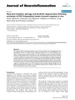
The SEM comparison of the e lectrode materials fabri-
cated on the Ag thick film paste with and without use
of catalyst is shown in Figure 1B. It clearly follows from
the results obtained that both pastes are covered with
vertically aligned MWCNTs.
Detection of nucleic acids
Further, three carbon composite electrodes with differ-
ent conte nt of carbon particles with various shapes and
sizes were prepared and tested on measuring of nucleic
acids. The first carbon composite electrode (called as
“microcarbon” ) was made of 90% “ glassy carbon pow-
der,” where the particles are from glassy carbon material
and they have spherical shape 2 μm(w/w,Sigma-
Aldrich, USA) and 10% mineral o il (m/w,Sigma-
Aldrich; free of DNase, RNase, and protease). The sec-
ond one (called as “ nanocarbon” )wasmadeof60%
“ glassy carbon powder” (w/w, Sigma-Aldrich), 30%
powdered cylinder carbon nanotubes (w/w,Sigma-
Aldric h) and 10% mineral oil (w/w, Sigma-Aldrich). The
third one was made of (called as “nanocarbon II”) 60%
“ glassy carbon powder” (w/w, Sigma-Aldrich), 30%
above prepared MWCNTs and 10% mineral oil. These
prepared materials were housed in a teflon body having
a 2.5-mm-diameter disk surface. Before measurements,
the electrode surface was renewed by polishing with a
soft filter paper in preparation for measurement [9-11],
which was ca rried out in the presence of 0.2 M acetate
buffer (5.0). The prepared carbon composite electrodes
were used in the following experiments, in which geno-
mic DNA isolated from salmon (genomic salmon DNA)
and oligonucleotide single strand from influenz a (ODN
influenza; 5’ -CAG TCG CAA GGA CTA ATC TGT
TTG-3’) were a nalysed. Carbon and/or graphite are of
particular interest but its voltammetric response is com-
plex as a result of its heterogeneous surface structure,
where it exhibits both edge and basal plane sites and,
depending upon how the graphite is aligned, the elec-
trode may be predominantly basal or edge plane in
character [12]. Numerous authors have been utilizing
AB
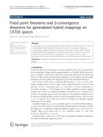
Figure 1 Prepara tion and characterization of MWCNTs.(A) Set-up of the apparatus for PECVD MWCNTs direct deposition (according to [8]).
(B) Surface-enhanced micrographs of the electrodes fabricated on the Ag-based thick film paste (1) without and (2) with use of 10 nm Fe
catalyst (Tescan, Czech Republic).
Prasek et al. Nanoscale Research Letters 2011, 6:385
/>Page 2 of 5
carbon nanotubes for the electro-oxidation of DNA [13].
The edge plane sites on graphite are generally accepted
to exhibit far greater rates of electron transfer as com-
pared to the basal plane sites. Further, the adsorption of
species on the graphite surf aces also differs at the two
sit es [14]. Therefore, the versatility of the electrode was
tested. Cyclic voltammetry was used for the detection of
two nucleic acids’ samples mentioned above and the
basic electrochemical behaviour of nucleic acids at the
surface of the above prepared carbon composite el ectro-
des were studied. It is known that that cytosine, adenine,
thymine and guanine give signals at carbon electrodes
[15-17]. We found that both the nucleic acid samples
gave all four signals corresponding to single bases at the
tested electrodes. Adenine gave the highest signal; how-
ever, the sequence of height of the other bases measured
on the electrodes differed. Guanine was the second-most
electroactive bases at nanocarbon electrode followed by
thymine and cytosine as w ell as at nanocarbon II
electrode, but the height of cytosine was higher com-
pared with thym ine. At the surface of microcarbon elec-
trode, the height of bases decreased in the following
order thymine, guanine and cytosine. These changes can
be associated with different surfaces and its affinity to
single bases, which can subsequently influence the redox
processes. To study the behaviour of bases on the sur-
face of the electrodes, the dependences of adenine peak
height on scan rate (50, 100, 200, 400, 600 and 800 mV/
s) were determined. The logarithmic dependences are
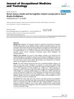
shown in Figure 2A-C for microcarbon, nanocarbon and
nanocarbon II electrodes, respectively. It clearly follows
from the results obtained that O DN influenza g ave
higher signal compared with genomic salmon DNA
except ODN influenza signal measured under 50 mV/s
at nanocarbon II. If we compared the sharpness of the
dependencies, then the sharpest were those measured at
nanocarbon II electrode followed by nanocarbon elec-
trode and microcarbon electrode. Moreover, the
10
7 9
ABC
m
i
crocarbon
nanocarbon
nanocarbon II
ODN influenza
ODN influenza ODN influenza
7
8
9
108
1.12
4
5
6
1.12
6
7
8
1.15
i
gnal (nA))
V
)
g
nal (nA))
g
nal (nA))
Genomic salmon DNA
Genomic salmon DNA Genomic salmon DNA
4
5
6
3.5 4.5 5.5 6.5 7.5
1
1.04
1
.
08
0 1,000
2
3
4
35
45
55
65
75
1.04
1.08
0 500 1000
3
4
5
35
45
55
65
75
1.00
1.05
1.10
0 500 1000
ln (s
i
Scan rate (mV/s)
Potential (
V
ln (si
g
Scan rate (mV/s)
Potential (V)
ln (si
g
Scan rate (mV/s)
Potential (V)
1 nA
Genomic salmon DNA – nanocarbon II
G
T
C
5 nA
A
Influenzas’ oligonucleotide – nanocarbon II
T
G
3
.
5
4
.
5
5
.
5
6
.
5
7
.
5
3
.
5
4
.
5
5
.
5
6
.
5
7
.
5
ln (scan rate (mV/s)) ln (scan rate (mV/s)) ln (scan rate (mV/s))
DE
3 A
1.23 1.27
E / V
300 nA
A
A
1.23 1.27
E / V
200 nA
C
1 A
T
0.95 1.05 1.15 1.25 1.35 1.45
E / V
0.76 0.80 0.84
E / V
10 A
30
A
0.95 1.05 1.15 1.25 1.35 1.45
E / V
0.76 0.80 0.84
E / V
0.5 0.75
1.0
1.25
1.5
scan
salmon
electrolyte
E/V
0.5 0.75 1.0 1.25 1.5
E / V
influenza
electrolyte
scan
E/V
Figure 2 Cyclic voltammetry. The dependences of adenine peak height on ln of scan rates (50, 100, 200, 400, 600 and 800 mV/s) measured at
(A) microcarbon, (B) nanocarbon and (C) nanocarbon II. In insets: dependencies of adenine peak potentials on ln of scan rate. Square wave
voltammetry. SW voltammograms of (D) single strand oligonucleotide influenza (13 μg/mL), and (E) double strand genomic DNA (15 μg/mL). In
insets: signals of all nucleic acid bases after baseline correction and smoothing of raw data.
Prasek et al. Nanoscale Research Letters 2011, 6:385
/>Page 3 of 5
dependencies of adenine peak potentials on scan rate
were determined and are shown in insets in Figure 2A-
C for microcarbon, nanocarbon and nanocarbon II elec-
trodes, respectively ( R
2
higher than 0.998). Based on
both the logarithmic and linear dependencies, we found
that the redox electrode process at all electrodes was
diffusion-limited. Moreover, b ased on the Randles-Sev-
cik equation for a reversible and diffusion-controllable
process, we estimated that the reaction exhibited nearly
heterogeneo us one-electron t ransfer. Moreov er, the
dependences of adenine peak height on concentration of
nucleic acid sample were measured. The dependences
were strictly linear for both nucleic acid samples with
R
2
higher than 0.996. The slo pe of the obtained curves
enhanced as follows: nanocarbon II > nanocarbon >
microcarbon. It clearly follows from the results obtained
that carbon composite electrode prepared from the mix-
ture of glassy and spherical carbon powder and
MWCNTs had the highest sensitivity to nucleic acids.
Based on these results, nanocarbon II electrodes w ere
further utilized for detection of both the nucleic acids’
samples (25 μg/mL) using square wave voltammetry
(not shown). We were interested in the issue whether
we could detect signals of all bases due to such high
sensitivity. Both the nucleic acids samples (15 μg/mL)
were detected, and all purine and pyrimidine bases sig-
nals were observed (Figure 2D, E, for ODN influenza
and genomic salm on DNA, respectively); peak potential
about G = 0.8 V; A = 1.05, T = 1.25 and C = 1.35 V.
Signals of the genomic DNA were high er (approx. 30%)
in comparison with the oligonucleotide. Another inter-
estingresultisthefactthatwewereabletoclearlydis-
tinguish signals for all bases by using baseline correction
and smoothing (insets in Figure 2D, E).
Conclusions
Based on these promising milestones of electroanalysis
of nucleic acids together with the fa ct that electrochem-
istry is still one of the most sensitive analytical techni-
que voltammetric methods can be considered as a
suitable tool for detection of nucleic acids. We show the
successfu l application of modern nano-technologies not
only for detecting of nucleic acids but also for distin-
guishing of all bases signals.
Methods
Chemicals
All chemicals of ACS purity used and parafilm were
purchased from Sigma-Aldrich Chemical Corp. (USA)
unless noted otherwise. Salmon sperm DNA was bought
from Applied Biosystems (USA). Synthetic oligonucleo-
tides, which were purified using high performance liquid
chromatography, were obtained from Sigma-Aldrich
with following sequence: influenza HPI 5’-CAG TCG
CAAGGACTAATCTGTTTG-3’.Stockstandard
solutions of the oligonucleotides (100 μg/mL) were pre-
pared with water of ACS purity (Sigma-Aldrich) and
stored in dark at -20°C. The concentrations of oligonu-
cleotides and DNA were determined spectrophotometri-
cally at 260 nm using spectrometer Specord 210
(Analytic Jena, Germany). Deionised water u nderwent
demineralization by reverse osmosis using the instru-
ment Aqua Osmotic 02 (Aqua Osmotic, Czech Repub-
lic), and then it was subsequently purified using
Millipore RG (Millipore Corp., USA, 18 MΩ) - MiliQ
water. The pH value was measured using inoLab con-
trolled by the personal computer program (MultiLab
Pilot; WTW, Germany).
Electrochemical analysis
Electrochemical measurements were performed using
AUTOLAB PGS30 Analyzer (EcoChemie, Netherlands)
connected to VA-Stand 663 (Metrohm, S witzerland),
using a standard cell with three electrodes. Carbon com-
posite electrodes were employed as the working elec-
trode. An Ag/AgCl/3 M KCl electrode served as the
reference el ectrode. Glassy carbon electrode was used as
the auxiliary electrode. For smoothing and baseline cor-
rection, the software GPES 4.9 supplied by EcoChemie
was employed. Cyclic voltammetric parameters were as
follows: potential step 5 mV; scan rates: 50, 100, 200,
400, 600 and 800 mV/s. Cyclic and square wave voltam-
metric measurements were carried out in the presence
of acetate buffer pH 5. Square wave voltammetry para-
meters: potential step 5 mV, frequency 280 Hz. The
samples measured by square wave voltammetry were
deoxygenated before me asurements by purging with
argon (99.999%) saturated with water for 120 s. The
temperature of supporting electrolyte was maintained by
the flow electrochemical cell coupled with thermostat
JULABO F12/ED (Labortechnik GmbH, Germany) and
was 25°C [18].
Abbreviations
CVD: chemical vapour deposition; MWCNTs: multiwalled carbon nanotubes.
Acknowledgements
The study was supported by the Czech grant projects GACR 102/09/P640,
NANIMEL GACR 102/08/1546 and GACR P205/10/1374.
Author details
1
Department of Microelectronics, Brno University of Technology, Technicka
10, CZ-61600 Brno, Czech Republic
2
Department of Chemistry and
Biochemistry, Mendel University in Brno, Zemedelska 1, CZ-61300 Brno,
Czech Republic
3
Department of Physical Electronics, Masaryk University,
Kotlarska 2, CZ-61137 Brno, Czech Republic
Authors’ contributions
JP prepared the screen-printed alumina substrates for MWCNT s deposition,
characterised MWCNTs using SEM and participated on paper drafting. DH
carried out electrochemical measurements. OJ physically prepared MWCNTs.
LZ participated on physically preparation of MWCNTs and on their
Prasek et al. Nanoscale Research Letters 2011, 6:385
/>Page 4 of 5
characterization. LT treated electrochemical data and participated in
preparation of the manuscript. VA participated in the design of the study
and performed the analysis of the data. RK conceived of the study, and
participated in its design. JH participated in design and coordination of the
study and drafted manuscript.
Competing interests
The authors declare that they have no competing interests.
Received: 27 October 2010 Accepted: 16 May 2011
Published: 16 May 2011
References
1. Sadaf JR, Israr MQ, Kishwar S, Nur O, Willander M: White
electroluminescence using ZnO nanotubes/GaN heterostructure light-
emitting diode. Nanoscale Res Lett 2010, 5:957-960.
2. Wang SQ, Li GH, Du GD, Li L, Jiang XY, Feng CQ, Guo ZP, Kim S: Synthesis
and characterization of cobalt-doped WS2 nanorods for lithium battery
applications. Nanoscale Res Lett 2010, 5:1301-1306.
3. Prasek J, Adamek M, Hubalek J, Adam V, Trnkova L, Kizek R: New
hydrodynamic electrochemical arrangement for cadmium ions detection
using thick-film chemical sensor electrodes. Sensors 2006, 6:1498-1512.
4. Albareda-Sirvent M, Merkoci A, Alegret S: Configurations used in the
design of screen-printed enzymatic biosensors. A review. Sens Actuator B
Chem 2000, 69:153-163.
5. Lisboa P, Valsesia A, Colpo P, Rossi F, Mascini M: Nanopatterned surfaces
for bio-detection. Anal Lett 2010, 43:1556-1571.
6. Jian SR, Chen YT, Wang CF, Wen HC, Chiu WM, Yang CS: The influences of
H-2 plasma pretreatment on the growth of vertically aligned carbon
nanotubes by microwave plasma chemical vapor deposition. Nanoscale
Res Lett 2008, 3:230-235.
7. Klosova K, Hubalek J: Advanced electrodes with nanostructured surfaces
for electrochemical microsensors. Phys Status Solidi A Appl Mater 2008,
205:1435-1438.
8. Zajickova L, Jasek O, Elias M, Synek P, Lazar L, Schneeweiss O, Hanzlikova R:
Synthesis of carbon nanotubes by plasma-enhanced chemical vapor
deposition in an atmospheric-pressure microwave torch. Pure Appl Chem
2010, 82:1259-1272.
9. Kizek R, Masarik M, Kramer KJ, Potesil D, Bailey M, Howard JA, Klejdus B,
Mikelova R, Adam V, Trnkova L, Jelen F: An analysis of avidin, biotin and
their interaction at attomole levels by voltammetric and
chromatographic techniques. Anal Bioanal Chem 2005, 381:1167-1178.
10. Masarik M, Kizek R, Kramer KJ, Billova S, Brazdova M, Vacek J, Bailey M,
Jelen F, Howard JA: Application of avidin-biotin technology and
adsorptive transfer stripping square-wave voltammetry for detection of
DNA hybridization and avidin in transgenic avidin maize. Anal Chem
2003, 75:2663-2669.
11. Petrlova J, Krizkova S, Supalkova V, Masarik M, Adam V, Havel L, Kramer KJ,
Kizek R: The determination of avidin in genetically modified maize by
voltammetric techniques. Plant Soil Environ 2007, 53:345-349.
12. Banks CE, Compton RG: Edge plane pyrolytic graphite electrodes in
electroanalysis: an overview. Anal Sci 2005, 21:1263-1268.
13. Erdem A: Nanomaterial-based electrochemical DNA sensing strategies.
Talanta 2007, 74:318-325.
14. Ji XB, Kadara RO, Krussma J, Chen QY, Banks CE: Understanding the
physicoelectrochemical properties of carbon nanotubes: current state of
the art. Electroanalysis 2010, 22:7-19.
15. Oliveira-Brett AM, Piedade JAP, Silva LA, Diculescu VC: Voltammetric
determination of all DNA nucleotides. Anal Biochem 2004, 332:321-329.
16. Adam V, Huska D, Hubalek J, Kizek R: Easy to use and rapid isolation and
detection of a viral nucleic acid by using paramagnetic microparticles
and carbon nanotubes-based screen-printed electrodes. Microfluid
Nanofluid 2010, 8:329-339.
17. Huska D, Adam V, Babula P, Hrabeta J, Stiborova M, Eckschlager T,
Trnkova L, Kizek R: Square-wave voltammetry as a tool for investigation
of doxorubicin interactions with DNA isolated from neuroblastoma cells.
Electroanalysis 2009, 21:487-494.
18. Huska D, Hubalek J, Adam V, Vajtr D, Horna A, Trnkova L, Havel L, Kizek R:
Automated nucleic acids isolation using paramagnetic microparticles
coupled with electrochemical detection. Talanta 2009, 79:402-411.
doi:10.1186/1556-276X-6-385
Cite this article as: Prasek et al.: Carbon composite micro- and nano-
tubes-based electrodes for detection of nucleic acids. Nanoscale
Research Letters 2011 6:385.
Submit your manuscript to a
journal and benefi t from:
7 Convenient online submission
7 Rigorous peer review
7 Immediate publication on acceptance
7 Open access: articles freely available online
7 High visibility within the fi eld
7 Retaining the copyright to your article
Submit your next manuscript at 7 springeropen.com
Prasek et al. Nanoscale Research Letters 2011, 6:385
/>Page 5 of 5