báo cáo hóa học: " Neuronal oxidative damage and dendritic degeneration following activation of CD14-dependent innate immune response in vivo" doc
Bạn đang xem bản rút gọn của tài liệu. Xem và tải ngay bản đầy đủ của tài liệu tại đây (651.55 KB, 7 trang )
BioMed Central
Page 1 of 7
(page number not for citation purposes)
Journal of Neuroinflammation
Open Access
Review
Neuronal oxidative damage and dendritic degeneration following
activation of CD14-dependent innate immune response in vivo
Dejan Milatovic, Snjezana Zaja-Milatovic, Kathleen S Montine, Feng-
Shiun Shie and Thomas J Montine*
Address: Department of Pathology, University of Washington, Harborview Medical Center, Seattle Washington 98104, USA
Email: Dejan Milatovic - ; Snjezana Zaja-Milatovic - ;
Kathleen S Montine - ; Feng-Shiun Shie - ;
Thomas J Montine* -
* Corresponding author
Abstract
The cause-and-effect relationship between innate immune activation and neurodegeneration has
been difficult to prove in complex animal models and patients. Here we review findings from a
model of direct innate immune activation via CD14 stimulation using intracerebroventricular
injection of lipopolysaccharide. These data show that CD14-dependent innate immune activation
in cerebrum leads to the closely linked outcomes of neuronal membrane oxidative damage and
dendritic degeneration. Both forms of neuronal damage could be blocked by ibuprofen and alpha-
tocopherol, but not naproxen or gamma-tocopherol, at pharmacologically relevant concentrations.
This model provides a convenient method to determine effective agents and their appropriate dose
ranges for protecting neurons from CD14-activated innate immunity-mediated damage, and can
guide drug development for diseases, such as Alzheimer disease, that are thought to derive in part
from CD14-activated innate immune response.
Introduction
Activated innate immunity is associated with several
degenerative and destructive brain diseases including
Alzheimer disease (AD), HIV-associated dementia (HAD),
ischemia, head trauma, stroke, cerebral palsy, and axonal
degeneration in multiple sclerosis [1]. In this complex
response, some aspects are proposed to be neurotrophic,
others neurotoxic, and each potentially a consequence
rather than a contributor to neurodegeneration. Indeed, a
severe limitation to understanding the precise role of
innate immunity in these diseases and their correspond-
ing animal models is that innate immunity is activated
simultaneously with multiple other stressors and
responses to injury, thereby greatly confounding any clear
conclusion about cause-and-effect relationships. For these
reasons we have adopted a simple but highly specific
model of isolated innate immune activation: intracere-
broventricular (ICV) injection of low dose lipopolysac-
charide (LPS).
LPS specifically activates innate immunity in peripheral
organs through a well-described Toll-like receptor (TLR)-
dependent signaling pathway [2,3]. There are 9 known
human plasma membrane-spanning TLRs expressed in
many cell types throughout the body that have been dis-
covered in the context of innate immune response to
micro-organisms. TLR-mediated innate immune response
can be considered in three phases: initial signal
Published: 21 October 2004
Journal of Neuroinflammation 2004, 1:20 doi:10.1186/1742-2094-1-20
Received: 6 October 2004
Accepted: 21 October 2004
This article is available from: />© 2004 Milatovic et al; licensee BioMed Central Ltd.
This is an open-access article distributed under the terms of the Creative Commons Attribution License ( />),
which permits unrestricted use, distribution, and reproduction in any medium, provided the original work is properly cited.
Journal of Neuroinflammation 2004, 1:20 />Page 2 of 7
(page number not for citation purposes)
transduction cascade, secondary signaling cascades, and
effectors. The initial signaling cascade starts with ligand
activating one of the 9 plasma membrane TLRs. All of
these receptors require the adaptor protein MyD88 for
immediate response to LPS and initiate a bifurcated signal
transduction cascade that culminates in altered gene tran-
scription, primarily via NF-κB activation but also through
c-Fos/c-Jun-dependent pathways. Some of the activated
gene transcripts encode directly for receptor ligands while
others are enzymes that catalyze the formation of receptor
ligands that in turn activate secondary autocrine and para-
crine signaling cascades. These signaling events culminate
in the generation of effector molecules including bacteri-
ocidal molecules, primarily free radicals generated by
NADPH oxidase and myeloperoxidase (MPO), as well as
cytokines and chemokines that can attract an adaptive
immune response. Although originally identified as part
of the response to exogenous antigens from micro-organ-
isms, a broader pathophysiologic role for TLR-dependent
signaling in response to endogenous ligands in now clear.
Indeed, from this perspective, the effectors at the culmina-
tion of these signaling pathways are more appropriately
viewed as cytocidal rather than specifically bacteriocidal.
The precise agents responsible for cytocidal activity are
not clearly established but likely include free radicals gen-
erated principally by NADPH oxidase, MPO, and induci-
ble nitric oxide synthase (iNOS) in combination with
cytokines and chemokines.
TLR-4 is the receptor for LPS in peripheral organs [2,3].
However, another protein, CD14 is critical to LPS activa-
tion of TLR-4. Membrane-anchored CD14 is now thought
to act a co-receptor for LPS but not to initiate intracellular
signaling cascades. It is important to note that CD14
serves a similar function with TLR-2, although the activat-
ing agents here are bacterial products other than LPS [4].
Within minutes to hours of exposure to LPS, there is
increased gene transcription and subsequent translation
of cytokines and chemokines, prominently including
tumor necrosis factor, interleukin-1, and interferons, as
well as several enzymes; important among these are iNOS
and cyclooxygenase 2 (COX-2) that catalyze the forma-
tion of NO and prostaglandin (PG) H
2
, respectively [4].
While NO is a potent cell signaling molecule, PGH
2
has
relatively low receptor binding affinity but is rapidly and
efficiently converted to multiple PGs or thromboxane A2,
each of which are potent activators of a large family of G
protein-coupled receptors [5]. The combination of these
initial and secondary signaling cascades produces a robust
innate immune response. This same response can occur in
response to endogenous ligands that also activate the
CD14/TLR-4 pathway [2,3]. Indeed, several endogenous
CD14/TLR ligands have received increasing attention for
their potential roles in human diseases [6], and polymor-
phisms in TLR-4 are associated with risk for atherosclero-
sis and asthma, as well as other human diseases [7]. With
respect to AD, amyloid beta (A ) fibrils have been shown
to activate the microglial innate immune response
through CD14-dependent mechanisms [8]. Relevant to a
broader range of neurodegenerative diseases, novel pep-
tides and neoantigens exposed by apoptotic cells [9] also
activate CD14-dependent innate immune response in
macrophages. While none of these data point to CD14 or
innate immune response as etiological in neurodegenera-
tive disorders, these findings from in vitro and cell culture
experiments raise the possibility that CD14-dependent
signaling may be a common process shared in the patho-
genesis of neurodegenerative diseases, especially AD.
Here we present our results from studies that have identi-
fied the molecular and pharmacologic determinants of
ICV LPS-initiated cerebral neuronal damage in vivo. It is
important to stress that several laboratories have shown
that glia, predominantly microglia, are activated by LPS
but that neurons do not respond to LPS because they lack
the appropriate receptors [10,11]. We measured two main
endpoints; one biochemical and one structural. Since free
radicals are a primary mechanism of cytocidal activity
from innate immune response, we used a stable isotope
dilution method with gas chromatography and negative
ion chemical ionization mass spectrometry to quantify
compounds formed by free radical attack on the neuronal
membrane-enriched fatty acid, docosohexaenoic acid
(DHA); we have termed these molecules F
4
-neuropros-
tanes (F
4
-NeuroPs) [12]. In addition to this biochemical
marker of neuronal oxidative damage, we directly quanti-
fied neuron number as well as dendrite length and spine
density in pyramidal neurons of hippocampal sector CA1
using the Golgi impregnation technique followed by
quantitative morphometry with Neurolucida (Micro-
BrightField, VT) [13].
Lack of adaptive immune response, fever, or structural
damage to brain following ICV LPS
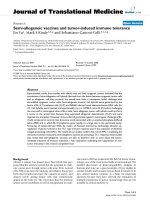
Despite the expectation that LPS would produce a febrile
response with widespread damage to brain and an acute
encephalitis, we observe that ICV LPS does not yield any
of these outcomes (Figure 1) [14]. Indeed, others who
injected similar amounts of LPS directly into brain paren-
chyma also do not observe behavioral changes, tissue
damage, or acute inflammatory infiltrate in young wild
type (wt) mice [14-18]. We pursued this further by stereo-
logical counting of hippocampal CA1 pyramidal neurons
24 and 72 hr following ICV LPS and observed no change
in neuron number from untreated controls [14]. These
data show that, at least over 3 days following ICV LPS,
there is no gross structural damage to brain, no detectable
adaptive immune response, and no loss of pyramidal neu-
rons from hippocampal sector CA1.
β
Journal of Neuroinflammation 2004, 1:20 />Page 3 of 7
(page number not for citation purposes)
Neuronal oxidative damage
Numerous methods exist to determine free radical-medi-
ated damage to cells. While most of these function well in
vitro, important limitations arise in living systems where
extensive, highly active enzymatic pathways have evolved
to metabolize many of the commonly measured products,
such as 4-hydroxynonenal [19]. One method that has
been highly replicated as a robust quantitative means of
measuring free radical damage in vivo is measuring F
2
-iso-
prostanes (F
2
-IsoPs) [20], products generated from free
radical damage to arachidonic acid (AA), that are not
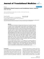
extensively metabolized in situ (Figure 2). Since AA is
present throughout brain and in different cells in brain at
roughly equal concentrations, measurement of cerebral
F
2
-IsoPs, like all other measures of oxidative damage,
reflects damage to brain tissue but not necessarily to neu-
rons. For these reasons, we developed an assay to measure
the analogous products generated from DHA, F
4
-NeuroPs
[12]. Since DHA is highly concentrated in neuronal mem-
branes, F
4
-NeuroPs offer a unique window into free radi-
cal damage to neuronal membranes in vivo [21].
We first determined the time course of F
4
-NeuroP accu-
mulation in cerebrum of wt mice exposed to ICV LPS and
observed a delayed, transient elevation that peaks at
approximately 24 hr after exposure and then returns to
baseline by 72 hr post exposure [14]. It is important to
note that while detectable neuronal oxidative damage is
delayed several hours following ICV LPS, others have
shown that altered gene transcription and increased
cytokine secretion occur rapidly and peak within a few
hours of LPS exposure. As with oxidation of lipoproteins,
it is likely that this delay in neuronal oxidative damage is
related, at least in part, to the time required to deplete
anti-oxidant defenses. Thus, despite the lack of tissue
damage, adaptive immune cell infiltrate, or detectable
neuron loss, there is significant, reversible free radical
damage to neuronal membranes following ICV LPS.
NeuN immunohistochemistry of mouse hippocampusFigure 1
NeuN immunohistochemistry of mouse hippocampus. Phot-
omicrograph (× 40) of NeuN immunoreactivity in mouse hip-
pocampus and adjacent structures 24 hr after ipsilateral ICV
LPS injection. Note normal density and distribution of neu-
rons without a cellular infiltrate.
Diagram showing the formation of F
2
-IsoPs and F
4
-NeuroPsFigure 2
Diagram showing the formation of F
2
-IsoPs and F
4
-NeuroPs.
AA (20:4ω6) in all cells DH A (22:6ω3) concentrated in neurons
Free Radical Attack and O
2
Insertion
OH
OH
OH
COOH
OH
OH
OH
COOH
F
2
-Iso P F
4
-N eu roP
Journal of Neuroinflammation 2004, 1:20 />Page 4 of 7
(page number not for citation purposes)
We next used a series of mice, all on the C57Bl/6 genetic
background, lacking specific genes to establish the deter-
minants of neuronal oxidative damage in this model. Our
results showed that genetic ablation of one co-receptor
(CD14), the required adaptor (MyD88), or one arm of the
initial signal cascade (the p50 subunit of NF-κB) each
completely blocks an LPS-induced increase in cerebral F
4
-
NeuroPs (Table 1). Further investigation of mice lacking
iNOS, an element of secondary signaling pathways, also
completely blocks ICV LPS-induced neuronal oxidative
damage. Finally, mice lacking prostaglandin E
2
receptor
subtype 2 (EP2), one of four prostaglandin E
2
(PGE
2
)
receptors expressed in brain and one of the two PGE
2
receptors expressed by microglia, have no neuronal oxida-
tive damage in response to ICV LPS [16]. There are some
important points to consider when interpreting these
data. First, not only glia but neurons also will be exposed
to LPS in this model. However, we and others have repeat-
edly shown that primary neurons enriched in cell culture
do not respond to LPS [10,11,22-24]; indeed, neurons do
not express CD14 and TLR-4 in vivo [25,26]. Second,
genetic ablation was not specific to cell type. While this
limits interpretation of data from some mice, such as p50
-/- and EP2-/- mice because these proteins are expressed
by both neurons and glia [27-32], it does not influence
interpretation of data from CD14 -/- mice because CD14
expression in vivo is restricted to microglia among paren-
cymal cells in brain [25,26]. Thus, these data strongly
imply that LPS-activated microglial-mediated paracrine
oxidative damage to neurons in vivo is dependent on
CD14, MyD88, p50 of NF-κB, iNOS, and EP2.
Dendritic degeneration
These data left us with an apparent conflict. We have
clearly demonstrated neuronal oxidative damage to
mouse cerebrum following ICV LPS that is of a magnitude
comparable to diseased regions of AD brain [33]. How-
ever, there is no apparent structural damage to brain in
our study or in others' following ICV or intraparenchymal
LPS. We viewed this as a serious potential challenge to the
significance of oxidative damage in neurodegeneration.
There are differences, of course, between the acute stress of
ICV LPS stress and the presumably chronic stress of AD;
nevertheless, these data force at least consideration of the
question: could oxidative damage to neurons occur in vivo
to the extent that is observed in AD brain without any
neurodegeneration?
Table 1: Neuronal oxidative damage and dendritic degeneration in various knockout mice. Effects of ICV LPS treatment determined
at 24 hr in mice homozygous deficient (knockout) for different genes or wildtype (wt) mice all on the C57Bl/6 genetic background (*P
< 0.001 by Bonferroni-corrected repeated pair comparisons with ICV saline-exposed mice).
Knockout Function Endpoints*
F
4
-NeuroPs Dendrite Length Spine Density
None (wt) N/A 352 + 53* 32 + 4* 37 + 6*
CD14 Receptor 87 + 14 101 + 8 92 + 11
TLR-2 Receptor 37 + 5* 51 + 8*
MyD88 Adaptor 98 + 10 96 + 9 102 + 7
p50 Initial Signal Cascade 108 + 11 105 + 7 106 + 10
iNOS Secondary Signaling 92 + 12 103 + 8 97 + 6
EP2 Secondary Signaling 89 + 9 102 + 12 109 + 5
*% ICV saline-exposed; n > 5 in each group
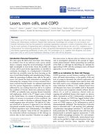
Dendritic degeneration of CA1 pyramidal neurons in mouse hippocampusFigure 3
Dendritic degeneration of CA1 pyramidal neurons in mouse
hippocampus. Neurolucida renderings of CA1 pyramidal neu-
rons stained by Golgi method; blue is soma and first order
dendrites, red is second order dendrites, green is third order
dendrites, yellow is fourth order dendrites, brown is fifth
order dendrites, and pink is sixth order dendrites. A. Typical
pyramidal neuron 24 hr after ipsilateral ICV Saline injection. B
and C. Pyramidal neurons following ipsilateral ICV LPS injec-
tion showing moderate (B) to severe (C) dendrite shortening
and spine loss.
Journal of Neuroinflammation 2004, 1:20 />Page 5 of 7
(page number not for citation purposes)
To address this question, we decided to examine directly
the dendritic compartment of neurons, which is largely
transparent to the standard histological techniques used
so far to investigate ICV LPS-induced damage. Using Golgi
impregnation and Neurolucida-assisted morphometry of
hippocampal CA1 pyramidal neurons [13], we first deter-
mined the time course of dendritic structural changes
following ICV LPS in wt mice. Our results show a time
course similar to neuronal oxidative damage with maxi-
mal reduction in both dendrite length and dendritic spine
density at approximately 24 hr post LPS and, remarkably,
a return to near baseline levels by 72 hr [14] (Figure 3).
We next pursued the molecular determinants of ICV LPS-
induced dendritic degeneration using the same genetically
altered mice that we used above (Table 1). We observed
perfect concordance between these results in that lack of a
gene that protected cerebrum from neuronal oxidative
damage also protected hippocampal CA1 pyramidal neu-
rons from dendritic degeneration and vice versa [14].
Importantly, we had the opportunity to add TLR-2 knock-
out mice to our analysis. TLR-2, like TLR-4, is one of the
plasma membrane TLRs that may be activated by LPS and
that also uses CD14 as a co-receptor. Our results show that
lack of TLR-2 does not protect hippocampal CA1 pyrami-
dal neurons from ICV LPS-induced neurodegeneration,
while lack of CD14 completely protects the dendritic tree
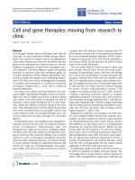
of these neurons. Further, it is interesting to note that in
mice receiving ICV saline, pyramidal neuron dendrite
length (Figure 4), but not spine density, is significantly
greater in CD14-/- mice than in wt or MyB88-/- mice, sug-
gesting that even in the absence of specific stimuli like ICV
LPS, lack of CD14 perhaps has a net neuroprotective or
neurotrophic effect.
Pharmacologic interventions
Considerable controversy surrounds the effective in vivo
neuroprotective doses of nonsteroidal anti-inflammatory
drugs and anti-oxidants that are being evaluated as poten-
ital protectants from AD. Indeed, a major criticism leveled
against nonsteroidal anti-inflammatory drugs (NSAIDs) is
that the concentrations that appear to be neuroprotective
in epidemiologic studies are lower than those that classi-
cally considered anti-inflammatory doses. Moreover,
there is some data suggesting that some NSAIDs, such as
ibuprofen and naproxen, that may differ in their effective-
ness as AD protectants despite being equivalent anti-
inflammatory agents in peripheral assays of inflammation
suggesting alternative mechanisms of action in AD [34].
Therefore, we determined the dose-response relationship
for ibuprofen and naproxen in our ICV LPS model utiliz-
ing a two-week pre-treatment with each NSAID in drink-
ing water (with concentration expressed as µg/ml
drinking water) followed by ICV LPS injection [14]. Nei-
ther NSAID alone alters basal levels of cerebral F
4
-Neu-
roPs. For ibuprofen, the EC
50
for suppressing ICV LPS-
induced F
4
-NeuroPs is between 0.1 and 0.5 µg/ml and the
maximal effect is reached by 1.4 µg/ml, considerably
lower than the classic anti-inflammatory dose. In contrast,
naproxen is without effect up to 1.4 µg/ml and thus an
EC
50
cannot be calculated from these data. As with F
4
-
NeuroPs, ibuprofen completely protects both dendrite
length and spine density (Figure 5) from the degenerative
consequences of ICV LPS; in contrast, naproxen is not sig-
nificantly protective even at the highest dose. These results
are intriguing because some have suggested that ibupro-
fen may be more effective than naproxen in lowering the
risk for AD [34]. The basis for the differing results with
these NSAIDs in our experiments are not entirely clear but
may derive from pharmacokinetic differences or pharma-
codynamic differences in actions other than COX
inhibition.
Next, we extended our studies to tocopherols, natural
antioxidant products with a number of proposed actions
[35] including both anti-oxidant and anti-inflammatory
activities [36]. As with NSAIDs, α-tocopherol (AT) or γ-
tocopherol (GT) alone does not alter basal F
4
-NeuroP lev-
els or dendritie arbor (not shown). AT partially suppresses
ICV LPS-induced F
4
-NeuroPs at 10 mg/kg and completely
suppresses F
4
-NeuroP formation and both reduction in
dendrite length and reduction in spine density at 100 mg/
kg (Figure 5). GT, an isomer of AT that has one-tenth its
Dendritic arbor in CA1 pyramidal neurons of hippocampus from knockout miceFigure 4
Dendritic arbor in CA1 pyramidal neurons of hippocampus
from knockout mice. Adult (6 to 8 week old) wt C57Bl/6,
CD14-/-, or MyD88-/- mice received ICV saline 24 hr prior
to sacrifice. Tissue sections of hippocampus and surrounding
structures were processed for Golgi stain and then evaluated
by Neurolucida. Data are dendrite length for CA1 hippocam-
pal pyramidal neurons (n > 15 neurons for each group). One-
way ANOVA had P < 0.0001 with Bonferroni-corrected
repeated pair comparisons having *P < 0.001 for wt vs.
CD14-/- and CD14-/- vs. MyD88-/
Journal of Neuroinflammation 2004, 1:20 />Page 6 of 7
(page number not for citation purposes)
anti-oxidant activity in vitro and lacks a specific trans-
porter in vivo, does not, as expected, protect from neuro-
nal oxidative damage or dendritic degeneration at the
same dose.
Conclusions
Our data show that CD14-dependent activation of cere-
bral innate immunity leads to an acute, transient increase
in oxidative damage to neuronal membranes that coin-
cides with reversible dendritic degeneration. Although we
did not directly test TLR-4 deficient mice in our studies,
given what is know about LPS receptor activation and the
fact that TLR-2-/- mice were not protected from neuronal
damage caused by ICV LPS, these data argue strongly for
CD14/TLR-4-dependent neuronal damage in our model.
Moreover, using a wide array of genetically altered mice,
we observed complete concordance between dendritic
degeneration and neuronal membrane oxidative damage.
In combination, these data suggest that these two events
are mechanistically related, perhaps with neuronal
membrane oxidative damage being a proximate contribu-
tor to dendritic degeneration in the context of innate
immune activation.
One obvious, commonly voiced criticism of the model
described here is that it produces an acute stress that does
not correspond to chronic neurodegenerative diseases.
However, it has yet to be shown whether the stress to
individual neurons in these protracted diseases truly is
chronic or instead the integration of innumerable micro-
scopic acute stresses over many years. Finally, to the extent
that CD14-dependent innate immunity activation
contributes to neurodegenerative diseases, such as AD and
HAD, the model described here provides a convenient
means to screen experimental therapeutics and rapidly
optimize dosing and timing parameters before moving to
more complex animal models or clinical trials.
List of abbreviations used
AA: arachidonic acid; AD: Alzheimer disease; AT: α-toco-
pherol; Aβ: amyloid beta; COX-2: cyclooxygenase 2; DHA:
docosohexaenoic acid; EP2: prostaglandin E
2
receptor
subtype 2; F
2
-IsoPs: F
2
-isoprostanes; F
4
-NeuroPs: F
4
-neu-
roprostanes; GT: γ-tocopherol; HAD: HIV-associated
dementia; ICV: intracerbroventricular; iNOS: inducible
nitric oxide synthase; LPS: lipopolysaccharide; MPO: mye-
loperoxidase; NSAIDs: nonsteroidal anti-inflammatory
drugs; PG: prostaglandin; PGE
2
: prostaglandin E
2
; TLR:
Toll-like receptor; wt: wild type.
Competing Interests
The authors declare that they have no competing interests.
Acknowledgements
This work was supported by the Alvord Endowed Chair in Neuropathology
as well as grants from the NIH including AG05144, AG05136, and
AG24011.
References
1. Polazzi E, Contestabile A: Reciprocal interactions between
microglia and neurons: from survival to neuropathology. Rev
Neurosci 2002, 13:221 -2242.
2. Imler JL, Hoffmann JA: Toll receptors in innate immunity. Trends
Cell Biol 2001, 11:304-311.
3. Akira S: Toll-like receptor signaling. J Biol Chem 2003,
278:38105-38108.
4. Palsson-McDermott EM, O'Neill LA: Signal transduction by the
lipopolysaccharide receptor, Toll-like receptor-4. Immunology
2004, 113:153-162.
5. Hata AN, Breyer RM: Pharmacology and signaling of prostag-
landin receptors: Multiple roles in inflammation and
immune modulation. Pharmacol Ther 2004, 103:147-166.
6. Johnson GB, Brunn GJ, Platt JL: Activation of mammalian Toll-
like receptors by endogenous agonists. Crit Rev Immunol 2003,
23:15-44.
7. Cook DN, Pisetsky DS, Schwartz DA: Toll-like receptors in the
pathogenesis of human disease. Nat Immunol 2004, 5:975-979.
8. Fassbender K, Walter S, Kuhl S, Landmann R, Ishii K, Bertsch T, Stal-
der AK, Muehlhauser F, Liu Y, Ulmer AJ, Rivest S, Lentschat A, Gul-
bins E, Jucker M, Staufenbiel M, Brechtel K, Walter J, Multhaup G,
Penke B, Adachi Y, Hartmann T, Beyreuther K: The LPS receptor
Pharmacologic suppression of dendritic degeneration in CA1 pyramidal neurons of mouse hippocampusFigure 5
Pharmacologic suppression of dendritic degeneration in CA1
pyramidal neurons of mouse hippocampus. Adult (6 to 8
week old) wt C57Bl/6 mice received ICV saline or ICV LPS
24 hr prior to sacrifice. Tissue sections of hippocampus and
surrounding structures were processed for Golgi stain and
then evaluated by Neurolucida. Data are dendritic spine den-
sity for CA1 hippocampal pyramidal neurons (n > 6 neurons
for each group). Two-way ANOVA had P < 0.001 for ICV
saline vs. ICV LPS, effect of drugs, and interaction. Post hoc
one-way ANOVA showed that no effect of drugs in ICV
saline exposed mice. Ibuprofen and α-tocopherol completely
protected spine density from ICV LPS exposure (P < 0.01
compared to vehicle treated mice) while naproxen and γ-
tocopherol did not significantly protect (P > 0.05).
Publish with Bio Med Central and every
scientist can read your work free of charge
"BioMed Central will be the most significant development for
disseminating the results of biomedical research in our lifetime."
Sir Paul Nurse, Cancer Research UK
Your research papers will be:
available free of charge to the entire biomedical community
peer reviewed and published immediately upon acceptance
cited in PubMed and archived on PubMed Central
yours — you keep the copyright
Submit your manuscript here:
/>BioMedcentral
Journal of Neuroinflammation 2004, 1:20 />Page 7 of 7
(page number not for citation purposes)
(CD14) links innate immunity with Alzheimer's disease.
FASEB J 2004, 18:203-205.
9. Moffatt OD, Devitt A, Bell ED, Simmons DL, Gregory CD: Macro-
phage recognition of ICAM-3 on apoptotic leukocytes. J
Immunol 1999, 162:6800-6810.
10. Minghetti L, Levi G: Induction of prostanoid biosynthesis by
bacterial lipopolysaccharide and isoproterenol in rat micro-
glial cultures. J Neurochem 1995, 65:2690-2698.
11. Fiebich BL, Schleicher S, Spleiss O, Czygan M, Hull M: Mechanisms
of prostaglandin E2-induced interleukin-6 release in astro-
cytes: possible involvement of EP4-like receptors, p38
mitogen-activated protein kinase and protein kinase C. J
Neurochem 2001, 79:950-958.
12. Roberts L J, 2nd, Montine TJ, Markesbery WR, Tapper AR, Hardy P,
Chemtob S, Dettbarn WD, Morrow JD: Formation of isopros-
tane-like compounds (neuroprostanes) in vivo from docosa-
hexaenoic acid. J Biol Chem 1998, 273:13605 -136012.
13. Leuner B, Falduto J, Shors TJ: Associative memory formation
increases the observation of dendritic spines in the
hippocampus. J Neurosci 2003, 23:659 -6665.
14. Milatovic D, Zaja-Milatovic S, Montine KS, Horner PJ, Montine TJ:
Pharmacologic suppression of neuronal oxidative damage
and dendritic degeneration following direct activation of
glial innate immunity in mouse cerebrum. J Neurochem 2003,
87:1518-1526.
15. Stern EL, Quan N, Proescholdt MG, Herkenham M: Spatiotempo-
ral induction patterns of cytokine and related immune signal
molecule mRNAs in response to intrastriatal injection of
lipopolysaccharide. J Neuroimmunol 2000, 106:114-129.
16. Montine TJ, Milatovic D, Gupta RC, Valyi-Nagy T, Morrow JD, Breyer
RM: Neuronal oxidative damage from activated innate
immunity is EP2 receptor-dependent. J Neurochem 2002,
83:463-470.
17. Nadeau S, Rivest S: Endotoxemia prevents the cerebral inflam-
matory wave induced by intraparenchymal lipopolysaccha-
ride injection: role of glucocorticoids and CD14. J Immunol
2002, 169:3370-3381.
18. Nadeau S, Rivest S: Glucocorticoids play a fundamental role in
protecting the brain during innate immune response. J
Neurosci 2003, 23:5536-5544.
19. Montine TJ, Neely MD, Quinn JF, Beal MF, Markesbery WR, Roberts
L J, 2nd, Morrow JD: Lipid peroxidation in aging brain and
Alzheimer's disease. Free Radic Biol Med 2002, 33:620-626.
20. Morrow JD, Roberts LJ: The isoprostanes: unique bioactive
products of lipid peroxidation. Prog Lipid Res 1997, 36:1 -21.
21. Montine KS, Quinn JF, Zhang J, Fessel JP, Roberts L J, 2nd, Morrow
JD, Montine TJ: Isoprostanes and related products of lipid per-
oxidation in neurodegenerative diseases. Chem Phys Lipids 2004,
128:117-124.
22. Liu B, Gao HM, Wang JY, Jeohn GH, Cooper CL, Hong JS: Role of
nitric oxide in inflammation-mediated neurodegeneration.
Ann NY Acad Sci 2002, 962:318-331.
23. Xie Z, Wei M, Morgan TE, Fabrizio P, Han D, Finch CE, Longo VD:
Peroxynitrite mediates neurotoxicity of amyloid beta-
peptide1-42- and lipopolysaccharide-activated microglia. J
Neurosci 2002, 22:3484-3492.
24. Rivest S: Molecular insights on the cerebral innate immune
system. Brain Behav Immun 2003, 17:13 -119.
25. Beschorner R, Nguyen TD, Gozalan F, Pedal I, Mattern R, Schluesener
HJ, Meyermann R, Schwab JM: CD14 expression by activated
parenchymal microglia/macrophages and infiltrating mono-
cytes following human traumatic brain injury. Acta Neuropathol
(Berl) 2002, 103:541-549.
26. Lehnardt S, Massillon L, Follett P, Jensen FE, Ratan R, Rosenberg PA,
Volpe JJ, Vartanian T: Activation of innate immunity in the CNS
triggers neurodegeneration through a Toll-like receptor 4-
dependent pathway. Proc Natl Acad Sci U S A 2003, 100:8514-8519.
27. Sugimoto Y, Shigemoto R, Namba T, Negishi M, Mizuno N, Narumiya
S, Ichikawa A: Distribution of the messenger RNA for the pros-
taglandin E receptor subtype EP3 in the mouse nervous
system. Neuroscience 1994, 62:919 -9928.
28. Dumont I, Peri KG, Hardy P, Hou X, Martinez-Bermudez AK, Mol-
otchnikoff S, Varma DR, Chemtob S: PGE2, via EP3 receptors,
regulates brain nitric oxide synthase in the perinatal period.
Am J Physiol 1998, 275:R1812 - R1821.
29. Zhang J, Rivest S: Distribution, regulation and colocalization of
the genes encoding EP2 and EP4 PGE2 receptors in the rat
brain and neuronal responses to inflammation. Eur J Neurosci
1999, 11:2651 -22668.
30. Ek M, Arias C, Sawchenko P, Ericsson-Dahlstrand A: Distribution of
the EP3 prostaglandin E2 receptor subtype in the rat brain:
relationship to sites of interleukin-1-induced cellular
responsiveness. J Comp Neurol 2000, 428:5 -20.
31. Nakamura K, Kaneko T, Yamashita Y, Hasegawa H, Katoh H, Negishi
M: Immunohistochemical localization of prostaglandin EP3
receptor in the rat central nervous system. J Comp Neurol 2000,
421:543 -5569.
32. McCullough L, Wu L, Haughey N, Liang X, Hand T, Wang Q, Breyer
RM, Andreasson K: Neuroprotective function of the PGE2 EP2
receptor in cerebral ischemia. J Neurosci 2004, 24:257-268.
33. Reich EE, Markesbery WR, Roberts L J, 2nd, Swift LL,, Morrow JD,
Montine TJ: Brain regional quantification of F-ring and D/E-
ring isoprostanes and neuroprostanes in Alzheimer's
disease. Am J Pathol 2001, 158:293 -2937.
34. Weggen S, Eriksen JL, Das P, Sagi SA, Wang R, Pietzik CU, Findlay KA,
Smith TE, Murphy MP, Butler T, Kang DE, Sterling N, Golde TE, Koo
EH: A subset of NSAIDs lower amyloidogenic Abeta42 inde-
pendently of cyclooxygenase activity. Nature 2001, 414:212
-2216.
35. Brigelius-Flohe R, Traber MG: Vitamin E: function and
metabolism. FASEB J 1999, 13:1145-1155.
36. Li Y, Liu L, Barger SW, Mrak RE, Griffin WS: Vitamin E suppres-
sion of microglial activation is neuroprotective. J Neurosci Res
2001, 66:163-170.