Báo cáo hóa học: " CdTe quantum dots with daunorubicin induce apoptosis of multidrug-resistant human hepatoma HepG2/ADM cells: in vitro and in vivo evaluation" pptx
Bạn đang xem bản rút gọn của tài liệu. Xem và tải ngay bản đầy đủ của tài liệu tại đây (2.87 MB, 11 trang )
CdTe quantum dots with daunorubicin induce
apoptosis of multidrug-resistant human hepatoma
HepG2/ADM cells: in vitro and in vivo evaluation
Zhang et al.
Zhang et al. Nanoscale Research Letters 2011, 6:418
(13 June 2011)
NANO EXPRESS Open Access
CdTe quantum dots with daunorubicin induce
apoptosis of multidrug-resistant human
hepatoma HepG2/ADM cells: in vitro and in vivo
evaluation
Gen Zhang
1
, Lixin Shi
2
, Matthias Selke
2
and Xuemei Wang
1*
Abstract
Cadmium telluride quantum dots (Cdte QDs) hav e received significa nt attention in biomedical research because of
their potential in disease diagnosis and drug delivery. In this study, we have investigated the interaction
mechanism and synergistic effect of 3-mercap topropionic acid-capped Cdte QDs with the anti-cancer drug
daunorubicin (DNR) on the induction of apoptosis using drug-resistant human hepatoma HepG2/ADM cells.
Electrochemical assay revealed that Cdte QDs readily facilitated the uptake of the DNR into HepG2/ADM cells.
Apoptotic staining, DNA fragmentation, and flow cytometry analysis further demonstrated that compared with
Cdte QDs or DNR treatment alone, the apoptosis rate increased after the treatment of Cdte QDs together with
DNR in HepG2/ADM cells. We observed that Cdte QDs treatment could reduce the effect of P-glycoprotein while
the treatment of Cdte QDs together with DNR can clearly activate apoptosis-related caspases protein expression in
HepG2/ADM cells. Moreover, our in vivo study indicated that the treatment of Cdte QDs together with DNR
effectively inhibited the human hepatoma HepG2/ADM nude mice tumor growth. The increased cell apoptosis rate
was closely correlated with the enhanced inhibition of tumor growth in the studied animals. Thus, Cdte QDs
combined with DNR may serve as a possible alternative for targeted therapeutic approache s for some cancer
treatments.
Introduction
Multidrug resistance, a phenomenon of resistance of can-
cer cells to structurally diverse and mechanically unre-
lated anti-cancer drugs, is a major obstacle to successful
cancer chemotherapy [1]. Canc er c ells are different in
their sensitivity and response upon treatment with anti-
cancer drugs [2]. Anti-cancer drugs have little activity
and produce a low p ercentage of response percentag e to
treatment with drug-resistant cells . Over-ex pression o f
P-glycoprotein (P-gp) is the most frequent event causing
multidrug resistance [3]. CdTe quantum dots (Cdte QDs)
have primarily receiv ed attentions in biological and bio-
medical fields due to their high luminescence efficiency,
photostability, and broad absorption and narro w
emission spectra [4]. They have also attracted consider-
able interest because the y exert tumor-inhibiting effects
by a mode of action different from other organic com-
pounds [5]. Potential biologically active Cdte QDs have
been extensively involved in potential new-type drug
design because of their more specific properties.
Liver cancer is one of the most common tumors world-
wide and a primary malignancy of the liver. HepG2 cell
line has been widely used as the human hepatom a model
cell line in the development of new anti-tumor medicines
[6]. The classical Topo II inhibitor daunorubicin (DNR)
is known as one of the most e ffective a nti-cancer drugs
on the market today [ 7]. Its anti-tumor activity has been
reported in clinical trials against a wide variety of tumors.
One of the biggest shortcomings of this drug, however, is
its low anti-tumor activity against drug-resistant cells, for
example adriamycin-resistant human hepatoma HepG2
cells.
* Correspondence:
1
State Key Lab of Bioelectronics (Chien-Shiung Wu Lab), Department of
Biological Science and Medical Engineering Southeast University, Nanjing,
210096, PR China
Full list of author information is available at the end of the article
Zhang et al. Nanoscale Research Letters 2011, 6:418
/>© 2011 Zhang et al; licensee Springer. This is an Open Access article distributed under the terms of the Creative Commons Attr ibution
License (http://c reativecommons.org/licenses/by/2.0), which permits unrestrict ed use, distribution, a nd reproduction in any medium,
provided the original work is properly cited.
Cdte QDs possess good bio compatibility and l ow to xi-
city; some recent observations illustrate that Cdte QDs
with DNR treatment may ind eed lead to improved selec-
tivity toward leukemia cancer cells and facilitate inhibi-
tion of the proliferation of targeted cells. Binding the
positively charged DNR molecule to a negatively charged
surface of Cdte QDs may enhance drug uptake. In this
study, we report the biological effects of Cdte QDs
capped wi th negativ ely charged surface stabilizers (i.e.,
capped with 3-mercaptopropionic acid) alone or com-
bined with anti-cancer drug DNR treating adriamycin-
resistant human hepatoma HepG2 cells, as well as nude
mice as model animal systems. We found that Cdte QDs
greatly increased the DNR sen sitivity against cancer cells.
The in vivo study also revealed that Cdte QDs with DNR
showed a good activity to inhibit tumor growth.
Apoptosis is an important biological process in many
systems and can be triggered by a variety of stimuli
received by the cells [8]. It is well known that apoptosis
can be triggered via two principal signaling pathways:
the death recepto r-mediated extrinsic apoptotic path-
way, and the mitochondrion-mediated (cytochrome c,
caspase-9) intrinsic apoptotic pathway [9]. Western blot-
ting was used in this study to explore the mechanism of
anti-cancer activity after cell treatment by Cdte QDs
with DNR. We found cell a poptosis with a rapid induc-
tion of cytochrome c, cleaved caspase-9 and caspase-3
activity, and stimulat ed proteolytic cleav age of poly-
(ADP-ribose) polymerase (PARP) activation, which
demonstrate that synergistic effects of Cdte QDs with
DNR to induce apoptosis can be through mitochon-
drion-mediated intrinsic apoptotic pathway.
Experimental section
Reagents
The drugs DNR and adriamycin were purchased from
Sigma-Aldrich(St.Louis,MO,USA).TheRPMI1640
cell culture medium was obtained from Gibco BRL
(GrandIsland,NY,USA).Thefetalcalfserum(FCS)
was from HyClone (South Logan, UT, USA). Penicillin,
streptomycin, 3-(4,5-dimethyl-2-thiazolyl)-2,5-diphenyl-
2H-tetrazolium bromide (MTT), acridine o range/ethi-
dium bromide was a ll purchased f rom Sigma-Aldrich
(St. Louis, MO, USA).
Preparation of Cdte QDs
CdteQDswerepreparedasdescribed elsewhere [10].
The water-soluble Cdte QDs capped with negatively
charged 3-mercaptopropionic acid. The morphology of
the Cdte QDs was characterized by JEM-2100 high-reso-
lution transmission electron microscopy (HRTEM).
Dynamic light scattering measurement was carried out
(ELS-8000L, Otsuk a Electronics Co. Ltd., Osaka, Japan).
Emission spectra of the Cdte QDs were measured by a
Hitachi-7000 fluorescent spectrometer.
Cell culture and development of multidrug resistance
Human hepatoma HepG2 cells were purchased from the
Institute of Hematology of Tianjin, Chinese Academy of
Medical Sciences (Tianjin, China). To de velop the drug-
resistant cell line (HepG2/ADM), adriamycin was added to
HepG2 cells in a stepwise increasing concentration, from
0.05 to 2 μg/ml over 8 months described [11]. Western
blotting was used to assess the MDR1 levels of HepG2 and
HepG2/ADM cells. The drug-resistant HepG2/ADM cells
were cultured in the cell culture medium containing 1 μg/
mL adriamycin (Sigma). Both cell lines were maintained in
RPMI-1640 medium containing 10% FCS, 100 U/ml of
penicillin, and 100 μg/ml of streptomycin at 37°C with 5%
CO2.
Cytotoxicity assays (MTT assay)
HepG2/ADM Cells (2 × 10
3
/well) were plated in 96-well
plates. After overnight i ncubation, HepG2/ADM cells
were treated with various concentrations of DNR and var-
ious concentrations of Cdte QDs, or 4 μM Cdte QDs with
various concentrations of DNR, respectively. After cells
were treated for 36 h, 20 μL MTT solution (5 mg/ml) was
added to each well. After 4-h incubation, the supernatant
was removed and 100 μL DMSO was added to each well.
Samples were then shaken for 15 min. The optical density
(OD) was read at the wavelength of 540 nm. All e xperi-
ments were performed in triplicates. Relative inhibition
of cell growth was expressed as follows: Percentage (%) =
(1 - [OD]test/[OD]control) × 100%.
Fluorescence microscopic studies
HepG2/ADM cells were treated with 4 × 10
-6
mol/L of
DNR, 4 μMCdteQDs+4×10
-6
mol/L DNR. Untreated
were ta ken a s co ntrol s. All samples we re maintained for
2 h at 37°C. The fluorescence was captured by IX71
inverted fluorescence microscope (Olympus America
Inc., Melville, NY, USA) with the excitation wavelength
at 488 nm and emission wavelength at 530 nm.
Electrochemical analysis of drug uptake
Differential pulse voltammetry was performed on a
CHI660b elect rochemical workstat ion to detect the elec-
trochemical response of Cdte QDs and DNR to cells. All
measurements were carried out in a three-component
electrochemical cell consisting of a glassy carbon electrode
as working electrode, a Pt wire as the counter electrode
and an A g wire electrode as the reference electrode. The
HepG2/ADM cells were separated from suspension by
centrifugation and washed twice; after that, the 1 × 10
6
cells wer e cultured with 4 × 10
-6
mol/L DNR, 4 μM Cdte
Zhang et al. Nanoscale Research Letters 2011, 6:418
/>Page 2 of 10
QDs + 4 × 10
-6
mol/L DNR in PBS for 2 h at 37°C in a 5%
CO2 incubator. The control was treated with PBS.
Acridine orange/ethidium bromide (AO/EB) staining to
detect apoptosis
HepG2/ADM cells were incubated with Cdte QDs +
DNR for 48 h. To stain apoptotic cells, the cells were
trypsinized for 5 min before adding l μlofAO/EBdye
mixture (100 μg/ml acridi ne orange and 100 μg/ml ethi-
dium bromide) to each well. Cells were viewed under the
fluorescent light microscope.
Flow cytometry analysis
Cells were seeded in 12-well plates at 1 × 10
5
cells/well.
After incubation for 72 h at 37°C, 5% CO2, HepG2/ADM
cells were treated with relative DNR, Cdte QDs, or Cdte
QDs + DNR for 48 h. “Annexin-V-FITC apoptosis detec-
tion kit” (Keygen, Biotech Co., Ltd, Nanjing, China) was
used to determine apoptosis. Flow cytometric analysis
was conducted using a BD FACSCanto flow cytometer
(BD Biosciences, Franklin Lakes, NJ, USA).
DNA fragmentation assay
HepG2/ADM cells were incubated with DNR, Cdte QDs,
or Cdte QDs + DNR for 72 h, respectively. The untreated
cells served as controls. DNA was extracted from HepG2/
ADM cells using Apoptotic DNA ladder isolation kit
(YuanPingHao Biotechnology Co., Ltd, Beijing, China), and
then loaded onto 1% agarose gel. The DNA ladders stained
with ethidium bromide were visualized under UV light.
Immunofluorescence microscopy
After Cdt e Q Ds + DNR treatments, HepG2/ADM cells
were washed with PBS and fixed in 100% methanol for
10 mi n. Cell monolayers were blocked in 5% BSA in PBS
for 45 m in and incubated fo r 1 h at room temperature
with P-gp antibodies (Invitrogen, Beijing, China), followed
by incubation for 1 h with secondary antibodies. The
fluorescence was captured by an IX7 1 inverted fluores-
cence microscope (Olympus)
Western blotting analysis in vitro
HepG2/ADM cells (1 × 10
5
/well) were plated in 2 mL med-
ium/well in six-well plates. After 72-h treatment of relevant
DNR, Cdte QDs, or Cdte QDs + DNR, HepG2/ADM cells
lysates were prepared from treatment using modified RIPA
lysis buffer. The lysates were subjected to SDS-PAGE/Wes-
tern blot analysis. The following antibodies were used: anti-
cytochrome c, anti-cleaved caspase-9, anti-cleaved caspase-
3, PARP (cell signaling, China), GAPDH levels were mea-
sured to ensure equal loa ding of protein. To determine if
Cdte QDs + DNR reduced HepG2/ADM cells over-expres-
sion P-gp, after 72-h treatment of Cdte QDs + DNR, anti-
P-gp antibody was used too.
Experimental animals
Nude mice were provided by the Animal Feeding Farm
of National Institute for the Control of Pharmaceutical
and Biological Products (People’ s Republic of China).
All mice were housed in the animal facility and animal
experiments were conducted foll owing the guidelines of
the Animal Research Ethics Board of Southeast Univer-
sity. HepG2/ADM cells (4-5 × 10
6
) were suspended in
100 μL of culture medium and subcutaneously inocu-
lated into the right flank of mice using a 1.0 mL syringe.
Intravenous injection of reagents and tumor growth
inhibition study
The nude mice inoculated with HepG2/ADM cells were
divided into four groups with seven mice in each group:
(1) control; (2) DNR; (3) C dte QDs; (4) Cdte QDs +
DNR. When the tumor volume became around 50 mm
3
after 1 week of inoculation, treatment was injected for
each group. Injection was intravenously administered by
tail vein at day 0, 2, 4, 6, 8, 10, 12, 14, 16, and 18. The
tumor volume o f nude mice were measured and calcu-
lated at the 20th days after treatment. The tu mor volume
calculation was performed using the formula V = π/6 ×
[(a + b)/2]
3
,wherea is the largest and b is the smallest
diameter of the tumor.
In situ apoptosis by TUNEL staining
Apoptotic cell deat h in deparaffinized tumor tissue sec-
tions was detected using terminal deoxynucleotidyl trans-
ferase-mediated dUTP nick end-labeling (TUNEL) with
the Klenow DNA fragmentation detection kit (Roche,
Indianapolis, IN, USA). Sections were permeabilized with
20 μg/mL protease K, and e ndogenous peroxidase was
inactivated by 3% H2O2 in methanol. Apopto sis was
detected by labeling the 3’-OH e nds of the fragmented
DNA with biotin-dNTP using Klenow at 37°C for 1.5 h.
The tumor slides were then incubated with streptavidin
horseradish peroxidase conjugate, followed by incubation
with 3,3’ -diaminobenzidine and H2O2. A poptotic cells
were identified by the dark brown nuclei observed under
light microscope.
Statistical analysis
Results were presented as mean ± SD. A t test was per-
formed in each group for each time point. A value of p <
0.05 was considered statistically significant.
Results and discussion
Results
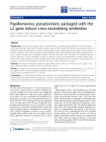
Characterization of CdTe quantum dots
The water-soluble Cdte QDs capped with negatively
charged 3-mercaptopropionic acid were prepared
according to the procedure a s reported previously. Our
TEM study illustrates that the average size of Cdte QDs
Zhang et al. Nanoscale Research Letters 2011, 6:418
/>Page 3 of 10
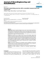
was about 4 nm, as shown in Figure 1A, and an
HRTEM indiv idual nanocrystal of Cdte QDs (Figur e 1A
a, HRTEM). The Cdte QDs in cell culture medium were
about 5 nm, as characterized with dynamic light scatter-
ing (Figure 1B). The typical fluorescence spectrum of
the Cdte QDs was shown in Figure 1C.
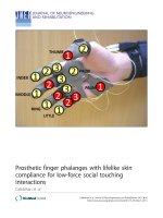
Cytotoxicity of Cdte QDs with DNR on HepG2/ADM cells
The MTT assay was carried out to explore the r elative
inhibition fo r the proliferation of the cells. The cells were
treated with different concentrations of DNR or Cdte
QDs, or treated by different concentrations of DNR com-
bined with Cdte QDs for 36 h. Since HepG2/ADM cells
are drug-resistant cell line, the high-concentration DNR
treatment only causes low growth inhibition for HepG2/
ADM cells (as shown in Figure 2). However, the growth
inhibition rate was significantly increa sed when HepG2/
ADM c ells we re treated by DNR combined with Cdte
QDs. Therefore, it is evident that the significant enhance-
ment of the cell proliferation inhibition may be facilitated
due to a synergistic effect of Cdte QDs with DNR to the
drug-resistant HepG2/ADM cells.
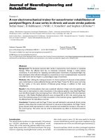
Fluorescence microscopy and electrochemical assay of
cellular drug uptake
Based on the above study, bio-imaging of DNR in
HepG2/ADM cell lines were assayed with inverted fluor-
escence microscopy. For the control cells without
Figure 1 TEM images of Cdte QDs:(A) the low magnification images Cdte QDs, (a) HRTEM image of an individual nanocrystal of Cdte QDs. (B)
Size of Cdte QDs suspended in cell culture medium was analyzed by dynamic light scattering. (C) Emission spectrum of Cdte QDs, excitation
wavelength at 330 nm.
Zhang et al. Nanoscale Research Letters 2011, 6:418
/>Page 4 of 10
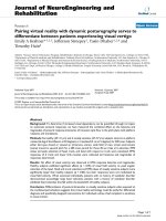
treatment, we observed almost no intracellular fluores-
cence HepG2/ADM cells (Figure 3 A a). DNR treatment
showed relatively low fluorescence in HepG2/ADM cells
(Figure 3A b). However, the intracellular fluorescence in
HepG2/ADM cells increased dramatically upon treat-
ment with DNR bound to the negatively charged surface
of QDs (Figure 3A c). To understand the mechanism of
this effect, electrochemical study was used to detect the
interaction between DNR and HepG2/ADM cells. The
results revealed that after treatment by Cdte QDs and
DNR for 2 h , the peak current of the DNR residue out-
side HepG2/ADM cells decreased more significantly
than that with DNR treatment alone, suggesting that
more significant decrease of the DNR residue outside
HepG2/ADM cells occurs with the treatment of Cdte
QDs and DNR (Figure 3B). These observations indicate
that Cdte QDs could readily facilitate the uptake of the
DNR into HepG2/ADM cells.
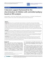
Staining and flow cytometry analysis to detect apoptosis
Using acridine orange/ethidium bromide (AO/EB) dye
mixture staining f or apoptotic cells, apoptotic nuclei
were identified by their distinctively marginated and frag-
mented appearance under the fluorescence microscope.
The a poptotic nuclei of He pG2/ADM cells (Figure 4A,
apoptosis nuclei) at 72 h co uld be identified by their dis-
tinctively marginated and fragmented appearance. For
the control cells without treatment, cells nuclei were nor-
mal as shown in (Figure 4A, control nuclei). Figure 4B
shows that Annexin-V-FITC apoptosis detection, Cdte
QDs + DNR induced a much higher HepG2/ADM cell
apoptosis rate than that of DNR, Cdte QDs, or untreated
control. We found that t he percentage of apoptotic cells
was 67.4%, 26.8%, 15.2%, 8.5% for the treatment with
Cdte QDs + DNR, Cdte QDs, DNR, untreatment, respec-
tively (Figure 4C).
DNA fragmentation assay
The DNA fragmentations were examined. When
HepG2/ADM cells were treated with Cdte QDs + DNR,
the intensity of fragmented chromosomal DNA bands
was m uch higher than that observed from cells treated
with Cdt e QDs, or DNR alo ne (Figure 5). These results
provideevidencethattheremarkableenhancementof
apoptosis was induced by synergistic effects of Cdte
QDs and DNR on HepG2/ADM cells.
Signal pathway of treatments in HepG2/ADM Cells
Treatment of human He pG2/ADM cells with Cdte QDs
+ DNR for 72 h caused decrease in the amount of P-gp
protein expression compared with control treatment
(Figure 6A). Cdte QDs + DNR treated cell monolayers
and immunostaining signals of P-gp protein were
reducedanddisrupted(Figure6B).Tofurtherunder-
stand the molecular mechanisms underlying the synergis-
tic effects of Cdte QDs + DNR-mediated apopt osis in
HepG2r/ADM cells, we investigated apoptosis-related
protein expression in the cells (Figure 6C). DNR or Cdte
QDs c annot induce apoptosis strongly in HepG2r/ADM
cells due to multidrug resistan ce. Interestingly, combined
treatment of Cdte QDs + DNR strongly caused cyto-
chrome c to be released into the cy tosol and significantly
activated caspase-9 and caspase-3 and induced de grada-
tion of its substrates, PARP. These data suggest that Cdte
QDs with DNR treatment involve the release of cyto-
chrome c from the mitochondr ia, which subsequently
causes apoptosis by activation of caspase-9, 3 in HepG2r/
ADM cells.
Tumor growth inhibition study
The nude mice were inoculated with HepG2/ADM cells
and the subsequent tumor grow th was recorded after
various treatments. From Figure 7A, the HepG2/ADM
nude mice, the tumor volume of the control group was
enlarged to almost 4970 mm
3
(Figure 7A, group 1).
Treatment with D NR or Cdte QDs alone has mild inhi-
bitory effect on the tumor growth in the HepG2/ADM
mice due to multidrug resistan ce of the HepG2/ADM
cell system (groups 2 and 3, respectively). In the group
treated with Cdte QDs + DNR (group 4), tumor growth
was significantly inhibited.
Analysis of cell apoptosis in HepG2/ADM xenograft tumors
The synergistic effect of Cdte QDs + DNR on the apop-
tosis induction in the xenograft tumors excised from
HepG2/ADM nude mice, the apoptotic rate in the con-
trol group was around 8.2% (Figure 7B). Cdte QDs +
DNR treatment causes a striking increase in the number
of TUNEL-positive nuclei , compared t o DNR or Cdte
QDs treatment alone. The result of apoptosis rate was
well correlated with the r esult of tumor growth inhibi-
tion in the studied animals.
Figure 2 MTT assay of the growth inhibition rate of HepG2/
ADM cells after various cellular treatments. The HepG2/ADM cells
were treated with 1 × 10
-6
,4×10
-6
,16×10
-6
,64×10
-6
, 12.8 × 10
-5
,
and 51.2 × 10
-5
mol/L of DNR; 1, 2.5, 5, 10, 20, and 40 μM Cdte QDs;
or 4 μM Cdte QDs with 1 × 10
-6
,4×10
-6
,16×10
-6
,64×10
-6
, 12.8 ×
10
-5
, and 51.2 × 10
-5
mol/L of DNR, respectively. *p < 0.05, indicates
the significant difference in comparison to no treatment.
Zhang et al. Nanoscale Research Letters 2011, 6:418
/>Page 5 of 10
Discussion
Clinical efficacy of many anti-cancer drugs is limited by
the develo pment of drug resistance [12]. In this study,
daunorubicin was not effective against HepG2/ADM
tumors. This is in agreement with previous studies,
which have shown that HepG2/ADM tumor cells overex-
press P-gp, and exhibit multidrug-resistant phenotype.
We demonstrated that a combination of Cdte QDs and
DNR where the DNR is bound to the Cdte QDs surface
by electrostatic interaction will improve the accumulation
of daunorubicin in tumor cells. The same or even certain
high concentration of DNR did not cause a significant
reduction in cell viability in HepG2/ADM cells. However,
when HepG2/ADM cells were treated with Cdte QDs
and DNR, we observed a remarkable enhancement of cell
growth inh ibition (Figure 2). The results suggest that the
synergistic effect of Cdte QDs with DNR can induce cell
growth inhibition of drug-resis tant HepG 2/ADM cells
in vitro.
We demonstrate that DNR taken in by cellular behavior
with synergistic effect of Cdte QDs was significantly higher
than that with only DNR treatment. Over-expression of P-
glycoprotein is the most frequent event causing multidrug
resistance. With Cdte QDs + DNR treatment, the expres-
sion of P-glycoprotein was remarkably reduced when com-
pared with the control treatment. It is already known that
nanoparticles can cause the formation of “ holes” on the
surface of cell membranes, which may increase the perme-
ability of the respective cell membranes and thus facilitate
uptake of the anti-cancer drug into cancer cells and
Figure 3 Measurement of cellular f luorescence and drug uptake.(A) Inverted fluorescence microscopy of HepG2/ADM cells; (a) control,
(b) 4 × 10
-6
mol/L DNR, and (c) 4 μM Cdte QDs + 4 × 10
-6
mol/L DNR; bar, 100 μm. (B) Differential pulse voltammetry study of DNR residue
outside HepG2/ADM cells after cell treatment for 2 h. (a) PBS; (b) 4 μM Cdte QDs + 4 × 10
-6
mol/L DNR treatment and cells for 2 h; and (c)
4 μM DNR. Pulse amplitude, 0.05 V; pulse width, 0.05 s; and pulse period, 0.2 s.
Zhang et al. Nanoscale Research Letters 2011, 6:418
/>Page 6 of 10
enhance drug accumulation in target cells [13]. This may
be the two reasons why Cdte QDs + DNR increase the
intracellular drug concent ration dramatically and thus
enhance the inhibition of the proliferation to target drug-
resistan t cancer cells. Furthermore, Cdte QDs with ne ga-
tively charged surface may combine with anti-cancer
drugs such as DNR which is positively charged through
electrostatic interaction.
Two major types of cel l death are recogniz ed: apopto-
sis and necrosis [14]. Apoptosis is a regulated process
that can be triggered by different stimuli and is
mediated by a cascade of enzymes. Necrosis is a cata-
strophic form of cell death which does not involve the
regulated action of enzymes. Studies have demonstrated
that the presence of smaller DNA fragments are
believed to reflect the release of nucleosomes from
apoptotic cells and higher molecu lar weight DNA mole-
cules are believed to reflect release from necrotic cells
[15]. Apoptosis results in fragmentation of cells into
apoptotic bodies which are engulfed by neighboring
cells and macrophages [16]. However, uptake of necrotic
cells has been reported to be less efficient than phagocy-
tosis of apoptotic cells. So active anti-cancer drugs
induce apoptosis in malignant cells should be a m ain
way to clinical anti-tumor. Interestingly, we found that
Cdte QDs + DNR can induce drug-re sistant HepG2/
ADM cell apoptosis rate significantly higher than that of
Cdte QDs, or DNR alone treatment in vitro.Moreover,
we analyzed the cells apoptosis morphology from var-
ious assay, nuclei staining. When cells were treated with
Cdte QDs + DNR, they exhibited characteristic morpho-
logical features of apoptosis, such as chromosomal
Figure 4 Assay of cell apoptosis rate and morphological images:(A) Detectio n of apoptotic and normal cells by acridine orange staining.
Control cell nuclei, apoptotic nuclei from HepG2/ADM cells ware observed. (B) HepG2/ADM cells detected by flow cytometry using Annexin-V-
FITC method. (a) control treatment; (b) 4 × 10
-6
mol/L DNR treatment; (c) 4 μM Cdte QDs treatment; and (d) 4 μM Cdte QDs + 4 × 10
-6
mol/L
DNR for 36 h. (C) Quantitative analysis of apoptotic cells after various treatments shown in (B). *p < 0.05, compared to the control treatment.
Figure 5 DNA fragmentation in HepG2/ADM cells after
different treatments. Genomic DNA was isolated from HepG2/
ADM cells. DNA ladders were visualized under UV light with
ethidium bromide staining. HepG2/ADM cells treated with: control
treatment; 4 × 10
-6
mol/L DNR; 4 μM Cdte QDs; and 4 μM Cdte
QDs+4×10
-6
mol/L DNR for 72 h.
Zhang et al. Nanoscale Research Letters 2011, 6:418
/>Page 7 of 10
Figure 6 Signal pathway analysis.(A) Western blotting analysis of P-gp in HepG2/ADM cells. HepG2/ADM cells wit hout treatment were used
as control (lane 1). Lysates were prepared from the cells treated 4 μM Cdte QDs with 4 × 10
-6
mol/L DNR (lane 2). (B) The control cells without
any treatment (1). The images were taken from cells treated with 4 μM Cdte QDs with 4 × 10
-6
mol/L DNR for 72 h (2). Bar, 20 μm. (C) Western
blotting analysis of cytochrome c released in HepG2/ADM cells: group 1, control group (lane 1); group 2, 4 × 10
-6
mol/L DNR (lane 2); group 3, 4
μM Cdte QDs (lane 3); and group 4, 4 μM Cdte QDs with 4 × 10
-6
mol/L DNR (lane 4). The following antibodies were used: anti-cleaved caspase-
9, anti-cleaved caspase-3, and anti-PARP antibody. GAPDH was served as a loading control.
Figure 7 Inh ibit ion of tu mor gro wth in Hep G2/A DM nude mice with different treatments. (A) The different treatment eff ects on the
tumor growth inhibition in nude mice inoculated with HepG2/ADM cells: group 1, no treatment, served as a control group; group 2, 4 × 10
-6
mol/kg DNR; group 3, 4 μmol/kg Cdte QDs; and group 4, 4 μmol/kg Cdte QDs with 4 × 10
-6
mol/kg DNR. (B) Quantitative analysis of apoptotic
cells using TUNEL staining after various treatments. HepG2/ADM xenograft tumors treated as follows: group 1, control group; group 2, 4 × 10
-6
mol/kg DNR; group 3, 4 μmol/kg Cdte QDs; and group 4, 4 μmol/kg Cdte QDs with 4 × 10
-6
mol/kg DNR.
Zhang et al. Nanoscale Research Letters 2011, 6:418
/>Page 8 of 10
condensation and DNA fragment. With flow cytometry
assay, we analyzed quantitativ e apoptotic cells after var-
ious treatments, the Cdte QDs + DNR could be used as
inducing HepG2/ADM cells apoptosis with relatively
low concentration.
Apoptosis is a regulated process that can be triggered by
different stimuli and is mediated by a cascade of enzymes
[17]. The realization of mechanisms will enable optimiza-
tion of chemotherapy for the treatment of cancer [18]. To
further understand the molecular mechanisms underlying
the Cdte QDs + DNR t reatment-mediated apoptosis in
HepG2/ADM cells, we investigated apoptosis-related pro-
tein expression in HepG2/ADM cells. Cdte QDs + DNR
treatment induces cytochrome c release, causing caspase-9
activation. Cleaved caspase-9 activated caspase-3 that cor-
related w ith t he increased expression of cleaved PARP
after relevant treatments [19,20]. Subsequently, DNA frag-
mentation is induced during the cells apoptosis by cleaved
PARP expression. Compared to Cdte QDs or DNR treat-
ment, Cdte QDs + DNR treatment showed much stronger
inducing apoptosis effect.
As the above results il lustrated, we recognized the evi -
dence of apoptosis of HepG2/ADM cells in vitro.Itispos-
siblethatCdteQDs+DNRcouldplayacriticalrolein
inducing apoptosis in vivo. The tumor growth in group 4
nude mice (treated with Cdte QDs + DNR) was sup-
pressed most efficiently. Cdte QDs or DNR alone cannot
significantly inhibit the tumor growth in HepG2/ADM
mice due to multidrug resistance of this cell line. Our pre-
sent study also shows apoptosis in tumor cells was
induced by three kinds of t reatment with TUNEL assay.
TheresultsoftheTUNELassayareconsistentwiththe
tumor growth inhibition results. Our observations indicate
that the growth-inhibitory effect of Cdte QDs + DNR
treatment is related to its ability to induce apoptosis, as
evidenced by TUNEL assay. Taken together, our data sup-
port the thesis that Cdte QDs + DNR treatment plays an
important role in inducing dr ug-resistant HepG2/ADM
cell apoptosis and tumor suppression, and furthermore
suggest that Cdte QDs + DNR treatment therapy might
provide a powerful treatment for liver cancer.
Conclusion
In summary, in this study, we have investigated the inter-
action mechanism and synergistic effect of 3-mercapto-
propionic acid-capped Cdte QDs with the anti-cancer
drug DNR on the induction of apoptosis of drug-resistant
human hepatoma HepG2/ADM cells. Our observations
demo nstrate that Cdte QDs readily facilitated the uptake
of the D NR into HepG2/ADM cells by electrochemical
assay. Apoptotic staining, DNA fragmentation, and flow
cytometry analysis further demonstrate that treatment of
Cdte QDs together with DNR can clearly activate apopto-
sis in HepG2/ADM cells. Cdte QDs + DNR treatment
activated caspases protein expression. W hile the Cdte
QDs + DNR treatment could reduce the effect of P-
glycoprotein (P-gp). Moreover, our in vivo study indicates
that the treatment of Cdte QDs together with DNR effec-
tively inhibited the human hepatoma HepG2/ADM nude
mice tumor growth. The increased cell apoptosis rate
was closely correlated with the enhanced inhibition of
tumor growth in the studied animals. Thus, Cdte QDs
combined with DNR m ay serve as a new eff ective addi-
tive agent to overcome the drug resistance and thus as a
novel s trateg y to sensitive ly tr ack the respec tive cancer
cells for efficient cancer chemotherapy.
Acknowledgements
This work was supported by the National Basic Research Program of China
(no. 2010CB732404), National Natural Science Foundation of China
(90713023), National High Technology Research and Development Program
of China (2007AA022007), Doctoral Fund of Ministry of Education of China
(20090092110028), and the Natural Science Foundation of Jiangsu Province
(BK2008149) to XMW. LS and MS acknowledge support by the NSF-CREST
program.
Author details
1
State Key Lab of Bioelectronics (Chien-Shiung Wu Lab), Department of
Biological Science and Medical Engineering Southeast University, Nanjing,
210096, PR China
2
Department of Chemistry and Biochemistry, California
State University, Los Angeles, CA 90032, USA
Authors’ contributions
Respond: GZ carried out the cell biology and molecular studies. LS prepared
the Cdte QDs. MS participated in the design of the study. XW conceived of
the study, and participated in its design and coordination. All authors read
and approved the final manuscript.
Competing interests
The authors declare that they have no competing interests.
Received: 14 March 2011 Accepted: 13 June 2011
Published: 13 June 2011
References
1. Xu D, Lu QH, Hu X: Down-regulation of P-glycoprotein expression in
MDR breast cancer cell MCF-7/ADR by honokiol. Cancer Lett 2006,
243:274-280.
2. Cree IA: Chemosensitivity and chemoresistance testing in ovarian cancer.
Curr Opin Obstet Gynecol 2009, 21:39-43.
3. Efferth T, Volm M: Modulation of P-glycoprotein-mediated multidrug
resistance by monoclonal antibodies, immunotoxins or antisense
oligodeoxynucleotides in kidney carcinoma and normal kidney. Oncology
1993, 50:303-308.
4. Lim YT, Kim S, Nakayama A, Stott NE, Bawendi MG, Frangioni JV: Selection
of quantum dot wavelengths for biomedical assays and imaging. Mol
Imaging 2003, 2:50-64.
5. Zhou YY, Shi LX, Li QN, Jiang H, Lv G, Zhao J, Wu CH, Selke M, Wang XM:
Imaging and inhibition of multi-drug resistance in cancer cells via
specific association with negatively charged CdTe quantum dots.
Biomaterials 2010, 31:4958-4963.
6. Li WH, Lei P, Yu B, Wu S, Peng J, Zhao XP, Zhu HF, Kirschfink M, Shen G:
Screening and identification of a novel target specific for hepatoma cell
line HepG2 from the FliTrx bacterial peptide library. Acta Biochim Biophys
Sin (Shanghai) 2008, 40:443-451.
7. Felix CA: Leukemias related to treatment with DNA topoisomerase II
inhibitors. Med Pediatr Oncol 2001, 36:525-535.
8. Christakos S, Liu Y: Biological actions and mechanism of action of
calbindin in the process of apoptosis. J Steroid Biochem Mol Biol 2004, 89-
90(1-5):401-404.
Zhang et al. Nanoscale Research Letters 2011, 6:418
/>Page 9 of 10
9. Hu W, Kavanagh JJ: Anticancer therapy targeting the apoptotic pathway.
Lancet Oncol 2003, 4:721-729.
10. Gaponik N, Talapin DV, Rogach AL, Hoppe K, Shevchenko EV, Kornowski A,
Eychmüller A, Weller H: Thiol-capping of CdTe nanocrystals: An
alternative to organometallic synthetic routes. Journal of Physical
Chemistry B 2002, 106:7177-7185.
11. Zheng LH, Bao YL, Wu Y, Yu CL, Meng XY, Li YX: Cantharidin reverses
multidrug resistance of human hepatoma HepG2/ADM cells via down-
regulation of P-glycoprotein expression. Cancer Letters 2008, 272:102-109.
12. Kim DH, Vaccaro AR, Henderson FC, Benzel EC: Molecular biology of
cervical myelopathy and spinal cord injury: role of oligodendrocyte
apoptosis. Spine J 2003, 3:510-519.
13. Guo DD, Wu CH, Li XM, Jiang H, Wang XM, Chen BA: In vitro cellular
uptake and cytotoxic effect of functionalized nickel nanoparticles on
leukemia cancer cells. J Nanosci Nanotechnol 2008, 8:2301-2307.
14. Raffray M, Cohen GM: Apoptosis and necrosis in toxicology: a continuum
or distinct modes of cell death? Pharmacol Ther 1997, 75:153-177.
15. Linder S, Havelka AM, Ueno T, Shoshan MC: Determining tumor apoptosis
and necrosis in patient serum using cytokeratin 18 as a biomarker.
Cancer Lett 2004, 214:1-9.
16. Ojcius DM, Souque P, Perfettini JL, Varsat AD: Apoptosis of epithelial cells
and macrophages due to infection with the obligate intracellular
pathogen Chlamydia psittaci. J Immunol 1998, 161:4220-4226.
17. Katunuma N, Murata E, Le QT, Hayashi Y, Ohashi A: New apoptosis cascade
mediated by lysosomal enzyme and its protection by epigallo-catechin
gallate. Adv Enzyme Regul 2004, 44:1-10.
18. Li JY, Chen C, Wang XM, Gu ZZ, Chen BA: Novel Strategy to Fabricate
PLA/Au Nanocomposites as an Efficient Drug Carrier for Human
Leukemia Cells in Vitro. Nanoscale Res Lett 2011.
19. Nabeshi H, Yoshikawa T, Arimori A, Yoshida T, Tochigi S, Hirai T, Akase T,
Nagano K, Abe Y, Kamada H, Tsunoda S, Itoh N, Yoshioka Y, Tsutsumi Y:
Effect of surface properties of silica nanoparticles on their cytotoxicity
and cellular distribution in murine macrophages. Nanoscale Res Lett 2011.
20. Das J, Ghosh J, Manna P, Sil PC: Taurine suppresses doxorubicin-triggered
oxidative stress and cardiac apoptosis in rat via up-regulation of PI3-K/
Akt and inhibition of p53, p38-JNK. Biochem Pharmacol 2011, 81:891-909.
doi:10.1186/1556-276X-6-418
Cite this article as: Zhang et al.: CdTe quantum dots with daunorubicin
induce apoptosis of multidrug-resistant human hepatoma HepG2/ADM
cells: in vitro and in vivo evaluation. Nanoscale Research Letters 2011
6:418.
Submit your manuscript to a
journal and benefi t from:
7 Convenient online submission
7 Rigorous peer review
7 Immediate publication on acceptance
7 Open access: articles freely available online
7 High visibility within the fi eld
7 Retaining the copyright to your article
Submit your next manuscript at 7 springeropen.com
Zhang et al. Nanoscale Research Letters 2011, 6:418
/>Page 10 of 10